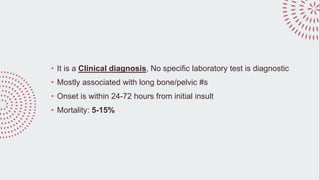
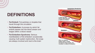
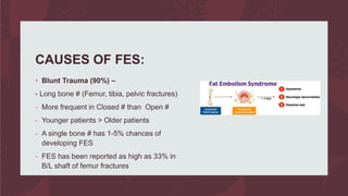
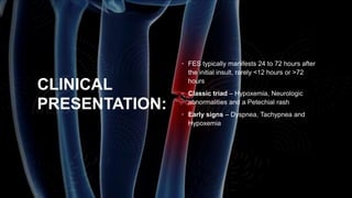
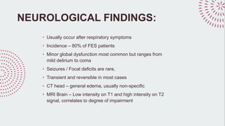
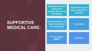
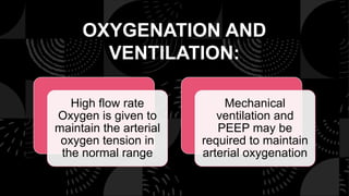
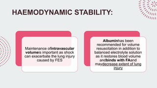
FAT EMBOLISM
BY,
DR VARUN KUMAR A
HISTORY
Zenker, a Pathologist, first identified Fat
embolism syndrome at an autopsy in 1862
1862
First diagnosed in 1873 byDr Von Bergmann
1873
FengerandSalisburypublished description of
FES in 1879
1879
• It is a Clinical diagnosis, No specific laboratory test is diagnostic
• Mostly associated with long bone/pelvic #s
• Onset is within 24-72 hours from initial insult
• Mortality: 5-15%
DEFINITIONS
• Fat Emboli: Fat particles or droplets that
travel through the circulation
• Fat Embolism: A process by which fat
emboli passes into the blood stream and
lodges within a blood vessel
• Fat Embolism Syndrome: Serious
manifestation of fat embolism occasionally
causing multi system dysfunction, the lungs
are always involved and next is the brain
CAUSES OF FES:
• Blunt Trauma (90%) –
- Long bone # (Femur, tibia, pelvic fractures)
- More frequent in Closed # than Open #
- Younger patients > Older patients
- A single bone # has 1-5% chances of
developing FES
- FES has been reported as high as 33% in
B/L shaft of femur fractures
CAUSES OF FES:
• Non-Trauma: Agglutination of chylomicrons and VLDL by high levels of
plasma CRP
- Disease related: Diabetes, acute pancreatitis, burns, SLE, sickle cell crisis
- Drug related: Parenteral lipid infusion
- Procedure related: Orthopaedic surgeries, liposuction
RISK FACTORS:
PATHOGENESIS:
Mechanical Theory:
Physical obstruction
of the pulmonary and
systemic vasculature
with embolized fat
Temporary rise in I/M
pressure - forces
marrow into injured
venous sinusoids
Cor pulmonale –
Inadequate
compensatory
pulmonary
vasodilatation
Microvascular
lodging – local
ischemia and
inflammation
Release of
inflammatory
mediators, platelet
aggregation and
vasoactive amines
Biochemical Theory:
Circulating FFAs – Directly
toxic to pneumocytes /
capillary endothelium in the
lung causing
interstitialhaemorrhage,
edema and chemical
pneumonitis
Coexisting shock,
hypovolemia and sepsis –
reduce liver flow, exacerbate
the toxic effects of FFAs
H/E STAIN OF
LUNG:
• Blood vessel with fibrinoid material
• Optical empty space - Lipid dissolved
during the staining process
CLINICAL
PRESENTATION:
• FES typically manifests 24 to 72 hours after
the initial insult, rarely <12 hours or >72
hours
• Classic triad – Hypoxemia, Neurologic
abnormalities and a Petechial rash
• Early signs – Dyspnea, Tachypnea and
Hypoxemia
PULMONARY:
• Hypoxia, rales, pleural friction rub
• ARDS may develop
• 50% of patients with FES require mechanical ventilation
• CXR is usually normal early on, later may show ‘snowstorm’ pattern – diffuse
bilateral infiltrates
• CT chest – Ground glass opacification with interlobular septal thickening
NEUROLOGICAL FINDINGS:
• Usually occur after respiratory symptoms
• Incidence – 80% of FES patients
• Minor global dysfunction most common but ranges from
mild delirium to coma
• Seizures / Focal deficits are rare,
• Transient and reversible in most cases
• CT head – general edema, usually non-specific
• MRI Brain – Low intensity on T1 and high intensity on T2
signal, correlates to degree of impairment
MRI:
• Foci of ischemia S/O FES
• Post-op day 2 showing multiple hyperintense areas consistent
with multiple emboli
• Post-op day 14 shows evolving cortical infarctions
DERMATOLOGICAL
FINDINGS:
• Petechial
• Usually on conjunctiva, neck, axillae
• Results from occlusion of dermal
capillaries by fat globules and then
extravasations of RBC
• Resolves in 5-7 days
• Pathognomonic but only present in 20-
50% of patients
OTHER FINDINGS:
• Retinopathy (exudates, cotton wool spots, haemorrhage)
• Lipiduria
• Fever
• DIC
• Myocardial depression
• Thrombocytopenia/Anemia
• Hypocalcemia
DIAGNOSIS:
FES is a CLINICAL diagnpsis, not
biochemical
High degree of suspicion is needed to
make diagnosis
In 50% of fracture patients, fat globules
was demonstrated in the serum, without
other symptoms of FES
Growing literature on the use of
bronchoscopy with bronchoalveolar
lavage to detect fat drplets in alveolar
macrophages as a means to diagnose
fat embolism (sensitivity and specifity
are unknown, being studied in Trauma
patients)
GURD’S CRITERIA:
LINDEQUE’S
CRITERIA:
FES = Femur fracture +/- tibia fracture + 1
feature
Based on respiratory parameters
SCHONFELD:
FAT
EMBOLISM
INDEX
FES = 5 or more points
SEVITT’S CLASSIFICATION:
SUBCLINICAL
FES
NON-
FULMINANT
FES
FULMINANT
FES
SUB CLINICAL
FES:
Around 3 days post trauma
Probably occurs in almost all long
bone fractures of the lowrer
extremity and fractures of the pelvis
Characterised by decreased PaO2,
decreased Hb% and decreased
platelets.
No clinical signs and symptoms of
respiratory insufficiency
NON-FULMINANT
FES
Any time upto 6 days post trauma
Clinical signs and symptoms are clearly
evident
Petechiae, tachycardia, respiratory
failure and signs of CNS embolism
Thrombocytopenia, anaemia and
coagulation abnormalities can be found
Pulmonary alveolar and interstitial
opacities on CXR
FULMINANT FES:
Occurs very suddenly and rapidly
after injury and progresses
quickly, often resulting in death
within few hours of initial trauma
Clinical features are acute
respiratory failure, acute cor-
pulmonale and embolic
neurological changes
Patients with multiple fractures are
particularly susceptible to this
form of FES, relatively rare but
significant because of its mortality
TREATMENT:
• Prophylaxis:
- Immobilisation and early internal fixation of fracture
- Fixation within 24 hours has been shown to yield 5 fold reduction in the
incidence of ARDS
- Continuous saturation monitoring in high-risk patients may help in detecting
desaturation early, allowing early institution of O2 and possible steroid
therapy
SUPPORTIVE
MEDICAL CARE:
Maintenance of
adequate
oxygenation and
ventilation
Maintenance of
haemodynamic
stability
Administration of
blood products
as indicated
Hydration
Prophylaxis
ofDVT
Nutrition
OXYGENATION AND
VENTILATION:
High flow rate
Oxygen is given to
maintain the arterial
oxygen tension in
the normal range
Mechanical
ventilation and
PEEP may be
required to maintain
arterial oxygenation
HAEMODYNAMIC STABILITY:
Maintenance ofintravascular
volumeis important as shock
can exacerbate the lung injury
caused by FES
Albuminhas been
recommended for volume
resuscitation in addition to
balanced electrolyte solution
as it restores blood volume
andbinds with FAand
maydecrease extent of lung
injury
STEROIDS:
• Steroid prophylaxis is controversial to prevent FES
• Blunting of inflammatory response and complement activation
• Prospective studies suggest prophylactic steroids benefit high
risk patients
• Once FES is established, steroids have not shown improved
outcomes
HEPARIN:
• Like steroids, controversial role in management
• Stimulates lipase to reduce lipemia
• Reverses the DIC picture
• S/E – increase in circulating FFAs, bleeding risks
• Routine use is not recommended
PROGNOSIS:
• Most deaths are contributed to pulmonary dysfunction
• Hard to determine exact mortality rate
• Difficult to predict duration of FES – As it is frequently
subclinical or overshadowed by other illness or injuries
• Increased alveolar-to-arterial oxygen gradient and neurologic
deficits, including coma may last for few days to weeks
SEQUELAE:
As in ARDS, pulmonary
sequelae usually
resolve almost
completely within 1 year
Residual subclinical
diffusion capacity
deficits may exist
Resisual neurologic
deficits may range from
non-existent to subtle
personality changes,
memory and cognitive
dysfunction
SUMMARY:
Clinical diagnosis, so high index of
suspicion
Most effective management is prevention
with rigid fixation of fractures within 24
hours
When developed management is
supportive
THANK YOU

FAT EMBOLISM.pptx

  • 1.
  • 2.
    HISTORY Zenker, a Pathologist,first identified Fat embolism syndrome at an autopsy in 1862 1862 First diagnosed in 1873 byDr Von Bergmann 1873 FengerandSalisburypublished description of FES in 1879 1879
  • 3.
    • It isa Clinical diagnosis, No specific laboratory test is diagnostic • Mostly associated with long bone/pelvic #s • Onset is within 24-72 hours from initial insult • Mortality: 5-15%
  • 4.
    DEFINITIONS • Fat Emboli:Fat particles or droplets that travel through the circulation • Fat Embolism: A process by which fat emboli passes into the blood stream and lodges within a blood vessel • Fat Embolism Syndrome: Serious manifestation of fat embolism occasionally causing multi system dysfunction, the lungs are always involved and next is the brain
  • 5.
    CAUSES OF FES: •Blunt Trauma (90%) – - Long bone # (Femur, tibia, pelvic fractures) - More frequent in Closed # than Open # - Younger patients > Older patients - A single bone # has 1-5% chances of developing FES - FES has been reported as high as 33% in B/L shaft of femur fractures
  • 6.
    CAUSES OF FES: •Non-Trauma: Agglutination of chylomicrons and VLDL by high levels of plasma CRP - Disease related: Diabetes, acute pancreatitis, burns, SLE, sickle cell crisis - Drug related: Parenteral lipid infusion - Procedure related: Orthopaedic surgeries, liposuction
  • 7.
  • 8.
    PATHOGENESIS: Mechanical Theory: Physical obstruction ofthe pulmonary and systemic vasculature with embolized fat Temporary rise in I/M pressure - forces marrow into injured venous sinusoids Cor pulmonale – Inadequate compensatory pulmonary vasodilatation Microvascular lodging – local ischemia and inflammation Release of inflammatory mediators, platelet aggregation and vasoactive amines
  • 9.
    Biochemical Theory: Circulating FFAs– Directly toxic to pneumocytes / capillary endothelium in the lung causing interstitialhaemorrhage, edema and chemical pneumonitis Coexisting shock, hypovolemia and sepsis – reduce liver flow, exacerbate the toxic effects of FFAs
  • 10.
    H/E STAIN OF LUNG: •Blood vessel with fibrinoid material • Optical empty space - Lipid dissolved during the staining process
  • 11.
    CLINICAL PRESENTATION: • FES typicallymanifests 24 to 72 hours after the initial insult, rarely <12 hours or >72 hours • Classic triad – Hypoxemia, Neurologic abnormalities and a Petechial rash • Early signs – Dyspnea, Tachypnea and Hypoxemia
  • 12.
    PULMONARY: • Hypoxia, rales,pleural friction rub • ARDS may develop • 50% of patients with FES require mechanical ventilation • CXR is usually normal early on, later may show ‘snowstorm’ pattern – diffuse bilateral infiltrates • CT chest – Ground glass opacification with interlobular septal thickening
  • 13.
    NEUROLOGICAL FINDINGS: • Usuallyoccur after respiratory symptoms • Incidence – 80% of FES patients • Minor global dysfunction most common but ranges from mild delirium to coma • Seizures / Focal deficits are rare, • Transient and reversible in most cases • CT head – general edema, usually non-specific • MRI Brain – Low intensity on T1 and high intensity on T2 signal, correlates to degree of impairment
  • 14.
    MRI: • Foci ofischemia S/O FES • Post-op day 2 showing multiple hyperintense areas consistent with multiple emboli • Post-op day 14 shows evolving cortical infarctions
  • 15.
    DERMATOLOGICAL FINDINGS: • Petechial • Usuallyon conjunctiva, neck, axillae • Results from occlusion of dermal capillaries by fat globules and then extravasations of RBC • Resolves in 5-7 days • Pathognomonic but only present in 20- 50% of patients
  • 16.
    OTHER FINDINGS: • Retinopathy(exudates, cotton wool spots, haemorrhage) • Lipiduria • Fever • DIC • Myocardial depression • Thrombocytopenia/Anemia • Hypocalcemia
  • 17.
    DIAGNOSIS: FES is aCLINICAL diagnpsis, not biochemical High degree of suspicion is needed to make diagnosis In 50% of fracture patients, fat globules was demonstrated in the serum, without other symptoms of FES Growing literature on the use of bronchoscopy with bronchoalveolar lavage to detect fat drplets in alveolar macrophages as a means to diagnose fat embolism (sensitivity and specifity are unknown, being studied in Trauma patients)
  • 18.
  • 19.
    LINDEQUE’S CRITERIA: FES = Femurfracture +/- tibia fracture + 1 feature Based on respiratory parameters
  • 20.
  • 21.
  • 22.
    SUB CLINICAL FES: Around 3days post trauma Probably occurs in almost all long bone fractures of the lowrer extremity and fractures of the pelvis Characterised by decreased PaO2, decreased Hb% and decreased platelets. No clinical signs and symptoms of respiratory insufficiency
  • 23.
    NON-FULMINANT FES Any time upto6 days post trauma Clinical signs and symptoms are clearly evident Petechiae, tachycardia, respiratory failure and signs of CNS embolism Thrombocytopenia, anaemia and coagulation abnormalities can be found Pulmonary alveolar and interstitial opacities on CXR
  • 24.
    FULMINANT FES: Occurs verysuddenly and rapidly after injury and progresses quickly, often resulting in death within few hours of initial trauma Clinical features are acute respiratory failure, acute cor- pulmonale and embolic neurological changes Patients with multiple fractures are particularly susceptible to this form of FES, relatively rare but significant because of its mortality
  • 25.
    TREATMENT: • Prophylaxis: - Immobilisationand early internal fixation of fracture - Fixation within 24 hours has been shown to yield 5 fold reduction in the incidence of ARDS - Continuous saturation monitoring in high-risk patients may help in detecting desaturation early, allowing early institution of O2 and possible steroid therapy
  • 26.
    SUPPORTIVE MEDICAL CARE: Maintenance of adequate oxygenationand ventilation Maintenance of haemodynamic stability Administration of blood products as indicated Hydration Prophylaxis ofDVT Nutrition
  • 27.
    OXYGENATION AND VENTILATION: High flowrate Oxygen is given to maintain the arterial oxygen tension in the normal range Mechanical ventilation and PEEP may be required to maintain arterial oxygenation
  • 28.
    HAEMODYNAMIC STABILITY: Maintenance ofintravascular volumeisimportant as shock can exacerbate the lung injury caused by FES Albuminhas been recommended for volume resuscitation in addition to balanced electrolyte solution as it restores blood volume andbinds with FAand maydecrease extent of lung injury
  • 29.
    STEROIDS: • Steroid prophylaxisis controversial to prevent FES • Blunting of inflammatory response and complement activation • Prospective studies suggest prophylactic steroids benefit high risk patients • Once FES is established, steroids have not shown improved outcomes
  • 30.
    HEPARIN: • Like steroids,controversial role in management • Stimulates lipase to reduce lipemia • Reverses the DIC picture • S/E – increase in circulating FFAs, bleeding risks • Routine use is not recommended
  • 31.
    PROGNOSIS: • Most deathsare contributed to pulmonary dysfunction • Hard to determine exact mortality rate • Difficult to predict duration of FES – As it is frequently subclinical or overshadowed by other illness or injuries • Increased alveolar-to-arterial oxygen gradient and neurologic deficits, including coma may last for few days to weeks
  • 32.
    SEQUELAE: As in ARDS,pulmonary sequelae usually resolve almost completely within 1 year Residual subclinical diffusion capacity deficits may exist Resisual neurologic deficits may range from non-existent to subtle personality changes, memory and cognitive dysfunction
  • 33.
    SUMMARY: Clinical diagnosis, sohigh index of suspicion Most effective management is prevention with rigid fixation of fractures within 24 hours When developed management is supportive
  • 34.