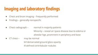
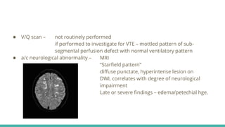
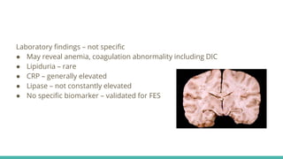
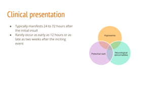
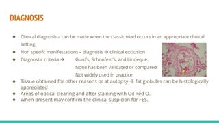
Fat embolism syndrome is a rare clinical syndrome caused by the presence of fat globules in the pulmonary circulation following trauma or other insults that release fat into the bloodstream. It is characterized by a clinical triad of hypoxemia, neurological abnormalities, and petechial rash that typically develops 24-72 hours after the initial injury. Diagnosis is clinical based on criteria sets, and treatment is supportive to address respiratory failure, shock, or other complications until it resolves spontaneously in days. Prevention focuses on early immobilization of fractures to reduce fat release and potential prophylactic corticosteroids, though their benefit is controversial.
This document discusses adrenal incidentalomas, which are adrenal masses discovered incidentally through imaging for unrelated conditions. It covers the relevant anatomy and physiology, defines incidentalomas and classifications, and describes the evaluation, management, and surgical treatment options. Functional adrenal tumors like Cushing's syndrome, Conn's syndrome, pheochromocytoma, and adrenocortical carcinoma are also discussed. Laboratory and imaging tests used in evaluation are outlined, along with signs, symptoms and screening tests for the functional tumors. Surgical resection is the treatment of choice for most cases.
Adrenal Gland Tumours and their ManagementFaisal Zia
The document discusses adrenal gland tumors and their management. It begins with the anatomy and physiology of the adrenal glands and outlines the classification of adrenal tumors. It then discusses specific tumor types like adrenocortical adenoma, adrenocortical carcinoma, pheochromocytoma, neuroblastoma, and ganglioneuroma. For each tumor, it covers clinical features, diagnosis, and management strategies including surgery, medication, and follow up. Surgical resection is the primary treatment for most benign functioning and non-functioning tumors, while malignant tumors may also require chemotherapy or radiation.
The document discusses the anatomy, physiology, and pathologies of the pituitary gland. It begins by describing the normal anatomy of the pituitary, including its location and that it is divided into anterior and posterior lobes. It then discusses the hormones produced by the anterior pituitary and various pituitary tumors and masses that can occur, such as prolactinomas, Cushing's disease, and acromegaly. It provides details on evaluating pituitary tumors, treatment options which include surgery, medication, and radiation, and management of specific tumor types.
MESENTERIC ISCHEMIA- GENERALISED ABDOMINAL PAIN
#surgicaleducator #epigastricabdominalpain #pepticulcerdisease #usmle #babysurgeon #surgicaltutor
• Dear Viewers,
• Greetings from “Surgical Educator”
• Today I have uploaded a video on Mesenteric Ischemia- a didactic lecture.
• It is one of the uncommon but life-threatening surgical problems you see in surgical wards.
• I have discussed the various causes for Generalised Abdominal Pain, epidemiology, etiology, pathology, clinical features, investigations, and treatment of Mesenteric Ischemia.
• I have also included a mind map and a treatment algorithm for Mesenteric Ischemia.
• I hope the video will be very useful and you will enjoy it.
• You can watch all my surgical teaching videos in the following link:
• youtube.com/c/surgicaleducator
• Thank you for watching the video.
The adrenal glands are paired endocrine organs located above the kidneys that are comprised of an outer cortex and inner medulla. The cortex secretes mineralocorticoids, glucocorticoids, and sex hormones. The medulla secretes catecholamines. Common non-neoplastic diseases include Cushing's syndrome, hyperaldosteronism, and adrenal insufficiency. Adrenal cortical tumors include adenomas, which can be functional or non-functional, and rare carcinomas. Pheochromocytomas arise from the medulla and secrete catecholamines, causing hypertension. Neuroblastomas are rare tumors seen in young children that often metastasize.
This document discusses Cushing's syndrome, which results from prolonged exposure to excess glucocorticoids. It can be caused by pituitary or adrenal tumors that overproduce cortisol or ACTH. The main clinical manifestations are central obesity, rounded face, fragile skin that bruises easily, and muscle weakness. Laboratory tests used for diagnosis include midnight cortisol levels, dexamethasone suppression tests, and measurements of ACTH. Treatment involves surgically removing the tumor or adrenal glands. Irradiation may also be used if the tumor cannot be fully removed. Lifelong steroid replacement is needed after adrenal gland removal.
This document discusses adrenal anatomy, physiology, and diseases. It covers adrenal tumors including adenomas, adrenocortical carcinoma (ACC), and primary adrenal lymphoma. ACC is typically larger than 5 cm, irregular, and shows necrosis/calcification. Diagnosis involves imaging like CT/MRI and biochemical testing. Treatment of ACC involves surgery with the goal of a complete resection followed by mitotane therapy. Prognosis depends on stage, resection margin, and markers like estrogen receptor status. Primary adrenal lymphoma is rare but has a better prognosis than ACC. Radiotherapy may be used for local recurrence. Ongoing research evaluates new adjuvant therapies.
This document provides information on the adrenal gland, including its embryology, anatomy, physiology, and various disorders. It discusses topics such as adrenal cortex and medulla disorders, adrenal tumors, adrenal insufficiency, and adrenal surgery. Specific conditions covered include Cushing's syndrome, pheochromocytoma, congenital adrenal hyperplasia, hyperaldosteronism, adrenocortical carcinoma, and adrenal incidentaloma. Diagnostic tests and treatment options are provided for each condition. Adrenal surgery techniques such as open and laparoscopic approaches are also summarized.
Adrenal Gland and its Disorders with surgical management.Manish Shetty
The document discusses adrenal gland tumours including adrenal cortical tumours, phaeochromocytoma, neuroblastoma, and Cushing's syndrome. Key points are:
1) Adrenal cortical tumours can be functioning or non-functioning adenomas. Incidentalomas are adrenal tumours found incidentally on imaging and require hormone evaluation.
2) Phaeochromocytomas arise from chromaffin cells and secrete catecholamines. They present with severe headaches and symptoms of sympathetic overactivity.
3) Neuroblastoma is the most common abdominal tumour in children that arises from adrenal medulla. It has different risk groups and stages that determine prognosis and treatment.
4
The document provides objectives and an overview of Cushing's syndrome, including its aetiology, pathophysiology, clinical manifestations, diagnosis, and nursing management. It aims to equip nursing students with knowledge of Cushing's syndrome and the management of a child presenting with the disorder through increased cortisol levels. A case study is presented to demonstrate nursing assessments and diagnoses for a teenage girl diagnosed with Cushing's syndrome.
This document discusses pituitary adenomas, benign tumors of the pituitary gland. It covers the anatomy and hormones of the pituitary gland. Pituitary adenomas can be secreting or non-secreting, and cause various clinical effects depending on the hormone secreted including visual field defects. Diagnosis involves endocrine and imaging evaluations. Treatment depends on the size and type of tumor, and may include medication, surgery, and radiation therapy. Long term monitoring of patients is important after treatment.
This document discusses the anatomy, physiology, and pathologies of the adrenal glands. It begins with an overview of adrenal gland structure and location. It then discusses the arterial supply, venous drainage, nerve supply, and lymphatic drainage. Histology and embryology are also covered. The document focuses on specific adrenal disorders including primary aldosteronism, Cushing's syndrome, adrenal tumors, adrenal metastases, and adrenocortical carcinoma. For each condition, the document discusses signs and symptoms, pathophysiology, diagnosis, subtypes if relevant, and treatment approaches.
This document provides information on transsphenoidal hypophysectomy for pituitary tumors. It discusses the classification, clinical features, indications for surgery, surgical approaches and techniques for transsphenoidal hypophysectomy. The transnasal, transseptal and transethmoidal routes are described. Preoperative workup and preparation are also outlined.
Splenic trauma - Causes, Complications, ManagementVikas V
The document discusses splenic trauma, including anatomy, mechanisms of injury, signs and symptoms, diagnostic modalities, grading systems, and treatment approaches. It notes that non-operative management is the preferred treatment for hemodynamically stable patients, regardless of injury grade. Operative management may be required for hemodynamic instability, failure of non-operative management, or high-grade injuries involving major vessels. Splenectomy is performed as a last resort, and vaccination and antibiotic prophylaxis are important after splenectomy to prevent infection.
The document discusses surgical diseases of the adrenal gland. It covers the anatomy and physiology of the adrenal glands, as well as conditions like pheochromocytoma, Conn's syndrome, Cushing's syndrome, and adrenal cortical carcinoma. It also discusses the evaluation and management of incidental adrenal masses found on imaging, noting that the majority are benign adenomas. Pheochromocytoma is highlighted as a functional tumor of the adrenal medulla that presents with symptoms of elevated catecholamines like hypertension. Surgical removal of pheochromocytoma requires pre-operative management to control blood pressure with medications.
Pituitary adenomas are tumors that arise from the pituitary gland. They are the most common cause of optic chiasm compression. Pituitary adenomas are classified based on their size, hormonal activity, and staining pattern. Symptoms vary depending on the type of adenoma but can include headaches, vision problems, and hormonal imbalances. Investigations include blood tests and MRI to determine tumor size and effects. Treatment options are surgery to remove the tumor, medication to suppress hormone secretion, and radiation therapy.
Addison and cushing syndrome and its managementPuja Gupta
Here is the nursing management for patients with Addison's disease and Cushing syndrome in 5 points each:
Addison's disease nursing management (5 marks):
1. Monitor vital signs closely and watch for signs of adrenal crisis like hypotension.
2. Administer glucocorticoid and mineralocorticoid replacement therapy as prescribed, such as hydrocortisone and fludrocortisone.
3. Provide a low-stress environment and rest periods to prevent fatigue.
4. Encourage high sodium and high carbohydrate diet to prevent hypoglycemia and hyponatremia.
5. Monitor fluid balance closely and weigh patient daily to watch for dehydration or fluid overload.
The document discusses several indications for splenectomy including immune thrombocytopenic purpura (ITP), hereditary spherocytosis, hemoglobinopathies, malignancy, splenic abscess, cysts, and vein thrombosis. For ITP, splenectomy is considered if thrombocytopenia is refractory to steroids, relapse occurs after treatment, or platelet levels remain low during pregnancy. It has a 65% success rate for improving thrombocytopenia. Hereditary spherocytosis and hemoglobinopathies can cause hemolytic anemia treated with splenectomy. Splenectomy may also be used for staging or treatment of certain lymphomas and metastases to the spleen. It is often recommended for
This document summarizes Cushing's syndrome, including its causes, signs and symptoms, screening tests, and diagnostic process. Cushing's syndrome can be ACTH-dependent or independent and is caused by excessive cortisol secretion. Screening tests include urine cortisol and low-dose dexamethasone suppression tests. If screening indicates Cushing's, further tests are needed to determine the source and guide treatment, such as MRI, PET, and bilateral inferior petrosal sinus sampling. Long-term complications include diabetes, high blood pressure, bone loss, and psychological issues if not treated.
Acute mesenteric ischemia is a life-threatening condition caused by interrupted blood flow to the intestines. It has a high mortality rate of 60-80% if not treated promptly. The document discusses the definition, causes, clinical presentation, diagnostic tests and management of acute mesenteric ischemia. Key diagnostic tests include bloodwork, abdominal imaging like CT angiography and angiography. Treatment involves fluid resuscitation, antibiotics, stopping vasoconstrictors, and often emergency surgery to revascularize the intestines or resect non-viable bowel segments. Prompt diagnosis and treatment are critical given the rapid progression of intestinal tissue damage from ischemia.
An extracranial aneurysm is a permanent dilatation of an artery outside the skull. The document defines and classifies different types of aneurysms including aortic, carotid, iliac, and popliteal aneurysms. Abdominal aortic aneurysms are the most common type and usually occur below the renal arteries due to atherosclerosis. They may be asymptomatic, symptomatic but unruptured, or ruptured with high mortality. Treatment involves surveillance of small aneurysms and surgery such as open repair or endovascular stent grafting for larger or symptomatic aneurysms to prevent rupture.
Cushing syndrome occurs when your body has too much of the hormone cortisol over time. This can result from taking oral corticosteroid medication. Or your body might produce too much cortisol.
Too much cortisol can cause some of the hallmark signs of Cushing syndrome — a fatty hump between your shoulders, a rounded face, and pink or purple stretch marks on your skin. Cushing syndrome can also result in high blood pressure, bone loss and, on occasion, type 2 diabetes.
Treatments for Cushing syndrome can return your body's cortisol levels to normal and improve your symptoms. The earlier treatment begins, the better your chances for recovery.
1) The document discusses mesenteric ischemia, describing the anatomy, classification, clinical presentation, evaluation and management. It covers both acute and chronic mesenteric ischemia.
2) Acute mesenteric ischemia is often caused by embolism or thrombosis and presents with severe abdominal pain. Chronic mesenteric ischemia is usually due to atherosclerosis and presents with post-prandial abdominal pain and weight loss.
3) Evaluation involves laboratory tests, imaging studies like CT angiography, and invasive angiography. Treatment depends on the severity and includes medical management, endovascular procedures like thrombectomy and stenting, and surgery to revascularize or resect ischemic bowel. The goal is to promptly restore blood flow
This document discusses adrenal incidentalomas, which are adrenal masses greater than 1cm discovered incidentally on imaging. It covers the epidemiology, risks of progression, imaging techniques, and assessment of hormonal functionality. For hormonally inactive incidentalomas, the risks of malignancy and developing hormonal hypersecretion are low. Dedicated adrenal imaging can help characterize lesions and determine need for follow up. Biochemical testing assesses for hormonal hypersecretion from conditions like pheochromocytoma, Cushing's syndrome, and primary hyperaldosteronism. Subclinical Cushing's syndrome is defined and testing approaches are outlined. Surgical resection may be considered for larger lesions or biochemically active
A pituitary tumor is caused by an abnormal growth in the pituitary gland located at the base of the brain. Symptoms can include headaches, vision changes, and hormonal imbalances. Treatment may involve surgery to remove the tumor through the nose or skull, radiation therapy, or medications to shrink the tumor. Complications can include blindness if the optic nerve is damaged or permanent hormone imbalances.
Presentation2, radiological imaging of diaphagmatic hernia.Abdellah Nazeer
This document discusses radiological imaging of diaphragmatic hernias. It describes various types of diaphragmatic hernias including congenital diaphragmatic herniation (CDH), which accounts for a small proportion but is one of the most common non-cardiac fetal anomalies. CDH occurs in around 1 in 2000-4000 live births and can be detected on antenatal ultrasound or after birth. Imaging techniques discussed for evaluating CDH include ultrasound, MRI, CT and plain radiography. Ultrasound findings suggestive of CDH include absent bowel loops in the abdomen and intra-thoracic herniation of organs. MRI and CT may further assess hernia contents and complications like pulmonary hypoplas
1. Mesenteric ischemia results from a reduction in blood flow to the intestines, insufficient to meet metabolic demands. It has a mortality rate of 24-96%, increasing with age.
2. The mesenteric vasculature includes the celiac axis, superior mesenteric artery, and inferior mesenteric artery. Collaterals exist between these arteries.
3. Mesenteric ischemia can be caused by arterial disease (occlusive or non-occlusive) or vein thrombosis. Diagnosis involves bloodwork, imaging like CT scans, and arteriography.
4. Treatment depends on the severity and includes medical management, endovascular procedures, and surgery to revascularize or re
Fat embolism occurs when fat particles enter the bloodstream after long bone fractures or other orthopedic injuries and procedures. This can lead to fat embolism syndrome (FES), a condition with respiratory, neurological, and dermatological symptoms. Diagnosis is based on clinical criteria like Gurd and Wilson's criteria. Treatment focuses on oxygenation, hydration, and preventing further emboli through early fracture stabilization. Outcomes are generally good, with most neurological deficits resolving.
This document discusses fat embolism syndrome and crush syndrome. It begins by describing a case of a 24-year-old male who was in a motor vehicle accident and sustained closed femur fractures. Two days later, the patient's condition deteriorated with decreased oxygen levels and blood pressure. The document then defines fat embolism syndrome and crush syndrome and discusses their causes, presentations, diagnoses, and management, which primarily involves supportive care focused on oxygenation and hemodynamics. The key differences between fat embolism syndrome and crush syndrome (rhabdomyolysis) are also outlined.
This document discusses fat embolism syndrome, which occurs when fat globules enter the bloodstream and lungs after long bone fractures or other major trauma. It presents most commonly 24-72 hours after injury. The classic triad of symptoms includes respiratory distress, neurological abnormalities, and a petechial rash. Diagnosis is based on clinical criteria and supportive investigations. Treatment is supportive, focusing on oxygenation, ventilation, and prevention of complications. Prognosis is good with proper care, though mortality can be 5-15% in severe cases.
Fat embolism syndrome occurs when fat droplets from bone marrow or adipose tissue embolize into the pulmonary circulation or systemic circulation following trauma or surgery. It is characterized by a clinical triad of respiratory issues, neurological abnormalities, and petechial rash. Diagnosis is based on clinical criteria and supportive investigations include imaging, blood tests, and identification of fat globules. Management is supportive with oxygen supplementation, ventilation if needed, intravenous fluids, diuretics, steroids, and anticoagulants to prevent further emboli and reduce inflammation. Outcomes depend on severity but most patients recover fully with treatment.
Fat embolism syndrome is a serious complication of long bone fractures and other trauma involving bone marrow. Fat droplets released from the bone marrow can cause pulmonary and neurological dysfunction. While diagnosis is clinical, imaging may show a "snowstorm" pattern on chest x-ray or fat globules in blood viewed through a Swan-Ganz catheter. Treatment is supportive, focusing on oxygenation, ventilation, and prevention of deep vein thrombosis. Outcomes are often good if respiratory failure can be managed, though fulminant cases can be fatal. Prevention prioritizes early fracture stabilization to reduce intramedullary pressure.
Fat embolism syndrome is a condition where fat particles travel through the bloodstream and lodge in blood vessels, potentially causing organ dysfunction. It most commonly occurs after long bone fractures or pelvic fractures. While fat emboli occur in most patients with these injuries, less than 10% exhibit clinical symptoms. Symptoms usually begin 1-3 days after injury and involve the lungs, brain, and skin. Diagnosis is clinical and treatment is supportive, focusing on oxygenation, ventilation, circulation, and early surgical fixation of fractures when possible. Prognosis is generally good, though neurological deficits can occasionally persist long-term.
Fat embolism syndrome is a clinical diagnosis characterized by a triad of hypoxemia, neurological abnormalities, and petechial rash that develops within 24-72 hours of long bone fractures or other trauma. Fat particles released from the bone marrow can cause pulmonary and systemic complications by lodging in the lungs and other organs. While supportive care focuses on oxygenation, ventilation, and hemodynamic stability, the most effective prevention method is early surgical fixation of fractures within 24 hours to reduce risk. Outcomes range from full recovery to respiratory failure and death, depending on severity.
This document discusses fat embolism syndrome (FES), including its history, causes, mechanisms, clinical presentation, diagnosis, and treatment. Some key points:
- FES was first identified in 1862 and occurs when fat enters the bloodstream after long bone fractures or other trauma, causing respiratory, neurological and dermal symptoms.
- The exact mechanism is unknown but theories include fat globules entering blood mechanically from bone marrow or biochemically through free fatty acid release.
- Clinical diagnosis is based on respiratory issues, neurological abnormalities like confusion, and a petechial rash occurring 1-3 days post-injury. Treatment focuses on respiratory support.
1. Fat embolism syndrome is a serious manifestation of fat embolism that can cause multi-system dysfunction, most commonly affecting the lungs and brain.
2. It occurs most often after long bone fractures, especially femur fractures, when fat droplets enter the bloodstream and lodge in the pulmonary capillaries or brain vasculature.
3. Clinical features include a triad of respiratory distress, neurological changes like confusion, and petechial rash. Diagnosis is based on clinical criteria and imaging may show changes in the lungs and brain. Treatment is supportive with oxygen, ventilation if needed, IV fluids and steroids. Prognosis is generally good if respiratory failure can be prevented.
- Fat embolism syndrome is a serious complication that can occur after major trauma like long bone fractures, where fat globules enter the bloodstream and become lodged in the lungs and other organs. It typically presents 1-3 days after injury with respiratory issues, neurological abnormalities, and a petechial rash.
- Diagnosis is based on meeting criteria involving the clinical presentation as well as imaging and lab findings. Treatment is supportive in nature, focusing on oxygenation, ventilation, hemodynamic stability and early stabilization of fractures to prevent further fat embolization. Corticosteroids and colloids may help reduce inflammation and expand plasma volume. Prognosis can be poor, with fatality rates up to 15
This document discusses fat embolism syndrome, which refers to a clinical syndrome following an insult that releases fat into the circulation, causing pulmonary and systemic symptoms. It presents 24-72 hours after injury and has a mortality rate of 10-20%. Risk factors include long bone fractures from blunt trauma. Pathophysiology involves fat globules lodged in the pulmonary vasculature and brain. The clinical triad includes pulmonary dysfunction, neurological signs, and dermatological petechiae. Diagnosis relies on criteria from Gurd or Schonfeld. Treatment focuses on supportive care, oxygenation, hemodynamics, and occasionally steroids.
This document provides background information on fat embolism syndrome (FES). It discusses that FES has a high incidence in patients with major trauma and causes multi-organ inflammation if not diagnosed and treated swiftly. While the exact mechanisms are not fully understood, FES is thought to be triggered by the release of fat emboli from bone fractures that occlude blood vessels and cause pulmonary and systemic inflammation. The diagnosis is mainly clinical based on respiratory, skin and neurological symptoms following trauma. Supportive care is the primary treatment approach through oxygenation, ventilation and symptom management while efforts focus on prevention, early diagnosis and surgical stabilization of fractures.
This document discusses coronary artery disease, including its causes, risk factors, symptoms, diagnostic studies, medical and nursing management. Coronary artery disease is caused by the accumulation of plaque in the coronary arteries. It develops in three stages: fatty streak, fibrous plaque, and complicated lesion. Risk factors that increase the risk of the condition include smoking, high blood pressure, diabetes, obesity, age, family history, and stress. The nursing management involves health history, physical examination, developing nursing diagnoses related to symptoms, and providing patient education on diet, stress management, and condition management.
Fat embolism syndrome (FES) is a serious complication that can occur after long bone fractures or other trauma involving bone marrow. Fat droplets released from the bone marrow can travel through the bloodstream and lodge in small blood vessels in the lungs, brain, and other organs. This can cause respiratory failure, neurological symptoms like confusion, and a characteristic rash. Diagnosis is based on clinical criteria involving these organ system dysfunctions. Treatment focuses on supportive care, oxygenation, ventilation if needed, and hemodynamic support. Outcomes depend on severity but mortality is typically under 10%. Prophylactic measures like early fracture fixation and steroids in high risk patients may help reduce risk of FES in trauma patients.
Fat embolism syndrome is a complication that can occur after long bone fractures or other trauma involving bone marrow. Fat droplets released from the bone marrow can travel through the bloodstream and lodge in the lungs and other organs. The classic triad of symptoms includes hypoxemia, neurological abnormalities, and petechial rash. Diagnosis is based on clinical criteria and there is no confirmatory lab test. Treatment is supportive and focuses on oxygenation, ventilation, hemodynamic stability, and occasionally corticosteroids are used for prevention. Prognosis depends on severity but most cases resolve without long term effects.
Fat embolism syndrome (FES) is a serious complication that can occur after long bone fractures or other trauma involving bone marrow. Fat droplets released from the bone marrow can travel through the bloodstream and lodge in small blood vessels in the lungs, brain, and other organs. This can cause respiratory failure, neurological symptoms like confusion, and a characteristic rash. FES is diagnosed clinically based on symptoms occurring within 72 hours of injury. Treatment focuses on supportive care, oxygenation, and preventing further fat emboli through early stabilization of fractures. While mortality was historically high, most cases are now successfully managed with supportive care alone.
This document discusses fat embolism syndrome, which occurs in 0.5-2% of patients following a long bone fracture. It is caused by the toxic effects of free fatty acids released into circulation from bone marrow. The syndrome is diagnosed clinically based on respiratory, cerebral and dermal manifestations appearing 12-72 hours after injury. Treatment is supportive to maintain respiratory function, as the condition is usually self-limiting. Risk is highest with multiple long bone fractures, and the pathophysiology involves mechanical obstruction of fat globules, biochemical toxicity of free fatty acids, and activation of coagulation pathways.
This document describes a case of cerebral fat embolism syndrome (FES) in a 25-year-old man admitted to the ICU. He fell into the sea from a ship and was submerged for 45 minutes, sustaining multiple long bone fractures. He later developed respiratory failure, seizures, and decreased consciousness consistent with FES. Imaging and labs supported the diagnosis. He was treated supportively in the ICU with ventilation, antibiotics, and steroids. His condition gradually improved and he was discharged from the ICU after three weeks of care. The document also provides background information on FES, including causes, pathophysiology, diagnostic criteria, management, and prognosis.
Fat embolism syndrome is caused by fat globules blocking blood vessels after trauma or fractures. It is characterized by respiratory distress, confusion, and a non-blanching rash. Diagnosis is clinical rather than chemical. Treatment focuses on managing respiratory failure and other symptoms. Preventative measures include early fracture immobilization to reduce intramedullary pressure and fat embolization.
TEST BANK For Basic and Clinical Pharmacology, 14th Edition by Bertram G. Kat...rightmanforbloodline
TEST BANK For Basic and Clinical Pharmacology, 14th Edition by Bertram G. Katzung, Verified Chapters 1 - 66, Complete Newest Version.
Here is the updated list of Top Best Ayurvedic medicine for Gas and Indigestion and those are Gas-O-Go Syp for Dyspepsia | Lavizyme Syrup for Acidity | Yumzyme Hepatoprotective Capsules etc
Does Over-Masturbation Contribute to Chronic Prostatitis.pptxwalterHu5
In some case, your chronic prostatitis may be related to over-masturbation. Generally, natural medicine Diuretic and Anti-inflammatory Pill can help mee get a cure.
- Video recording of this lecture in English language: https://youtu.be/kqbnxVAZs-0
- Video recording of this lecture in Arabic language: https://youtu.be/SINlygW1Mpc
- Link to download the book free: https://nephrotube.blogspot.com/p/nephrotube-nephrology-books.html
- Link to NephroTube website: www.NephroTube.com
- Link to NephroTube social media accounts: https://nephrotube.blogspot.com/p/join-nephrotube-on-social-media.html
Basavarajeeyam is a Sreshta Sangraha grantha (Compiled book ), written by Neelkanta kotturu Basavaraja Virachita. It contains 25 Prakaranas, First 24 Chapters related to Rogas& 25th to Rasadravyas.
Histololgy of Female Reproductive System.pptxAyeshaZaid1
Dive into an in-depth exploration of the histological structure of female reproductive system with this comprehensive lecture. Presented by Dr. Ayesha Irfan, Assistant Professor of Anatomy, this presentation covers the Gross anatomy and functional histology of the female reproductive organs. Ideal for students, educators, and anyone interested in medical science, this lecture provides clear explanations, detailed diagrams, and valuable insights into female reproductive system. Enhance your knowledge and understanding of this essential aspect of human biology.
Adhd Medication Shortage Uk - trinexpharmacy.comreignlana06
The UK is currently facing a Adhd Medication Shortage Uk, which has left many patients and their families grappling with uncertainty and frustration. ADHD, or Attention Deficit Hyperactivity Disorder, is a chronic condition that requires consistent medication to manage effectively. This shortage has highlighted the critical role these medications play in the daily lives of those affected by ADHD. Contact : +1 (747) 209 – 3649 E-mail : sales@trinexpharmacy.com
Local Advanced Lung Cancer: Artificial Intelligence, Synergetics, Complex Sys...Oleg Kshivets
Overall life span (LS) was 1671.7±1721.6 days and cumulative 5YS reached 62.4%, 10 years – 50.4%, 20 years – 44.6%. 94 LCP lived more than 5 years without cancer (LS=2958.6±1723.6 days), 22 – more than 10 years (LS=5571±1841.8 days). 67 LCP died because of LC (LS=471.9±344 days). AT significantly improved 5YS (68% vs. 53.7%) (P=0.028 by log-rank test). Cox modeling displayed that 5YS of LCP significantly depended on: N0-N12, T3-4, blood cell circuit, cell ratio factors (ratio between cancer cells-CC and blood cells subpopulations), LC cell dynamics, recalcification time, heparin tolerance, prothrombin index, protein, AT, procedure type (P=0.000-0.031). Neural networks, genetic algorithm selection and bootstrap simulation revealed relationships between 5YS and N0-12 (rank=1), thrombocytes/CC (rank=2), segmented neutrophils/CC (3), eosinophils/CC (4), erythrocytes/CC (5), healthy cells/CC (6), lymphocytes/CC (7), stick neutrophils/CC (8), leucocytes/CC (9), monocytes/CC (10). Correct prediction of 5YS was 100% by neural networks computing (error=0.000; area under ROC curve=1.0).
These lecture slides, by Dr Sidra Arshad, offer a quick overview of the physiological basis of a normal electrocardiogram.
Learning objectives:
1. Define an electrocardiogram (ECG) and electrocardiography
2. Describe how dipoles generated by the heart produce the waveforms of the ECG
3. Describe the components of a normal electrocardiogram of a typical bipolar lead (limb II)
4. Differentiate between intervals and segments
5. Enlist some common indications for obtaining an ECG
6. Describe the flow of current around the heart during the cardiac cycle
7. Discuss the placement and polarity of the leads of electrocardiograph
8. Describe the normal electrocardiograms recorded from the limb leads and explain the physiological basis of the different records that are obtained
9. Define mean electrical vector (axis) of the heart and give the normal range
10. Define the mean QRS vector
11. Describe the axes of leads (hexagonal reference system)
12. Comprehend the vectorial analysis of the normal ECG
13. Determine the mean electrical axis of the ventricular QRS and appreciate the mean axis deviation
14. Explain the concepts of current of injury, J point, and their significance
Study Resources:
1. Chapter 11, Guyton and Hall Textbook of Medical Physiology, 14th edition
2. Chapter 9, Human Physiology - From Cells to Systems, Lauralee Sherwood, 9th edition
3. Chapter 29, Ganong’s Review of Medical Physiology, 26th edition
4. Electrocardiogram, StatPearls - https://www.ncbi.nlm.nih.gov/books/NBK549803/
5. ECG in Medical Practice by ABM Abdullah, 4th edition
6. Chapter 3, Cardiology Explained, https://www.ncbi.nlm.nih.gov/books/NBK2214/
7. ECG Basics, http://www.nataliescasebook.com/tag/e-c-g-basics
2. INTRODUCTION
● FES – rare syndrome, when severe associated with resp. failure, neurocognitive deficit
and death
● Remains diagnostic challenge – prompt recognition is important – supportive Rx can be
instituted early
DEFINITION
● Fat embolism – ‘’presence of fat globules in pulmonary circulation’’
● FES – ‘’clinical syndrome that follows an identifiable insult which releases fat into the
circulation resulting in pulmonary and systemic symptoms’’
3. Conditions associated with fat embolism
Trauma related Non-trauma related
• Long bone fractures
• Pelvic fractures
• Fractures of other marrow-containing bones
• Orthopaedic procedures
• Soft tissue injuries (e.g. chest compression with
or without rib fractures)
• Burns
• Liposuction
• Bone marrow harvesting and transplant
• Pancreatitis
• Diabetes mellitus
• Osteomyelitis and panniculitis
• Bone tumour lyses
• Steroid therapy
• Sickle cell haemoglobinopathies
• Alcoholic (fatty) liver disease
• Lipid fusion
• Cyclosporine A solvent
4. ● Most commonly associated with fractures of long bones and the pelvis.
● More frequent in closed, rather than open fractures.
● Incidence increases with the number of fractures involved.
● Patients with a single long bone fracture 1–3%.
● Reported up to 33% of patients with bilateral femoral fractures.
● more common in men than in women.
● Incidence is highest in those between 10 and 40 years
○ reflecting the incidence of trauma in this age group
● An overall mortality - 5–15%
5. Pathogenesis
● Mechanism – not clearly understood
● Proposed mechanism - mechanical and
biochemical causes
Fat emboli by direct
entry of fat globules
Via disrupted adipose
tissue or bone marrow
into the bloodstream in
areas of trauma
(mechanical)
via production
of toxic
intermediaries
of fat present
in the plasma
(biochemical).
6. ‘Mechanical’ fat embolism (mechanical theory)
● Fat from disrupted bone marrow or adipose tissue forced into torn venules in
areas of trauma.
● Fractures of marrow-containing bone highest incidence of fat embolism
syndrome and cause the largest volume fat emboli.
● Disrupted venules in the marrow remain open by osseous attachments.
● The marrow contents may enter the venous circulation with little difficulty.
● Supported by the description of ‘echogenic material’ passing into the right heart
during orthopedic surgery.
● Produce an increase in pulmonary artery and right heart pressures.
7. ● Material can pass through a patent foramen ovale into the systemic circulation
paradoxical embolism.
● Some studies demonstrated the appearance of embolic material in the systemic
circulation in the absence of a patent foramen ovale.
● Depend on the embolized fat being sufficiently deformable forced through the
pulmonary capillaries by the raised right ventricular pressure microembolism
This theory does not sufficiently explain the 24–72 h delay in development after the
acute injury.
8. Production of toxic intermediaries (biochemical theory)
● A number of biochemical mechanisms involved
● Widely held – embolized fat is degraded in plasma to free fatty acids.
● Neutral fat, found in bone marrow, does not cause an acute lung injury.
● Hydrolyzed over the course of hours to several products, including free fatty acids
shown to cause ARDS in animal models.
● Free fatty acids associated with cardiac contractile dysfunction can be a
feature of fat embolism syndrome.
● The plasma lipase concentration is increased in some patients.
9. ● Serum from acutely ill patients - shown to have the capacity to agglutinate chylomicrons,
low-density lipoproteins, and liposomes of nutritional fat emulsions.
● C-reactive protein - elevated in these patients
○ responsible for lipid agglutination
○ may also participate in the mechanism of nontraumatic fat embolism syndrome.
● The delay in development of symptoms timescale required to produce these toxic
metabolites.
● The onset of symptoms may coincide with the agglutination and degradation of fat
emboli.
10. Clinical presentation
● Typically manifests 24 to 72 hours after
the initial insult
● Rarely occur as early as 12 hours or as
late as two weeks after the inciting
event
Hypoxemia
Neurological
abnormalities
Petechial rash
11. Signs and symptoms
Respiratory abnormalities
● Pulmonary manifestations are the most
common presenting features of FES.
● Hypoxemia, dyspnea, and tachypnea –
most frequent early findings.
● Hypoxemia present in 96 percent of
cases.
● ~50% of patients with FES due to long
bone fractures develop severe
hypoxemia and require mechanical
ventilation.
Neurologic abnormalities
● Common
● Typically manifest after respiratory
abnormalities,
● Rare case reports suggest neurological
symptoms can occur in isolation.
● Neurologic manifestations
○ Ranges from the development of an acute
confusional state
○ Altered level of consciousness
○ Seizures
○ Focal deficits
12. Petechial rash
● The characteristic red-brown petechial
● Last component of the triad to develop
● Occurs in only 20 to 50 percent (on average
one third) of cases (picture 1)
● found on the nondependent regions of the
body.
○ Head
○ Neck
○ Anterior thorax
○ Axillae
○ Sub-conjunctiva
13. Other clinical and laboratory findings —
Less common and nonspecific manifestations:
● Anemia and thrombocytopenia (one-third to two-thirds)
● Retinal scotomata (Purtscher's retinopathy)
● Lipiduria
● Fever
● Coagulation abnormalities, rarely disseminated intravascular coagulation (DIC)
● Myocardial depression
● Right ventricle dysfunction
● Hypotension
● Obstructive shock
14. Imaging and laboratory findings
● Chest and brain imaging – frequently performed
● Findings – generally nonspecific
● Chest radiograph – normal in majority patients
Minority – reveal air space disease due to edema or
alveolar hge, prominent in periphery and base
● CT chest – may be normal
b/l demarcated ground glass opacity
Ill defined centrilobular nodules
15. ● V/Q scan – not routinely performed
if performed to investigate for VTE – mottled pattern of sub-
segmental perfusion defect with normal ventilatory pattern
● a/c neurological abnormality – MRI
‘’Starfield pattern’’
diffuse punctate, hyperintense lesion on
DWI, correlates with degree of neurological
impairment
Late or severe findings – edema/petechial hge.
16. Laboratory findings – not specific
● May reveal anemia, coagulation abnormality including DIC
● Lipiduria – rare
● CRP – generally elevated
● Lipase – not constantly elevated
● No specific biomarker – validated for FES
17. DIAGNOSTIC EVALUATION
● Main goals: Exclude alternate diagnoses
Assess severity of disease
Determine the need for supportive care.
Initial assessment
● Suspected in those pts who are at risk – with signs of respiratory failure.
● Should be further increased when neurological or petechiae appears within 24 to 72 hrs
● When suspected, chest imaging, typically chest radiography and/or computed tomography (CT), should be performed.
● CT or MRI brain – those with neurologic symptoms.
● Routine laboratory studies – complete blood count and coagulation studies.
● Measuring free fatty acid
c-reactive protein levels not routinely performed since their diagnostic utility is unclear.
urine or sputum for the presence of fat
18. ● CT pulmonary angiography - not routinely performed for diagnosis.
may help exclude pulmonary
thromboembolism as an etiology for
hypoxemia
● Microbiology studies and echocardiography – help to rule out competing diagnoses
such as pneumonia and heart failure.
● most experts consider FES a clinical diagnosis - further testing is not usually
performed
● In most cases, this noninvasive approach is considered appropriate - the only
therapy that is available for FES is supportive.
19. Invasive testing
● Not routinely performed in most patients with
● Only performed when a competing diagnosis is suspected, for which available therapies could
alter the prognosis.
Pulmonary artery catheter:
● Not routinely placed for fat analysis – neither sensitive nor specific.
● Rarely done when PAC is inserted for alternate reason – eg shock, PHTN
● Absence of fat does not preclude the diagnosis.
Bronchoscopy
● Not routinely performed
● Some evidences – BAL can detect fat droplets within alveolar macrophages as am means of fat embolism
● Absence does not preclude the diagnosis.
● Fat globules present in alveolar macrophages – nonspecific – can be seen in sepsis and multiorgan dysfunction.
20. ● Study – trauma and acute chest syndrome in sickle cell disease
BAL may be useful – diagnostic criteria vary and lack standardization,
Sn, Sp – unknown
● Another study – trauma patients – FES asso. With high % of alveolar macrophage contain
lipid inclusion bodies (>30%)
some pts with non trauma related ARDS had same %
● Trans bronchial bx, video assisted biopsy – no role
22. DIAGNOSIS
● Clinical diagnosis – can be made when the classic triad occurs in an appropriate clinical
setting.
● Non specifc manifestations – diagnosis clinical exclusion
● Diagnostic criteria Gurd’s, Schonfeld's, and Lindeque.
None has been validated or compared
Not widely used in practice
● Tissue obtained for other reasons or at autopsy fat globules can be histologically
appreciated
● Areas of optical clearing and after staining with Oil Red O.
● When present may confirm the clinical suspicion for FES.
23. Gurd’s criteria
Major criteria
• Axillary or subconjunctival petechiae
• Hypoxaemia PaO2 <60 mm Hg; FIO2 = 0.4)
• Central nervous system depression
disproportionate to hypoxaemia
• Pulmonary oedema
Minor criteria
• Tachycardia <110 bpm
• Pyrexia <38.5oC
• Emboli present in the retina on fundoscopy
• Fat present in urine
• A sudden inexplicable drop in hematocrit or
platelet values
• Increasing ESR
• Fat globules present in the sputum
1 major and 4 minor criteria
Schonfeld’s criteria
Petechiae 5
Chest X-ray changes (diffuse alveolar infiltrates) 4
Hypoxaemia (Pao2, <9.3 kPa) 3
Fever (>38oC) 1
Tachycardia (>120 beats/min) 1
Tachypnoea (>30 bpm) 1
Cumulative score >5 required for diagnosis
Score > 5
Lindeque’s criteria
Sustained Pao2 <8 kPa
Sustained PCO2 of .7.3 kPa or a pH <7.3
Sustained respiratory rate >35 breaths/min, despite sedation
Increased work of breathing: dyspnea, accessory muscle use,
tachycardia, and anxiety
24. Treatment
● No definitive treatments for fat embolism syndrome
● Largely supportive – resolves spontaneously.
Treatment of the cause:
● Early correction of fractures may prevent FES
● Unknown whether or not this strategy works as a treatment for those with established FES,
● Most clinicians advocate for early treatment of the underlying cause as a rational approach to treating FES.
Supportive care:
● Supportive care – mainstay of therapy for clinically symptomatic FES.
● Involves fluid resuscitation, oxygenation, and when indicated, noninvasive or invasive mechanical ventilation.
● Rarely, patients require:
- intracranial pressure monitoring for massive cerebral involvement
- vasopressors, mechanical cardiac support devices, or extracorporeal membrane oxygenation for refractory
shock.
● Supportive therapy is continued until FES resolves or death occurs.
25. Systemic corticosteroids – controversial.
● The rationale for the administration - based upon their anti-inflammatory effects & evidence that supports their role in
preventing FES.
● For patients with life-threatening cases of FES, a limited trial (eg, 1 to 5 days) of systemic corticosteroids (eg,
hydrocortisone 100 mg three time daily intravenously or methylprednisolone 1 to 1.5 mg/kg/day) is appropriate.
● Administration should be weighed against the increased risk of steroid-associated infections.
Heparin: Routine administration - not advised.
● Proposed as an agent that may increase the clearance of intravascular lipids.
● Risk of hemorrhage
● Lack of trials demonstrating benefit and Theoretical harm from an increase in free fatty acid production – Prevents its
administration in the absence of a true indication for anticoagulation (eg, venous thromboembolism).
26. PROGNOSIS
● Fully recover spontaneously.
● In most cases findings are transient and fully reversible, within a few days,
● features may persist beyond one week in severe cases.
● Individual studies – mortality rates ranges from 5 to 15 %.
● True mortality – probably lower (suggested by a meta-analysis – 2 deaths among the 166
patients with FES who received supportive care alone (1.2 %).
● The etiology of death is typically related to severe respiratory failure, refractory shock, or
brain death.
27. Prevention
Early immobilization of fractures:
● reduces the incidence of FES.
● risk - further reduced by operative correction, rather than conservative management.
Intraosseous pressure limitation — reduces the intravasation of intramedullary fat and other debris.
● venting hole – between the greater and the lesser trochanter.
● Cementless fixation of hip prostheses
● Unreamed intramedullary femoral shaft stabilization
Prophylactic corticosteroids — controversial
● Should only be considered on a case-by-case basis.
● Always weigh the potential benefits of preventing FES against the risk associated with their administration.
● many surgeons do not use - associated adverse effects, most patients recover with supportive care alone.
● meta-analysis - reduced incidence of FES and hypoxemia -mortality was not decreased.