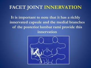
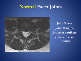
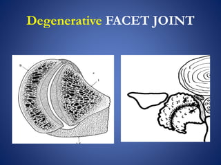
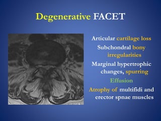
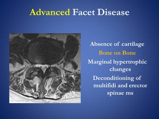
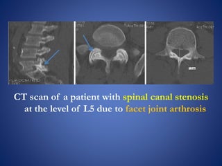
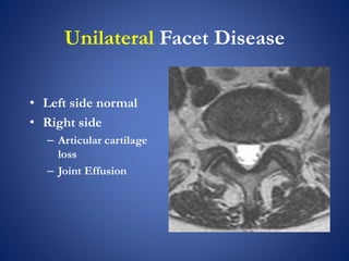
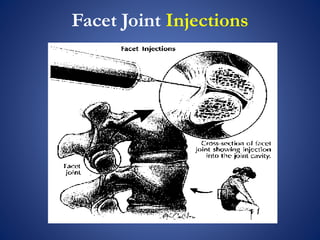
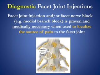
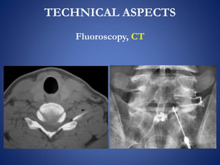
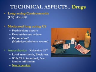
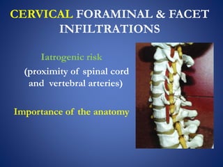
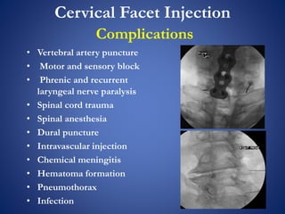
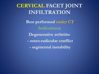
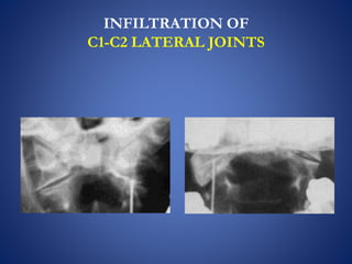
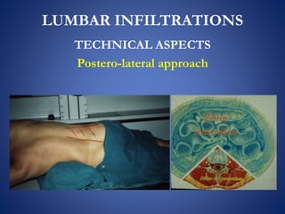
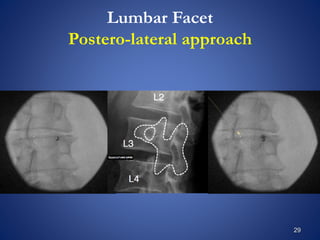
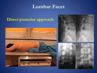
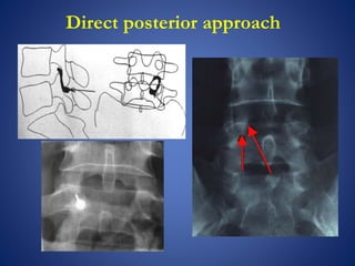
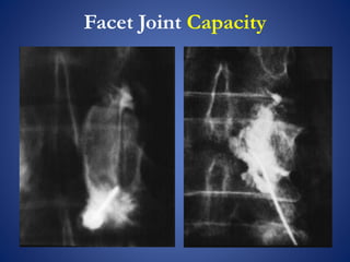
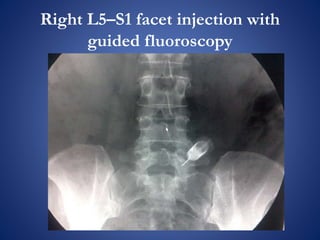
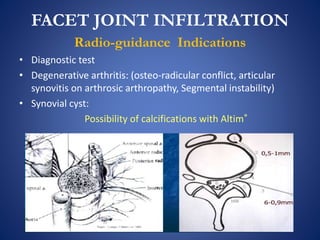
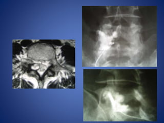
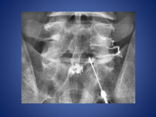
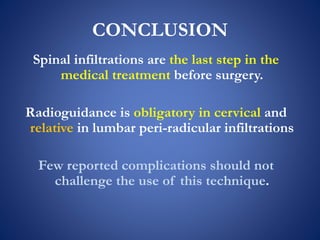
Facet Joint Injections
Comments
Mohamed M. Mohi Eldin,
Professor of Neurosurgery,
Faculty of Medicine,
Cairo University
One-Day Spine Clinic 1st workshop & hands-on
March 2nd 2016
FACET JOINT INNERVATION
It is important to note that it has a richly
innervated capsule and the medial branches
of the posterior lumbar rami provide this
innervation
Normal Facet Joints
Joint Space
Joint Margins
Articular cartilage
Periarticular soft
tissues
Degenerative FACET JOINT
Degenerative FACET
Articular cartilage loss
Subchondral bony
irregularities
Marginal hypertrophic
changes, spurring
Effusion
Atrophy of multifidi and
erector spnae muscles
Advanced Facet Disease
Absence of cartilage
Bone on Bone
Marginal hypertrophic
changes
Deconditioning of
multifidi and erector
spinae ms
CT scan of a patient with spinal canal stenosis
at the level of L5 due to facet joint arthrosis
Unilateral Facet Disease
• Left side normal
• Right side
– Articular cartilage
loss
– Joint Effusion
Unilateral Facet inflammation
• Articular cartilage
• Gadolinium
contrast
enhancement
– Medullary bone
marrow
– Periarticular soft
tissues
Facet Synovitis
• Prominent effusion
• Note: medial
ligamentum flavum
hypertrophy
• Paraspinous muscles
preservered
Facet Tropism
Paravertebral Soft Tissue
• Gadolinium
contrast
enhancement
– Periarticular soft
tissue
– Medullary bone
marrow
• High clinical
correlation with
pain generator
Facet Marrow
Edema
• Medullary bone
marrow edema
• Indicator of
inflammation
• High clinical
correlation with pain
generator
Facet Joint Injections
Clinical signs and radiologic appearance
in accordance
Diagnostic Facet Joint Injections
Facet joint injection and/or facet nerve block
(e.g. medial branch block) is proven and
medically necessary when used to localize
the source of pain to the facet joint
Diagnostic Facet Joint Injections
Is not recommended in patients with:
Neurologic abnormalities
More than one pain syndrome
Definitive specific diagnosis other than facet joint syndrome
Previous spinal surgery at the clinically suspected levels
Therapeutic facet joint injection
is unproven and not medically necessary for
the treatment of chronic spinal pain.
(pain lasting more than 3 months).
Facet block
Is an injection of local anesthetic and/or steroids into
or near the facet joint
Use of diagnostic blocks with injection of local
anesthesia into the facet joints or around the medial
branch nerves to identify the possible sources of
spinal pain is an established diagnostic procedure.
In a positive response, pain relief occurs but lasts
longer with the long-acting anesthetic.
Single lumbar facet joint diagnostic block had a 38%
false-positive rate.
TECHNICAL ASPECTS
Fluoroscopy, CT
TECHNICAL ASPECTS.. Drugs
• Long acting Corticosteroids
(CS): Altim®
• Moderated long acting CS
– Prednisolone acetate
– Dexaméthasone acétate
– Depomedrol®
(Methylprednisolone acetate).
• Anaesthesics : Xylocaïne 1%®
– Local anaesthesia, Block test.
– With CS in foraminal, facet
lumbar infiltration
– Not in cervical
CERVICAL FORAMINAL & FACET
INFILTRATIONS
Iatrogenic risk
(proximity of spinal cord
and vertebral arteries)
Importance of the anatomy
Cervical Facet Injection
Complications
• Vertebral artery puncture
• Motor and sensory block
• Phrenic and recurrent
laryngeal nerve paralysis
• Spinal cord trauma
• Spinal anesthesia
• Dural puncture
• Intravascular injection
• Chemical meningitis
• Hematoma formation
• Pneumothorax
• Infection
CERVICAL FACET JOINT
INFILTRATION
Best performed under CT
Indications:
Degenerative arthritis:
- osteo-radicular conflict
- segmental instability
INFILTRATION OF
C1-C2 LATERAL JOINTS
LUMBAR INFILTRATIONS
TECHNICAL ASPECTS
Postero-lateral approach
29
Lumbar Facet
Postero-lateral approach
Lumbar Facet
Direct posterior approach
Facet Joint Capacity
Facet joints have been shown to hold up to a
maximum of 1–2 mL of injected fluid by
anatomical studies.
Above 2 ml, rupture of the joint capsule occurs
and a resultant extravasation into the back and
epidural space lead to non-specific therapeutic
effects
Keeping in mind that contrast, steroid and local
anesthetic must subsequently be introduced.
Facet Joint Capacity
Right L5–S1 facet injection with
guided fluoroscopy
FACET JOINT INFILTRATION
Radio-guidance Indications
• Diagnostic test
• Degenerative arthritis: (osteo-radicular conflict, articular
synovitis on arthrosic arthropathy, Segmental instability)
• Synovial cyst:
Possibility of calcifications with Altim®
Facet joint injection in LBP
is its continued use justified?
The injection of local anaesthetic and/or
steroids into the facet joint is used
worldwide treating patients facetogenic
back pain;
However, the effectiveness of this therapy for
short- and long-term pain relief requires
evaluation
CONCLUSION
Spinal infiltrations are the last step in the
medical treatment before surgery.
Radioguidance is obligatory in cervical and
relative in lumbar peri-radicular infiltrations
Few reported complications should not
challenge the use of this technique.
Thank You

Facet joint injection

  • 1.
    Facet Joint Injections Comments MohamedM. Mohi Eldin, Professor of Neurosurgery, Faculty of Medicine, Cairo University One-Day Spine Clinic 1st workshop & hands-on March 2nd 2016
  • 2.
    FACET JOINT INNERVATION Itis important to note that it has a richly innervated capsule and the medial branches of the posterior lumbar rami provide this innervation
  • 3.
    Normal Facet Joints JointSpace Joint Margins Articular cartilage Periarticular soft tissues
  • 4.
  • 5.
    Degenerative FACET Articular cartilageloss Subchondral bony irregularities Marginal hypertrophic changes, spurring Effusion Atrophy of multifidi and erector spnae muscles
  • 6.
    Advanced Facet Disease Absenceof cartilage Bone on Bone Marginal hypertrophic changes Deconditioning of multifidi and erector spinae ms
  • 7.
    CT scan ofa patient with spinal canal stenosis at the level of L5 due to facet joint arthrosis
  • 8.
    Unilateral Facet Disease •Left side normal • Right side – Articular cartilage loss – Joint Effusion
  • 9.
    Unilateral Facet inflammation •Articular cartilage • Gadolinium contrast enhancement – Medullary bone marrow – Periarticular soft tissues
  • 10.
    Facet Synovitis • Prominenteffusion • Note: medial ligamentum flavum hypertrophy • Paraspinous muscles preservered
  • 11.
  • 12.
    Paravertebral Soft Tissue •Gadolinium contrast enhancement – Periarticular soft tissue – Medullary bone marrow • High clinical correlation with pain generator
  • 13.
    Facet Marrow Edema • Medullarybone marrow edema • Indicator of inflammation • High clinical correlation with pain generator
  • 14.
  • 15.
    Clinical signs andradiologic appearance in accordance
  • 16.
    Diagnostic Facet JointInjections Facet joint injection and/or facet nerve block (e.g. medial branch block) is proven and medically necessary when used to localize the source of pain to the facet joint
  • 17.
    Diagnostic Facet JointInjections Is not recommended in patients with: Neurologic abnormalities More than one pain syndrome Definitive specific diagnosis other than facet joint syndrome Previous spinal surgery at the clinically suspected levels
  • 18.
    Therapeutic facet jointinjection is unproven and not medically necessary for the treatment of chronic spinal pain. (pain lasting more than 3 months).
  • 19.
    Facet block Is aninjection of local anesthetic and/or steroids into or near the facet joint Use of diagnostic blocks with injection of local anesthesia into the facet joints or around the medial branch nerves to identify the possible sources of spinal pain is an established diagnostic procedure. In a positive response, pain relief occurs but lasts longer with the long-acting anesthetic. Single lumbar facet joint diagnostic block had a 38% false-positive rate.
  • 21.
  • 23.
    TECHNICAL ASPECTS.. Drugs •Long acting Corticosteroids (CS): Altim® • Moderated long acting CS – Prednisolone acetate – Dexaméthasone acétate – Depomedrol® (Methylprednisolone acetate). • Anaesthesics : Xylocaïne 1%® – Local anaesthesia, Block test. – With CS in foraminal, facet lumbar infiltration – Not in cervical
  • 24.
    CERVICAL FORAMINAL &FACET INFILTRATIONS Iatrogenic risk (proximity of spinal cord and vertebral arteries) Importance of the anatomy
  • 25.
    Cervical Facet Injection Complications •Vertebral artery puncture • Motor and sensory block • Phrenic and recurrent laryngeal nerve paralysis • Spinal cord trauma • Spinal anesthesia • Dural puncture • Intravascular injection • Chemical meningitis • Hematoma formation • Pneumothorax • Infection
  • 26.
    CERVICAL FACET JOINT INFILTRATION Bestperformed under CT Indications: Degenerative arthritis: - osteo-radicular conflict - segmental instability
  • 27.
  • 28.
  • 29.
  • 30.
  • 31.
  • 32.
    Facet Joint Capacity Facetjoints have been shown to hold up to a maximum of 1–2 mL of injected fluid by anatomical studies. Above 2 ml, rupture of the joint capsule occurs and a resultant extravasation into the back and epidural space lead to non-specific therapeutic effects Keeping in mind that contrast, steroid and local anesthetic must subsequently be introduced.
  • 33.
  • 34.
    Right L5–S1 facetinjection with guided fluoroscopy
  • 35.
    FACET JOINT INFILTRATION Radio-guidanceIndications • Diagnostic test • Degenerative arthritis: (osteo-radicular conflict, articular synovitis on arthrosic arthropathy, Segmental instability) • Synovial cyst: Possibility of calcifications with Altim®
  • 38.
    Facet joint injectionin LBP is its continued use justified? The injection of local anaesthetic and/or steroids into the facet joint is used worldwide treating patients facetogenic back pain; However, the effectiveness of this therapy for short- and long-term pain relief requires evaluation
  • 39.
    CONCLUSION Spinal infiltrations arethe last step in the medical treatment before surgery. Radioguidance is obligatory in cervical and relative in lumbar peri-radicular infiltrations Few reported complications should not challenge the use of this technique.
  • 40.