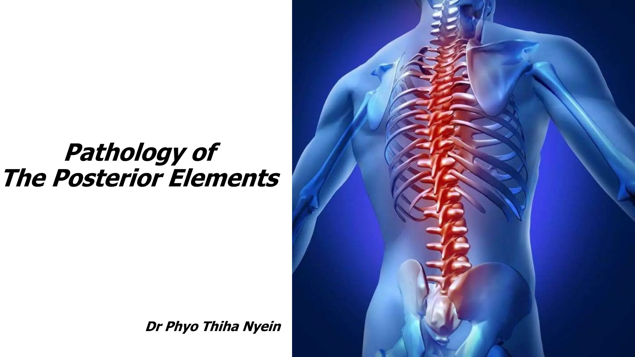
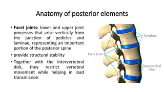
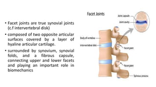
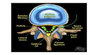
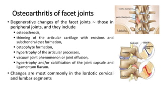
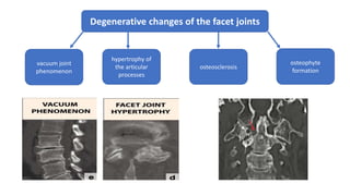
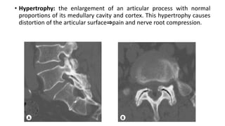
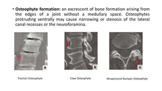
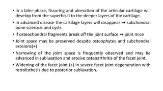
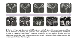
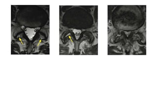
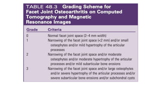
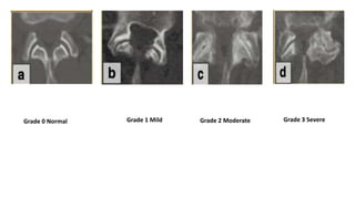
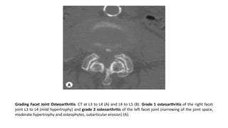
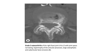
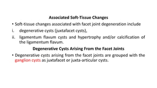
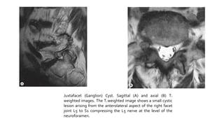
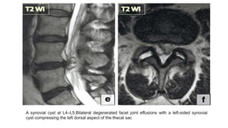
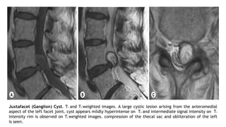
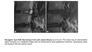
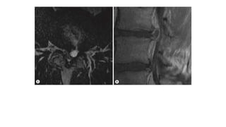
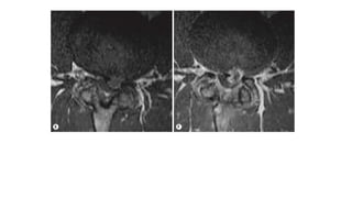
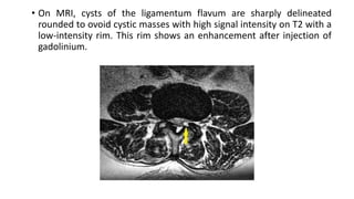
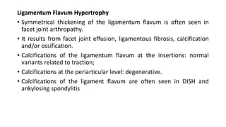
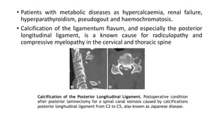
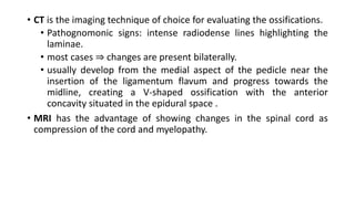
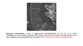
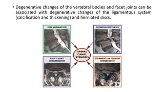
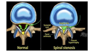
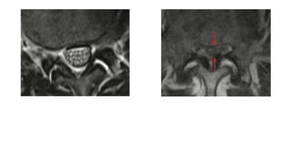
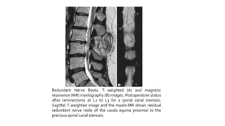
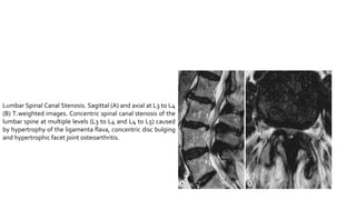
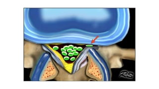
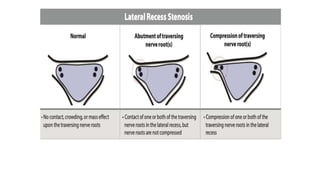
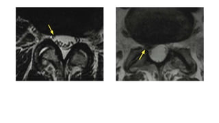
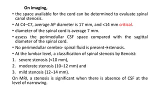
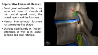
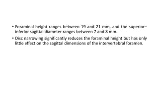
Pathology of
The Posterior Elements
Dr Phyo Thiha Nyein
Anatomy of posterior elements
• Facet joints: lower and upper joint
processes that arise vertically from
the junction of pedicles and
laminae, representing an important
portion of the posterior spine
• provide structural stability
• Together with the intervertebral
disk, they restrict vertebral
movement while helping in load
transmission
• Facet joints are true synovial joints
(c.f intervertebral disk)
• composed of two opposite articular
surfaces covered by a layer of
hyaline articular cartilage.
• surrounded by synovium, synovial
folds, and a fibrous capsule,
connecting upper and lower facets
and playing an important role in
biomechanics
• The entire posterior ligament
complex
i. facet joint capsule,
ii. ligamentum flavum, and
iii. Interspinous and supra
spinous ligaments
• keeps facet joints and
vertebrae in a stable position
with each other
• Injuries to this complex can
result in subluxation or
displacement of the facet
• Familiarity with the vascularization of facet joints is important because it
can help explain the pathophysiology of certain diseases.
• Arterial vascularization is provided by branches of the ascending cervical
artery (cervical spine), thoracic aorta (thoracic spine), and abdominal
aorta (lumbar spine)
• Venous drainage is provided by the internal and external venous
plexus, basivertebral veins, and intervertebral veins.
• Because of its avalvular nature, the venous plexus may become a path
to spread metastases and infections
• Articular cartilage surfaces of facet joints are aneural structures, while
capsules, subchondral bone, and synovium are richly innervated.
• Each facet joint receives a pair of nerves coming from the medial branch
of the spinal nerve with nociceptive, autonomic, and mechanoreceptor
fibers
• A medial branch runs through the transverse processes at a lower level in
relation to its origin, and the facet joint is innervated by a medial branch at the
same level and another medial branch at a higher level, a consequence of the
presence of a C8 nerve without the presence of C8 vertebra.
• Eg. C6-C7 facet joint ↤ medial branches of C6 and C7.
C7-T1 facet joint ↤ medial branches of C7 and C8.
T1-T2 facet joint ↤ medial branches of C8 andT1.
• With exception of the L5-S1 facet joint, which is innervated by the medial
branch of L4 and the dorsal branch of L5
• The medial branches ↦multifidus muscle, interspinous muscle and
ligament, and the periosteum of the neural arch
• Intermediate and lateral ↦ the rest of the paravertebral musculature
Pathology of Posterior elements
• Facet joint syndrome: a range of symptoms that result in diffuse pain
that does not follow a clear nerve root pattern.
• In the past the role of the facet joints as a cause of low back pain -
underestimated.
• As the synovial linings and joint capsules of the facet joints are richly
innervated, it can be an important source of pain.
• As the intervertebral disc and the facet joints work as a three-joint
complex, degenerative changes in the disc will change function and
anatomy of the posterior elements.
• On the one hand, facet joint osteoarthritis mostly occurs in the
presence of disc degeneration; on the other hand, disc degeneration
sometimes occurs without facet joint changes.
• Therefore it was suggested that disc degeneration occurs before facet
joint osteoarthritis, possibly secondary to mechanical changes in the
loading of the facet joints.
• Other factors contributing to facet joint degeneration include weight,
lordosis and scoliosis.
• Significant association: between sagittal orientation and osteoarthritis
of the lumbar facet joints, even in patients without degenerative
spondylolisthesis.
• In a study, asymptomatic patients ↦ more sagittally orientated facet
joints at the fourth and fifth lumbar vertebra were associated with
herniated discs and spondylolisthesis.
• Degenerative changes of the facet joints ∼ those in
peripheral joints, and they include
 osteosclerosis,
 thinning of the articular cartilage with erosions and
subchondral cyst formation,
 osteophyte formation,
 hypertrophy of the articular processes,
 vacuum joint phenomenon or joint effusion,
 hypertrophy and/or calcification of the joint capsule and
ligamentum flavum.
• Changes are most commonly in the lordotic cervical
and lumbar segments
Osteoarthritis of facet joints
Degenerative changes of the facet joints
vacuum joint
phenomenon
hypertrophy of
the articular
processes
osteosclerosis
osteophyte
formation
Degenerative changes of the facet joints
joint effusion
thinning of the articular cartilage
with erosions and subchondral
cyst formation
hypertrophy and/or calcification of
the joint capsule and ligamentum
flavum
• Hypertrophy: the enlargement of an articular process with normal
proportions of its medullary cavity and cortex. This hypertrophy causes
distortion of the articular surface⇒pain and nerve root compression.
• Osteophyte formation: an excrescent of bone formation arising from
the edges of a joint without a medullary space. Osteophytes
protruding ventrally may cause narrowing or stenosis of the lateral
canal recesses or the neuroforamina.
Traction Osteophyte Claw Osteophyte Wraparound Bumper Osteophyte
• In a later phase, fissuring and ulceration of the articular cartilage will
develop from the superficial to the deeper layers of the cartilage.
• In advanced disease the cartilage layers will disappear ↦ subchondral
bone sclerosis and cysts
• If osteochondral fragments break off the joint surface ↦ joint mice
• Joint space may be preserved despite osteophytes and subchondral
erosions(+)
• Narrowing of the joint space is frequently observed and may be
advanced in subluxation and erosive osteoarthritis of the facet joint.
• Widening of the facet joint (+) in severe facet joint degeneration with
retrolisthesis due to posterior subluxation.
Evolution of OA in facet joints. a, Axial CT (top) and axial MR (bottom) images show a normal facet
joint. b, Narrowing of the facet joint space and small osteophytes. c, Mild hypertrophy of the articular
process. d, Moderate osteophytes, moderate hypertrophy of the articular process, and mild
subarticular subchondral cysts. e, Large osteophytes, severe hypertrophy of the articular process, and
severe subarticular subchondral cysts.
Grade 0 Normal Grade 3 Severe
Grade 2 Moderate
Grade 1 Mild
Grading Facet Joint Osteoarthritis. CT at L3 to L4 (A) and L4 to L5 (B). Grade 1 osteoarthritis of the right facet
joint L3 to L4 (mild hypertrophy) and grade 2 osteoarthritis of the left facet joint (narrowing of the joint space,
moderate hypertrophy and osteophytes, subarticular erosion) (A).
Grade 3 osteoarthritis of the right facet joint L4 to L5 with joint space
narrowing, hypertrophy of the articular processes, large osteophytes
and subarticular bone erosions (B).
Associated Soft-Tissue Changes
• Soft-tissue changes associated with facet joint degeneration include
i. degenerative cysts (juxtafacet cysts),
ii. ligamentum flavum cysts and hypertrophy and/or calcification of
the ligamentum flavum.
Degenerative Cysts Arising From the Facet Joints
• Degenerative cysts arising from the facet joints are grouped with the
ganglion cysts as juxtafacet or juxta-articular cysts.
• Ganglion cysts have no connection to the joint and have no synovial
lining.
• Cysts are periarticular and are attached to the joint by a membrane.
• Walls are made of loose myxoid connective or fibrocollagenous tissue
with a synovial lining, and they are filled with yellow or clear
mucinous fluid. The consistency of the fluid may vary as haemorrhage
and inflammation can be seen.
• Juxtafacet cysts are most commonly seen in the lumbar spine and
especially at the level L4 to L5, which has the most motion in the
spine.
Juxtafacet (Ganglion) Cyst. Sagittal (A) and axial (B) T2
weighted images. The T2 weighted image shows a small cystic
lesion arising from the anterolateral aspect of the right facet
joint L5 to S1 compressing the L5 nerve at the level of the
neuroforamen.
• Synovial cysts of the facet joints are related to osteoarthritis of the
facet joint ⇐increased motion in the degenerative facet joints.
• Instability can cause herniation of the synovium through tears in the
facet joint capsule.
• An association between juxtafacet cyst and trauma, chondrocalcinosis
spondylolysis and Baastrup’s disease has been reported.
• Most juxtafacet cysts arise from the posterior aspect of the facet joint
(outside the vertebral canal) >4times anterior cysts
A synovial cyst at L4–L5.Bilateral degenerated facet joint effusions with a left-sided synovial
cyst compressing the left dorsal aspect of the thecal sac
Presentation
• In the lumbar spine, degenerative cysts of the facet joint may cause
pain and radicular symptoms by compression of the thecal sac and/or
nerve roots in the lateral recesses.
• Inflammatory reaction around the juxtafacet cysts may also cause
sciatica.
• Depending on the size and location of the cyst, spinal stenosis and/or
neurogenic claudication may occur.
• In the cervical spine facet joint cysts rarely cause radiculopathy.
Treatment
• In case of radiculopathy and myelopathy caused by a facet joint cyst,
surgical removal is indicated. (an effective and safe option)
• If no association with a spinal canal stenosis ⇒ a simple excision of
the cyst
• In associated spondylotic spinal canal stenosis⇒removal of the cyst
with decompression and laminectomy
• An alternative and less invasive technique ⇒ percutaneous injection
of local anesthetics and/or long-acting steroids.
CT
• a synovial cyst is a rounded mass adjacent to the facet joint with low
attenuation.
• This cyst may show eggshell calcification of the wall and gas inside.
MRI: high sensitivity and is the imaging technique of choice.
• Intraspinal synovial cysts: sharply marginated epidural masses near
the facet joint.
• Typical synovial cysts have a hypointense peripheral rim
• This rim enhances after the administration of gadolinium.
• SI of a synovial cyst is nearly equal to CSF on T1 and on T2. High signal
intensity on T1 and T2 indicates subacute blood degradation
products.
Juxtafacet (Ganglion) Cyst. T1 and T2 weighted images. A large cystic lesion arising from the anteromedial
aspect of the left facet joint. cyst appears mildly hyperintense on T2 and intermediate signal intensity on T1
intensity rim is observed on T2 weighted images. compression of the thecal sac and obliteration of the left
is seen.
• Differential diagnosis of a mass with this type of signal characteristics
in the posterior or lateral spinal canal is broad:
a) juxtafacet cysts,
b) discogenic cysts,
c) cysts of the ligamentum flavum,
d) sequestered disc fragments,
e) infectious cysts or arachnoid cysts and
f) neoplasms (cystic degenerated schwannoma or neurofibroma)
However, degenerative facet joints are the clue to the most likely
diagnosis.
• Facet joint arthrography and CT arthrography can be performed if
there is any doubt about communication with the adjacent facet
joint.
Discogenic Cyst With Narrowing of the Left Lateral Recess at L4 to L5. This lesion has an intermediate
signal intensity on T1 weighted images and rim enhancement after gadolinium injection. Secondarily, there
narrowing of the left lateral recess.
Cysts of the Ligamentum Flavum
• Cysts of the ligamentum flavum arise from or are partially embedded
in the ligamentum flavum, rather than being close to the facet joints.
• Seldom and their development may be related to necrosis or myxoid
degeneration in a hypertrophied ligamentum flavum.
• Chronic degenerative changes ⇉ repeated haemorrhage ⇉ small
degenerative cysts ⇉ one large cyst later.
• Typically located at the level L4 to L5, just as juxtafacet cysts.
• Cysts of the ligamentum flavum ∼imaging characteristics as facet joint
cysts.
ligamentum flavum cyst on right side at L4/L5 level, producing
posterior indentation upon thecal sac.
• Simple laminectomy is sufficient for treatment of the ligamentum
flavum cysts.
On imaging,
• an extradural, intraspinal mass in close relationship with the
ligamentum flavum
• On CT imaging, this cyst has a low density compared with the
ligamentum flavum.
• Unlike juxtafacet cysts, no rim calcification has been described.
• On MRI, cysts of the ligamentum flavum are sharply delineated
rounded to ovoid cystic masses with high signal intensity on T2 with a
low-intensity rim. This rim shows an enhancement after injection of
gadolinium.
Ligamentum Flavum Hypertrophy
• Symmetrical thickening of the ligamentum flavum is often seen in
facet joint arthropathy.
• It results from facet joint effusion, ligamentous fibrosis, calcification
and/or ossification.
• Calcifications of the ligamentum flavum at the insertions: normal
variants related to traction;
• Calcifications at the periarticular level: degenerative.
• Calcifications of the ligament flavum are often seen in DISH and
ankylosing spondylitis
Ligamentum Flavum Hypertrophy and Calcification at L4 to L5 and, to a less
extent, at L3 to L4, resulting in a spinal canal stenosis at L4 to L5.
• Patients with metabolic diseases as hypercalcaemia, renal failure,
hyperparathyroidism, pseudogout and haemochromatosis.
• Calcification of the ligamentum flavum, and especially the posterior
longitudinal ligament, is a known cause for radiculopathy and
compressive myelopathy in the cervical and thoracic spine
Calcification of the Posterior Longitudinal Ligament. Postoperative condition
after posterior laminectomy for a spinal canal stenosis caused by calcifications
posterior longitudinal ligament from C2 to C5, also known as Japanese disease.
• Calcification and/or ossification of the thoracic ligamentum flavum is
a rare disease mainly described in the Asian/Japanese literature, also
known as Japanese disease.
• mostly affects males <50 years
• cause spinal stenosis with/without myelopathy and/or radiculopathy
⇒ progressive myelopathy ⇒ spastic paraparesis.
• lower third of thoracic spine is the most commonly involved(cervical
spine is only rare)
• CT is the imaging technique of choice for evaluating the ossifications.
• Pathognomonic signs: intense radiodense lines highlighting the
laminae.
• most cases ⇒ changes are present bilaterally.
• usually develop from the medial aspect of the pedicle near the
insertion of the ligamentum flavum and progress towards the
midline, creating a V-shaped ossification with the anterior
concavity situated in the epidural space .
• MRI has the advantage of showing changes in the spinal cord as
compression of the cord and myelopathy.
• Baastrup’s phenomenon, also known as kissing spine
• Pain caused by irritation of the periosteum or adventitial bursae between
abutting spinous processes.
• Close approximation and contact of the adjacent spinous processes with
enlargement, flattening and reactive sclerosis of the opposing interspinous
surfaces.
• Interspinous bursitis may communicate with the facet joints and can be
treated with injection of steroids.
• Extension of the synovial cavity to the intraspinal space can result in cyst
formation⇒can enter the epidural space via midline cleft of LF ⇒
extradural compression.
Spinous Process Abnormalities and Associated Ligamentous
Changes
Baastrup’s Phenomenon. Grade 2 degenerative spondylolisthesis at the L5 to S1 level,
malalignment of the spinous processes with anterior slip of the L4 spinous process relative to L5
type 3 degenerative spondylolisthesis and allows differentiation from isthmic spondylolisthesis.
Degenerative Spondylolisthesis
• A spondylolisthesis, also known as an
anterolisthesis, is an anterior displacement
of a vertebral body relative to the vertebra
below.
• The reverse, when the vertebral body below
is displaced anteriorly relative to the
superior vertebra, is called a retrolisthesis.
• most popular grading method: Meyerding
grading system
• Six types of spondylolisthesis can be differentiated:
i. congenital dysplasia of the articular processes,
ii. defect of the pars articularis,
iii. degenerative changes of the facet joint,
iv. fracture of the neural arch,
v. weakening of the neural arch due to bone disorders and
vi. excessive removal of bone after spinal decompression.
•grade I: 0-25%
•grade II: 26-50%
•grade III: 51-75%
•grade IV: 76-100%
•grade V (spondyloptosis):
>100%
• > 50 yrs
• caused by degenerative changes of the facet joints(more sagittal
orientation)
• As the neural arch is intact, even small progression in the slip may
cause cauda equina syndrome.
• As facet joint osteoarthrosis is most commonly seen at the level L4–
L5, spondylolisthesis is also most common at this level.
• Incidence ↑ x4 times (sacralization and F>M)
Presentation
• Clinical symptoms include low back pain and leg pain as a result of
disc and facet joint degeneration, lateral recess and foraminal
stenosis leading to nerve root compression.
• With progression of the spondylolisthesis, from low back pain to
neurogenic claudication due to central canal stenosis.
On imaging,
• a lateral conventional radiograph shows anterolisthesis with
degenerative changes at the facet joint and disc space narrowing.
• Differentiation with isthmic spondylolisthesis can be made by the
malalignment of the spinous processes with anterior slip of the
spinous process relative to the one of the vertebral bodies below
CT and MRI will show osteoarthritis of the facet joints and associated
spondylolisthesis.
• The sagittal orientation of the facet joints, as well as disc
degeneration and/or disc bulging, can be evaluated.
• Sagittal images ⇒narrowing of the intervertebral foramina, the lateral
recess and/or the central canal with associated compression of the
cauda equina and exiting nerve roots.
• Anterior slip of the inferior articular process will narrow the inferior
aspect of the lateral recess and the intervertebral foramen.
• Associated thickening of the ligamentum flavum ⇒central canal and
lateral recess stenosis.
• On MRI, bone marrow changes in the pedicle are non-specific for
spondylolisthesis, and they are often observed in patients with facet
joint osteoarthritis.
DEGENERATIVE SPINAL STENOSIS
Degenerative Spinal Canal Stenosis
• Normal size of the spinal canal varies according to the location.
• Spinal canal stenosis may lead to neurological disorders associated
with compression of the nervous structures inside the spinal canal
(spinal cord, conus medullaris, cauda equina, nerve roots and
meninges).
• Acquired spinal canal stenosis is the most common type of spinal
stenosis at the cervical and lumbar level and less frequent in the
thoracic spine.
• Degenerative changes of the vertebral bodies and facet joints can be
associated with degenerative changes of the ligamentous system
(calcification and thickening) and herniated discs.
• Posterior and central marginal osteophytes can reduce the spinal
canal diameter centrally with possible cord compression ⇒
development of a cauda equina syndrome
• Bony protrusion at facet level may cause lateral radicular compression
by radicular entrapment in the lateral recess. This phenomenon is
more commonly seen at the lumbar level.
• Disc herniation can also be associated with narrowing of the lateral
recess.
• Compression of the dural sac leads to a reduced space for the nerve
roots of the cauda equina.
• In the case of multilevel disease the dural sac can have a ‘string of
pearls’-like appearance on myelography.
• The pressure of the subarachnoidal fluid increases, causing an
alteration in the venous drainage, which causes perineural venous
congestion and ischaemic damage➨ breakdown of the blood–nerve
barrier, inflammatory reaction ➨ the roots may show enhancement
on contrast-enhanced MRI
• Because of its vascular anatomy, the nerve roots of the cauda equina
have a higher ischaemic risk if compression is at more than one level.
The nerve roots L4, L5 and S1 are predominantly affected.
• With high-grade stenosis in the lumbar spine, the nerve roots
proximal to the spinal canal stenosis can be elongated, large and
tortuous; this phenomenon is called ‘redundant nerve roots’ .
Spinal Canal Stenosis With Redundant
Nerve Roots at L3 to L4 as a result of
anterolisthesis due to facet joint
(concentric spinal canal stenosis with
hypertrophy of the ligamenta flava and disc
bulging, redundant nerve roots of the cauda
equina
Redundant Nerve Roots. T2 weighted (A) and magnetic
resonance (MR) myelography (B) images. Postoperative status
after laminectomy at L2 to L3 for a spinal canal stenosis.
Sagittal T2 weighted image and the myelo-MR shows residual
redundant nerve roots of the cauda equina proximal to the
previous spinal canal stenosis.
Lumbar Spinal Canal Stenosis. Sagittal (A) and axial at L3 to L4
(B) T2 weighted images. Concentric spinal canal stenosis of the
lumbar spine at multiple levels (L3 to L4 and L4 to L5) caused
by hypertrophy of the ligamenta flava, concentric disc bulging
and hypertrophic facet joint osteoarthritis.
• Clinical symptoms of a spinal canal stenosis vary with the level of the
spinal stenosis. Patients often remain asymptomatic until an acute
event happens.
• Acute or chronic limb pain and paraesthesias are seen in patients with
lateral or foraminal radicular compression.
• Forward bending and supine position may relieve the symptoms due
to increase size of the spinal canal.
• Vascular claudication and degenerative changes of knee and hip
• Neurogenic bladder due to mechanic compression of the S2–S4 roots
On imaging,
• the space available for the cord can be determined to evaluate spinal
canal stenosis.
• At C4–C7, average AP diameter is 17 mm, and <14 mm critical.
• diameter of the spinal cord is average 7 mm.
• assess the perimedullar CSF space compared with the sagittal
diameter of the spinal cord.
• No perimedullar cerebro- spinal fluid is present→stenosis.
• At the lumbar level, a classification of spinal stenosis by Benoist:
1. severe stenosis (<10 mm),
2. moderate stenosis (10–12 mm) and
3. mild stenosis (12–14 mm).
On MRI, a stenosis is significant when there is absence of CSF at the
level of narrowing.
Degenerative Foraminal Stenosis
• Facet joint osteoarthritis is an
important cause of stenosis of
the central spinal canal, the
lateral recess and the foramen.
• Normal intervertebral foramen
has a teardrop-like shape
• changes significantly in flexion–
extension, as well as in lateral-
bending and axial rotation.
• Foraminal height ranges between 19 and 21 mm, and the superior–
inferior sagittal diameter ranges between 7 and 8 mm.
• Disc narrowing significantly reduces the foraminal height but has only
little effect on the sagittal dimensions of the intervertebral foramen.
• Instead of measuring the dimensions, Wildermuth et al. introduced a
qualitative scoring system.
• Grade 0: normal intervertebral foramina; normal dorsolateral border of the
intervertebral disc and normal form at the foraminal epidural fat (oval or
inverted pear shape).
• Grade 1: slight foraminal stenosis and deformity of the epidural fat, with
the remaining fat still completely surrounding the exiting nerve root.
• Grade 2: marked foraminal stenosis, with epidural fat only partially
surrounding the nerve root.
• Grade 3: advanced stenosis with obliteration of the epidural fat.
• Although lateral recess stenosis may be more common than foraminal
stenosis, foraminal stenosis with compression of the spinal nerve
within the intervertebral foramen ➜lateral spinal stenosis.
• Retrolisthesis and isthmic spondylolisthesis ➜foraminal stenosis,
while degenerative spondylolisthesis ➜ lateral or central stenosis by
slipping of the inferior articular processes.
• SUMMARY BOX: Pathology of the Posterior Elements
• Facet joint arthrosis plays an important role in low back pain as the
joint lining and capsule are richly innervated.
• Degenerative changes of the facet joints are similar to those in
peripheral joints.
• Juxtafacet cysts and hypertrophy of the ligamentum flavum are most
frequently seen in the lumbar spine and can cause nerve compression
and spinal canal stenosis.
• Baastrup’s phenomenon is a common cause of low back pain.
• A degenerative spondylolisthesis is mostly caused by facet joint
arthrosis in the lumbar spine at the level L4–L5 and can cause stenosis
of the spinal canal and transverse foramina.
• Degenerative stenosis of the spinal canal can be caused by
degenerative changes of vertebral bodies, facet joints, ligamentous
thickening and herniated discs.
• Spinal canal stenosis in the lumbar spine can cause the nerve roots to
elongate and is called ‘redundant nerve roots’.
• Stenosis of the transverse foramina can be caused by osteophytic
spurs of the vertebra, as well as an enlarged superior articular facet.
GetridofBackPain🙏🙏🙏

Pathology of posterior elements.pptx

  • 1.
    Pathology of The PosteriorElements Dr Phyo Thiha Nyein
  • 2.
    Anatomy of posteriorelements • Facet joints: lower and upper joint processes that arise vertically from the junction of pedicles and laminae, representing an important portion of the posterior spine • provide structural stability • Together with the intervertebral disk, they restrict vertebral movement while helping in load transmission
  • 3.
    • Facet jointsare true synovial joints (c.f intervertebral disk) • composed of two opposite articular surfaces covered by a layer of hyaline articular cartilage. • surrounded by synovium, synovial folds, and a fibrous capsule, connecting upper and lower facets and playing an important role in biomechanics
  • 4.
    • The entireposterior ligament complex i. facet joint capsule, ii. ligamentum flavum, and iii. Interspinous and supra spinous ligaments • keeps facet joints and vertebrae in a stable position with each other • Injuries to this complex can result in subluxation or displacement of the facet
  • 6.
    • Familiarity withthe vascularization of facet joints is important because it can help explain the pathophysiology of certain diseases. • Arterial vascularization is provided by branches of the ascending cervical artery (cervical spine), thoracic aorta (thoracic spine), and abdominal aorta (lumbar spine)
  • 7.
    • Venous drainageis provided by the internal and external venous plexus, basivertebral veins, and intervertebral veins. • Because of its avalvular nature, the venous plexus may become a path to spread metastases and infections
  • 8.
    • Articular cartilagesurfaces of facet joints are aneural structures, while capsules, subchondral bone, and synovium are richly innervated. • Each facet joint receives a pair of nerves coming from the medial branch of the spinal nerve with nociceptive, autonomic, and mechanoreceptor fibers
  • 9.
    • A medialbranch runs through the transverse processes at a lower level in relation to its origin, and the facet joint is innervated by a medial branch at the same level and another medial branch at a higher level, a consequence of the presence of a C8 nerve without the presence of C8 vertebra.
  • 10.
    • Eg. C6-C7facet joint ↤ medial branches of C6 and C7. C7-T1 facet joint ↤ medial branches of C7 and C8. T1-T2 facet joint ↤ medial branches of C8 andT1. • With exception of the L5-S1 facet joint, which is innervated by the medial branch of L4 and the dorsal branch of L5 • The medial branches ↦multifidus muscle, interspinous muscle and ligament, and the periosteum of the neural arch • Intermediate and lateral ↦ the rest of the paravertebral musculature
  • 11.
    Pathology of Posteriorelements • Facet joint syndrome: a range of symptoms that result in diffuse pain that does not follow a clear nerve root pattern. • In the past the role of the facet joints as a cause of low back pain - underestimated. • As the synovial linings and joint capsules of the facet joints are richly innervated, it can be an important source of pain.
  • 12.
    • As theintervertebral disc and the facet joints work as a three-joint complex, degenerative changes in the disc will change function and anatomy of the posterior elements. • On the one hand, facet joint osteoarthritis mostly occurs in the presence of disc degeneration; on the other hand, disc degeneration sometimes occurs without facet joint changes. • Therefore it was suggested that disc degeneration occurs before facet joint osteoarthritis, possibly secondary to mechanical changes in the loading of the facet joints.
  • 13.
    • Other factorscontributing to facet joint degeneration include weight, lordosis and scoliosis. • Significant association: between sagittal orientation and osteoarthritis of the lumbar facet joints, even in patients without degenerative spondylolisthesis. • In a study, asymptomatic patients ↦ more sagittally orientated facet joints at the fourth and fifth lumbar vertebra were associated with herniated discs and spondylolisthesis.
  • 14.
    • Degenerative changesof the facet joints ∼ those in peripheral joints, and they include  osteosclerosis,  thinning of the articular cartilage with erosions and subchondral cyst formation,  osteophyte formation,  hypertrophy of the articular processes,  vacuum joint phenomenon or joint effusion,  hypertrophy and/or calcification of the joint capsule and ligamentum flavum. • Changes are most commonly in the lordotic cervical and lumbar segments Osteoarthritis of facet joints
  • 15.
    Degenerative changes ofthe facet joints vacuum joint phenomenon hypertrophy of the articular processes osteosclerosis osteophyte formation
  • 16.
    Degenerative changes ofthe facet joints joint effusion thinning of the articular cartilage with erosions and subchondral cyst formation hypertrophy and/or calcification of the joint capsule and ligamentum flavum
  • 17.
    • Hypertrophy: theenlargement of an articular process with normal proportions of its medullary cavity and cortex. This hypertrophy causes distortion of the articular surface⇒pain and nerve root compression.
  • 18.
    • Osteophyte formation:an excrescent of bone formation arising from the edges of a joint without a medullary space. Osteophytes protruding ventrally may cause narrowing or stenosis of the lateral canal recesses or the neuroforamina. Traction Osteophyte Claw Osteophyte Wraparound Bumper Osteophyte
  • 19.
    • In alater phase, fissuring and ulceration of the articular cartilage will develop from the superficial to the deeper layers of the cartilage. • In advanced disease the cartilage layers will disappear ↦ subchondral bone sclerosis and cysts • If osteochondral fragments break off the joint surface ↦ joint mice • Joint space may be preserved despite osteophytes and subchondral erosions(+) • Narrowing of the joint space is frequently observed and may be advanced in subluxation and erosive osteoarthritis of the facet joint. • Widening of the facet joint (+) in severe facet joint degeneration with retrolisthesis due to posterior subluxation.
  • 21.
    Evolution of OAin facet joints. a, Axial CT (top) and axial MR (bottom) images show a normal facet joint. b, Narrowing of the facet joint space and small osteophytes. c, Mild hypertrophy of the articular process. d, Moderate osteophytes, moderate hypertrophy of the articular process, and mild subarticular subchondral cysts. e, Large osteophytes, severe hypertrophy of the articular process, and severe subarticular subchondral cysts.
  • 24.
    Grade 0 NormalGrade 3 Severe Grade 2 Moderate Grade 1 Mild
  • 25.
    Grading Facet JointOsteoarthritis. CT at L3 to L4 (A) and L4 to L5 (B). Grade 1 osteoarthritis of the right facet joint L3 to L4 (mild hypertrophy) and grade 2 osteoarthritis of the left facet joint (narrowing of the joint space, moderate hypertrophy and osteophytes, subarticular erosion) (A).
  • 26.
    Grade 3 osteoarthritisof the right facet joint L4 to L5 with joint space narrowing, hypertrophy of the articular processes, large osteophytes and subarticular bone erosions (B).
  • 27.
    Associated Soft-Tissue Changes •Soft-tissue changes associated with facet joint degeneration include i. degenerative cysts (juxtafacet cysts), ii. ligamentum flavum cysts and hypertrophy and/or calcification of the ligamentum flavum. Degenerative Cysts Arising From the Facet Joints • Degenerative cysts arising from the facet joints are grouped with the ganglion cysts as juxtafacet or juxta-articular cysts.
  • 28.
    • Ganglion cystshave no connection to the joint and have no synovial lining. • Cysts are periarticular and are attached to the joint by a membrane. • Walls are made of loose myxoid connective or fibrocollagenous tissue with a synovial lining, and they are filled with yellow or clear mucinous fluid. The consistency of the fluid may vary as haemorrhage and inflammation can be seen. • Juxtafacet cysts are most commonly seen in the lumbar spine and especially at the level L4 to L5, which has the most motion in the spine.
  • 29.
    Juxtafacet (Ganglion) Cyst.Sagittal (A) and axial (B) T2 weighted images. The T2 weighted image shows a small cystic lesion arising from the anterolateral aspect of the right facet joint L5 to S1 compressing the L5 nerve at the level of the neuroforamen.
  • 30.
    • Synovial cystsof the facet joints are related to osteoarthritis of the facet joint ⇐increased motion in the degenerative facet joints. • Instability can cause herniation of the synovium through tears in the facet joint capsule. • An association between juxtafacet cyst and trauma, chondrocalcinosis spondylolysis and Baastrup’s disease has been reported. • Most juxtafacet cysts arise from the posterior aspect of the facet joint (outside the vertebral canal) >4times anterior cysts
  • 31.
    A synovial cystat L4–L5.Bilateral degenerated facet joint effusions with a left-sided synovial cyst compressing the left dorsal aspect of the thecal sac
  • 32.
    Presentation • In thelumbar spine, degenerative cysts of the facet joint may cause pain and radicular symptoms by compression of the thecal sac and/or nerve roots in the lateral recesses. • Inflammatory reaction around the juxtafacet cysts may also cause sciatica. • Depending on the size and location of the cyst, spinal stenosis and/or neurogenic claudication may occur. • In the cervical spine facet joint cysts rarely cause radiculopathy.
  • 33.
    Treatment • In caseof radiculopathy and myelopathy caused by a facet joint cyst, surgical removal is indicated. (an effective and safe option) • If no association with a spinal canal stenosis ⇒ a simple excision of the cyst • In associated spondylotic spinal canal stenosis⇒removal of the cyst with decompression and laminectomy • An alternative and less invasive technique ⇒ percutaneous injection of local anesthetics and/or long-acting steroids.
  • 34.
    CT • a synovialcyst is a rounded mass adjacent to the facet joint with low attenuation. • This cyst may show eggshell calcification of the wall and gas inside. MRI: high sensitivity and is the imaging technique of choice. • Intraspinal synovial cysts: sharply marginated epidural masses near the facet joint. • Typical synovial cysts have a hypointense peripheral rim • This rim enhances after the administration of gadolinium. • SI of a synovial cyst is nearly equal to CSF on T1 and on T2. High signal intensity on T1 and T2 indicates subacute blood degradation products.
  • 36.
    Juxtafacet (Ganglion) Cyst.T1 and T2 weighted images. A large cystic lesion arising from the anteromedial aspect of the left facet joint. cyst appears mildly hyperintense on T2 and intermediate signal intensity on T1 intensity rim is observed on T2 weighted images. compression of the thecal sac and obliteration of the left is seen.
  • 37.
    • Differential diagnosisof a mass with this type of signal characteristics in the posterior or lateral spinal canal is broad: a) juxtafacet cysts, b) discogenic cysts, c) cysts of the ligamentum flavum, d) sequestered disc fragments, e) infectious cysts or arachnoid cysts and f) neoplasms (cystic degenerated schwannoma or neurofibroma) However, degenerative facet joints are the clue to the most likely diagnosis. • Facet joint arthrography and CT arthrography can be performed if there is any doubt about communication with the adjacent facet joint.
  • 38.
    Discogenic Cyst WithNarrowing of the Left Lateral Recess at L4 to L5. This lesion has an intermediate signal intensity on T1 weighted images and rim enhancement after gadolinium injection. Secondarily, there narrowing of the left lateral recess.
  • 41.
    Cysts of theLigamentum Flavum • Cysts of the ligamentum flavum arise from or are partially embedded in the ligamentum flavum, rather than being close to the facet joints. • Seldom and their development may be related to necrosis or myxoid degeneration in a hypertrophied ligamentum flavum. • Chronic degenerative changes ⇉ repeated haemorrhage ⇉ small degenerative cysts ⇉ one large cyst later. • Typically located at the level L4 to L5, just as juxtafacet cysts. • Cysts of the ligamentum flavum ∼imaging characteristics as facet joint cysts.
  • 42.
    ligamentum flavum cyston right side at L4/L5 level, producing posterior indentation upon thecal sac.
  • 43.
    • Simple laminectomyis sufficient for treatment of the ligamentum flavum cysts. On imaging, • an extradural, intraspinal mass in close relationship with the ligamentum flavum • On CT imaging, this cyst has a low density compared with the ligamentum flavum. • Unlike juxtafacet cysts, no rim calcification has been described.
  • 44.
    • On MRI,cysts of the ligamentum flavum are sharply delineated rounded to ovoid cystic masses with high signal intensity on T2 with a low-intensity rim. This rim shows an enhancement after injection of gadolinium.
  • 45.
    Ligamentum Flavum Hypertrophy •Symmetrical thickening of the ligamentum flavum is often seen in facet joint arthropathy. • It results from facet joint effusion, ligamentous fibrosis, calcification and/or ossification. • Calcifications of the ligamentum flavum at the insertions: normal variants related to traction; • Calcifications at the periarticular level: degenerative. • Calcifications of the ligament flavum are often seen in DISH and ankylosing spondylitis
  • 46.
    Ligamentum Flavum Hypertrophyand Calcification at L4 to L5 and, to a less extent, at L3 to L4, resulting in a spinal canal stenosis at L4 to L5.
  • 47.
    • Patients withmetabolic diseases as hypercalcaemia, renal failure, hyperparathyroidism, pseudogout and haemochromatosis. • Calcification of the ligamentum flavum, and especially the posterior longitudinal ligament, is a known cause for radiculopathy and compressive myelopathy in the cervical and thoracic spine Calcification of the Posterior Longitudinal Ligament. Postoperative condition after posterior laminectomy for a spinal canal stenosis caused by calcifications posterior longitudinal ligament from C2 to C5, also known as Japanese disease.
  • 48.
    • Calcification and/orossification of the thoracic ligamentum flavum is a rare disease mainly described in the Asian/Japanese literature, also known as Japanese disease. • mostly affects males <50 years • cause spinal stenosis with/without myelopathy and/or radiculopathy ⇒ progressive myelopathy ⇒ spastic paraparesis. • lower third of thoracic spine is the most commonly involved(cervical spine is only rare)
  • 49.
    • CT isthe imaging technique of choice for evaluating the ossifications. • Pathognomonic signs: intense radiodense lines highlighting the laminae. • most cases ⇒ changes are present bilaterally. • usually develop from the medial aspect of the pedicle near the insertion of the ligamentum flavum and progress towards the midline, creating a V-shaped ossification with the anterior concavity situated in the epidural space . • MRI has the advantage of showing changes in the spinal cord as compression of the cord and myelopathy.
  • 50.
    • Baastrup’s phenomenon,also known as kissing spine • Pain caused by irritation of the periosteum or adventitial bursae between abutting spinous processes. • Close approximation and contact of the adjacent spinous processes with enlargement, flattening and reactive sclerosis of the opposing interspinous surfaces. • Interspinous bursitis may communicate with the facet joints and can be treated with injection of steroids. • Extension of the synovial cavity to the intraspinal space can result in cyst formation⇒can enter the epidural space via midline cleft of LF ⇒ extradural compression. Spinous Process Abnormalities and Associated Ligamentous Changes
  • 51.
    Baastrup’s Phenomenon. Grade2 degenerative spondylolisthesis at the L5 to S1 level, malalignment of the spinous processes with anterior slip of the L4 spinous process relative to L5 type 3 degenerative spondylolisthesis and allows differentiation from isthmic spondylolisthesis.
  • 52.
    Degenerative Spondylolisthesis • Aspondylolisthesis, also known as an anterolisthesis, is an anterior displacement of a vertebral body relative to the vertebra below. • The reverse, when the vertebral body below is displaced anteriorly relative to the superior vertebra, is called a retrolisthesis. • most popular grading method: Meyerding grading system
  • 53.
    • Six typesof spondylolisthesis can be differentiated: i. congenital dysplasia of the articular processes, ii. defect of the pars articularis, iii. degenerative changes of the facet joint, iv. fracture of the neural arch, v. weakening of the neural arch due to bone disorders and vi. excessive removal of bone after spinal decompression.
  • 54.
    •grade I: 0-25% •gradeII: 26-50% •grade III: 51-75% •grade IV: 76-100% •grade V (spondyloptosis): >100%
  • 55.
    • > 50yrs • caused by degenerative changes of the facet joints(more sagittal orientation) • As the neural arch is intact, even small progression in the slip may cause cauda equina syndrome. • As facet joint osteoarthrosis is most commonly seen at the level L4– L5, spondylolisthesis is also most common at this level. • Incidence ↑ x4 times (sacralization and F>M)
  • 56.
    Presentation • Clinical symptomsinclude low back pain and leg pain as a result of disc and facet joint degeneration, lateral recess and foraminal stenosis leading to nerve root compression. • With progression of the spondylolisthesis, from low back pain to neurogenic claudication due to central canal stenosis.
  • 57.
    On imaging, • alateral conventional radiograph shows anterolisthesis with degenerative changes at the facet joint and disc space narrowing. • Differentiation with isthmic spondylolisthesis can be made by the malalignment of the spinous processes with anterior slip of the spinous process relative to the one of the vertebral bodies below
  • 58.
    CT and MRIwill show osteoarthritis of the facet joints and associated spondylolisthesis. • The sagittal orientation of the facet joints, as well as disc degeneration and/or disc bulging, can be evaluated. • Sagittal images ⇒narrowing of the intervertebral foramina, the lateral recess and/or the central canal with associated compression of the cauda equina and exiting nerve roots. • Anterior slip of the inferior articular process will narrow the inferior aspect of the lateral recess and the intervertebral foramen. • Associated thickening of the ligamentum flavum ⇒central canal and lateral recess stenosis. • On MRI, bone marrow changes in the pedicle are non-specific for spondylolisthesis, and they are often observed in patients with facet joint osteoarthritis.
  • 59.
    DEGENERATIVE SPINAL STENOSIS DegenerativeSpinal Canal Stenosis • Normal size of the spinal canal varies according to the location. • Spinal canal stenosis may lead to neurological disorders associated with compression of the nervous structures inside the spinal canal (spinal cord, conus medullaris, cauda equina, nerve roots and meninges). • Acquired spinal canal stenosis is the most common type of spinal stenosis at the cervical and lumbar level and less frequent in the thoracic spine.
  • 60.
    • Degenerative changesof the vertebral bodies and facet joints can be associated with degenerative changes of the ligamentous system (calcification and thickening) and herniated discs.
  • 62.
    • Posterior andcentral marginal osteophytes can reduce the spinal canal diameter centrally with possible cord compression ⇒ development of a cauda equina syndrome • Bony protrusion at facet level may cause lateral radicular compression by radicular entrapment in the lateral recess. This phenomenon is more commonly seen at the lumbar level. • Disc herniation can also be associated with narrowing of the lateral recess. • Compression of the dural sac leads to a reduced space for the nerve roots of the cauda equina. • In the case of multilevel disease the dural sac can have a ‘string of pearls’-like appearance on myelography.
  • 63.
    • The pressureof the subarachnoidal fluid increases, causing an alteration in the venous drainage, which causes perineural venous congestion and ischaemic damage➨ breakdown of the blood–nerve barrier, inflammatory reaction ➨ the roots may show enhancement on contrast-enhanced MRI • Because of its vascular anatomy, the nerve roots of the cauda equina have a higher ischaemic risk if compression is at more than one level. The nerve roots L4, L5 and S1 are predominantly affected.
  • 67.
    • With high-gradestenosis in the lumbar spine, the nerve roots proximal to the spinal canal stenosis can be elongated, large and tortuous; this phenomenon is called ‘redundant nerve roots’ . Spinal Canal Stenosis With Redundant Nerve Roots at L3 to L4 as a result of anterolisthesis due to facet joint (concentric spinal canal stenosis with hypertrophy of the ligamenta flava and disc bulging, redundant nerve roots of the cauda equina
  • 68.
    Redundant Nerve Roots.T2 weighted (A) and magnetic resonance (MR) myelography (B) images. Postoperative status after laminectomy at L2 to L3 for a spinal canal stenosis. Sagittal T2 weighted image and the myelo-MR shows residual redundant nerve roots of the cauda equina proximal to the previous spinal canal stenosis.
  • 69.
    Lumbar Spinal CanalStenosis. Sagittal (A) and axial at L3 to L4 (B) T2 weighted images. Concentric spinal canal stenosis of the lumbar spine at multiple levels (L3 to L4 and L4 to L5) caused by hypertrophy of the ligamenta flava, concentric disc bulging and hypertrophic facet joint osteoarthritis.
  • 74.
    • Clinical symptomsof a spinal canal stenosis vary with the level of the spinal stenosis. Patients often remain asymptomatic until an acute event happens. • Acute or chronic limb pain and paraesthesias are seen in patients with lateral or foraminal radicular compression. • Forward bending and supine position may relieve the symptoms due to increase size of the spinal canal. • Vascular claudication and degenerative changes of knee and hip • Neurogenic bladder due to mechanic compression of the S2–S4 roots
  • 75.
    On imaging, • thespace available for the cord can be determined to evaluate spinal canal stenosis. • At C4–C7, average AP diameter is 17 mm, and <14 mm critical. • diameter of the spinal cord is average 7 mm. • assess the perimedullar CSF space compared with the sagittal diameter of the spinal cord. • No perimedullar cerebro- spinal fluid is present→stenosis. • At the lumbar level, a classification of spinal stenosis by Benoist: 1. severe stenosis (<10 mm), 2. moderate stenosis (10–12 mm) and 3. mild stenosis (12–14 mm). On MRI, a stenosis is significant when there is absence of CSF at the level of narrowing.
  • 76.
    Degenerative Foraminal Stenosis •Facet joint osteoarthritis is an important cause of stenosis of the central spinal canal, the lateral recess and the foramen. • Normal intervertebral foramen has a teardrop-like shape • changes significantly in flexion– extension, as well as in lateral- bending and axial rotation.
  • 77.
    • Foraminal heightranges between 19 and 21 mm, and the superior– inferior sagittal diameter ranges between 7 and 8 mm. • Disc narrowing significantly reduces the foraminal height but has only little effect on the sagittal dimensions of the intervertebral foramen.
  • 78.
    • Instead ofmeasuring the dimensions, Wildermuth et al. introduced a qualitative scoring system. • Grade 0: normal intervertebral foramina; normal dorsolateral border of the intervertebral disc and normal form at the foraminal epidural fat (oval or inverted pear shape). • Grade 1: slight foraminal stenosis and deformity of the epidural fat, with the remaining fat still completely surrounding the exiting nerve root. • Grade 2: marked foraminal stenosis, with epidural fat only partially surrounding the nerve root. • Grade 3: advanced stenosis with obliteration of the epidural fat.
  • 81.
    • Although lateralrecess stenosis may be more common than foraminal stenosis, foraminal stenosis with compression of the spinal nerve within the intervertebral foramen ➜lateral spinal stenosis. • Retrolisthesis and isthmic spondylolisthesis ➜foraminal stenosis, while degenerative spondylolisthesis ➜ lateral or central stenosis by slipping of the inferior articular processes.
  • 82.
    • SUMMARY BOX:Pathology of the Posterior Elements • Facet joint arthrosis plays an important role in low back pain as the joint lining and capsule are richly innervated. • Degenerative changes of the facet joints are similar to those in peripheral joints. • Juxtafacet cysts and hypertrophy of the ligamentum flavum are most frequently seen in the lumbar spine and can cause nerve compression and spinal canal stenosis. • Baastrup’s phenomenon is a common cause of low back pain. • A degenerative spondylolisthesis is mostly caused by facet joint arthrosis in the lumbar spine at the level L4–L5 and can cause stenosis of the spinal canal and transverse foramina.
  • 83.
    • Degenerative stenosisof the spinal canal can be caused by degenerative changes of vertebral bodies, facet joints, ligamentous thickening and herniated discs. • Spinal canal stenosis in the lumbar spine can cause the nerve roots to elongate and is called ‘redundant nerve roots’. • Stenosis of the transverse foramina can be caused by osteophytic spurs of the vertebra, as well as an enlarged superior articular facet.
  • 84.