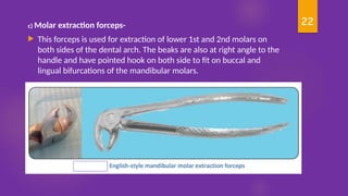
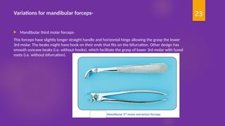
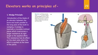
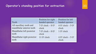
1
Extraction in pediatric dentistry
Dr Simran Vangani
2
Contents
 Introduction
 Indications for extraction
 Contraindication for extraction
 Preparation for extraction
 Extraction forceps
 Elevators
 Principles of extraction
 Techniques of extraction
 Operative Complications
3
Introduction-
 Exodontia is a branch of dentistry that deals with
the extraction/removal of teeth.
 Howe had described an ideal extraction as “the
painless removal of the whole tooth/ root with
minimum trauma to the investing tissues (hard
and soft), so that the wound heals uneventfully”.
 Removal of a tooth does not require large amount
of force, but fine and controlled force, so that
the tooth is gently lifted from alveolar process and
not pulled out.
4
Indication for extraction of teeth-
• Teeth affected by advanced caries and
its sequelae
• Teeth affected by periodontal disease
• Extraction of healthy teeth to correct
malocclusion
• Over-retained teeth
• Trauma to the teeth or jaws may cause
dislocation of a tooth from its socket
(avulsion)
• Extraction of teeth for esthetic reasons
• Extraction of teeth for prosthodontic reasons
• Impacted and supernumerary teeth
• Extraction of decayed 1st or 2nd molars to prevent
impaction of 3rd molars
• Teeth involved in fracture line
• Teeth involved in tumours or cysts
• Tooth as foci of infection
• Teeth affected by crown, abrasion, attrition or
hypoplasia
• Teeth affected by pulpal lesions e.g. pulpitis, pink
spot or pulp polyp
• Teeth in the area of direct therapeutic irradiation.
5
Contraindications of tooth extraction
 The removal of a tooth is contraindicated where acute symptoms of oral or systemic disease are manifested
in the child patient such as:
 Acute Systemic Infections After the acute stages of systemic infections, such as glomerulonephritis,
congenital heart disease, rheumatic fever, rheumatic heart disease, are reduced to chronicity, regimens of
chemoprophylaxis will be required before extractions.
 Blood Diseases -The hemophilic or leukemic child will require a well trained general dentist, a pediatric
dentist, or an oral surgeon along with a hematologist to perform satisfactorily the measures required during
tooth removal.
 Uncontrolled Diabetes Mellitus -Tooth removals should be avoided. Surgical wounds heal poorly, and
postoperative pain can be extreme. Recurrent hemorrhages may result.
 Irradiated Bone -Tooth removal should be avoided. If an extraction is necessary, it should be accomplished
before radiation therapy. Osteomyelitis usually develops following an extraction in an irradiated patient
because of osseous avascularity
 Acute Oral Infection -In the presence of oral infections, such as acute necrotizing ulcerative gingivitis,
acute herpetic stomatitis, acute dentoalveolar abscess, and other acute forms of oral disease , tooth removals
are definitely contraindicated until the infections are eliminated.
6
Special consideration while deciding whether a primary tooth
has to be extracted or retained:
 Child management: If the emergency is severe enough, the tooth has questionable prognosis and treatment may
require many appointments in a child who is difficult to manage then the tooth is extracted followed by placement
of a space maintainer .
 Degree of root resorption: If > ½ of the root is resorbed and the tooth requires pulp treatment then it is
extracted
 Space problems: If there is an existing space problem, early extraction of the tooth may allow space closure. In
such cases tooth has to be retained by pulp treatment
 Degree of parental concern: If parents exhibit an obvious lack of concern over the emergency situation related to
the injured tooth, the tooth is removed
 Habits: Deleterious oral habits if present will enhance the rate of space closure. In such cases tooth must be
retained
 Speech: Early loss of the anterior teeth may have a direct effect on the speech patterns and this is another
reason for maintaining the tooth in the arch .
 Esthetics: For psychological purpose it is better to postpone the extraction
7
PREPARATION FOR EXTRACTION
Preoperative Assessment-
A history of general disease, nervousness, or previous difficulty with extractions, will govern both the choice of
anesthesia and procedure of tooth extraction.
 The general cleanliness of the patient’s mouth and oral hygiene are observed.
 Pre-extraction scaling should be performed, especially in neglected mouths, at least one week prior to surgery.
 Sick or fatigued should rest before operative procedures.
 Highly apprehensive patient should receive some form of sedation before the operation.
 Patient undergoing general anaesthesia should be instructed to omit the previous meal and to take nothing by
mouth for at least 6 hours before extraction.
 Patient with inflamed or infected gingival should use an antiseptic mouth rinse before the extraction.
 Removable prostheses must be taken out of the patient’s mouth.
8
Pre-extraction Radiograph
The purpose of pre-extraction radiograph is to show the whole root structure and the alveolar bone
investing the tooth with IOPA, lateral oblique view, OPG. The following are the main indication for
preoperative radiographs:
 History of difficult or attempted extractions
 A tooth which is resistant to forceps extraction
 All mandibular and maxillary 3rd molars, in standing premolars or misplaced canines
 Pulp less teeth with resorbed roots
 Teeth affected by periodontal disease
 Traumatic teeth
 An isolated tooth
 Any partially erupted or unerupted tooth or retained root
 Retained deciduous tooth
 Submerged tooth
9
 Conditions which predisposes to dental or alveolar abnormality, e.g.
 Cleidocrania ldysostolia
 pseudo-anodontia
 Osteitisdeformans
 hypercementosed root
 Patient with therapeutic irradiation
 Osteopetrosia.
10
 Close relationship of tooth or root with
– Maxillary sinus
– Inferior alveolar canal
– Mental nerves
11
Choice of Anesthesia
 Teeth may be extracted under either local anesthesia or general
anesthesia and one should assess the indication and contraindications
of both before deciding which to use in a particular case. Most
extraction of tooth can be done with local anesthesia alone.
 To decrease the nervousness, relieve tension and control psychic
behaviour, sedation can be used in conjunction with the local
anesthesia. In young children, general anesthesia rather than local
anesthesia may be indicated to facilitate patient management.
 All patients with general anesthesia or local anesthesia should be
observed in a recovery area until they are able to go home unaided or
should be accompanied by adult .
12
Extraction Forceps
 The history of dental extraction forceps goes back to 322– 384 BC, at that time Aristotle
introduced the dental forceps.
 The anatomical design for the dental forceps was given by Sir John Toms in1841 and in
the 20th century, the current design of forceps and elevators was developed and it is still
in use till today.
 Each forceps has two handles: a hinge joint and two beaks
Forceps Design Styles-
 American pattern: Hinge is directed horizontally with the handles of the forceps.
 English pattern: Hinge is directed vertically to the handles of the forceps
 The forceps used for maxillary and mandibular teeth differ in design
13
Extraction Forceps
 These instruments are specifically designed for
removing teeth from their socket. In addition, the
extraction forceps can expand the alveolar bone
when used under controlled forces.
 Component of extraction forceps
1. Handle: This is the part that grasped by the
operator, on which the forces are applied. It
might be serrated to allow positive grip and
prevent slippage.
2. Hinge: It connects both parts of the forceps.
The hinge concentrates and delivers the forces
from the handle to the beaks.
3. Beaks: These are the functional component
that delivers the applied forces to the target
tooth. It specifically designed to fit the cervical
part of the tooth and varies according to
anatomy of the tooth to be extracted
14
Maxillary extraction forceps
a) Anterior extraction forceps-
These forceps are used to extract
the six anterior maxillary teeth
(i.e. from right canine to left
canine). The forceps is generally
straight when viewed from the
top and the side as well, having
the handle and the beaks at the
same level. The beaks are concave
from both sides to accommodate
the single root of these teeth.
15
b) Premolar extraction forceps
This forceps is used for extraction of upper 1st
and 2nd premolars on both sides of the dental
arch.
The forceps have slight S-shape configuration
(i.e. two curves), when viewed from the side. This
help to direct the forces to the long axis of the
tooth and also to accommodate the oral cavity,
avoiding trauma to opposite dentation and the
lower lip. The beaks are smooth and concave on
both sides to fit the root trunk of these teeth.
c) Molar extraction forceps-
 This forceps is used for extraction
maxillary 1st and 2nd molar teeth. Similar
to premolar forceps, it has an S-shape
configuration when viewed from the side.
 Since the maxillary molars have three
roots one palatal and two buccal, the
beaks are designed to fit on the buccal
bifurcation with pointed hook and a
smooth concave beak to fit on the single
palatal root. This requires that this
forceps to come in pairs right and left.
16
17
Variations for maxillary forceps-
 Maxillary third molar forceps-
This forceps is considered the longest among the maxillary extraction forceps due to the
posterior position of the 3rd molar. The beaks are smooth and concave to fit the wide
anatomical variations of 3rd molar roots
18
Maxillary cow-horn molar forceps-
 This forceps is used mainly for extraction of severely carious maxillary molar. It has
sharply pointed beaks that reach deep into the trifurcation. It can generate large
amount of forces that can, with uncontrolled use, fracture the buccal plate.
19
Maxillary root tip forceps-
This forceps have straight handle with angled, offset and narrow beaks. It is used
primarily for extraction upper retained roots
20
Mandibular extraction forceps:
1) Anterior extraction forceps-
These forceps are used to extract the six anterior mandibular teeth (i.e. from right canine
to left canine). The English-style forceps have vertical hinge with beaks 90° to handle (i.e.
at right angle). When being held in hand the beaks directed downward. The end of these
beaks are smooth and concave and in contact with each other, when the forceps is closed
21
2)Premolar extraction forceps-
This forceps is used for extraction of lower 1 st and 2nd premolars on both sides of the
dental arch. This forceps are similar to anterior extraction forceps except for that the
beaks are slightly broader and have slight spacing between each other when the
forceps is closed.
22
c) Molar extraction forceps-
 This forceps is used for extraction of lower 1st and 2nd molars on
both sides of the dental arch. The beaks are also at right angle to the
handle and have pointed hook on both side to fit on buccal and
lingual bifurcations of the mandibular molars.
23
Variations for mandibular forceps-
 Mandibular third molar forceps-
This forceps have slightly longer straight handle and horizontal hinge allowing the grasp the lower
3rd molar. The beaks might have hook on their ends that fits on the bifurcation. Other design has
smooth concave beaks (i.e. without hooks), which facilitate the grasp of lower 3rd molar with fused
roots (i.e. without bifurcation).
24
Mandibular cow-horn molar forceps-
 This forceps is variation of mandibular molar forceps. The beaks are semicircular with
sharp pointed ends that are designed to fit inside the bifurcation of lower 1st and 2nd
molar. The beaks use the buccal and lingual bone plates as fulcrum. When the operator
squeezes the handles, the tooth can be luxated from the socket. This forceps can also be
used to section carious lower molars by applying controlled forces on the bifurcation. This
forceps should be used with caution as it may generate great forces that might fracture the
alveolar bone.

25
Most pediatric dentists prefer the smaller pediatric extraction forceps; such
as the no. 150s and no. 151s for the following reasons:
• Their reduced size more easily allows placement in the smaller oral cavity of the child patient.
• The smaller pediatric forceps are more easily concealed by the operator’s hand
• The smaller working ends (beaks) more closely adapt to the anatomy of the primary teeth.
26
Proper Holding of the Forceps
 The handles of the forceps are held differently depending on the position of the tooth to be removed.
The forceps are picked up in the operator’s right hand with the thumb supporting it at its joint or just
below it and the forceps handles in the palm of the hand. The little finger should be inside the handle
during application to the tooth and during extraction, it should be on the handle. For maxillary
extractions, beaks are pointed towards superior direction, and for mandibular extractions, the beaks
are pointed downwards
27
Elevators
 Elevators are the instruments used for luxating (loosening) the teeth
before application of forceps making extraction easier, subsequently
avoiding complications like fracture of crowns, roots, and bone. They
are also used to remove fractured or surgically sectioned roots.
Elevators are single bladed instruments designed for specific purposes
delivering maximum mechanical advantage with minimum efforts.
 Elevators have three components-
Handle: It is of generous size for proper grip and delivering adequate but
controlled force. Handle can be a continuation of the shank or at a right
angle to the shank.
Shank: It connects the handle with the working end or blade of the
elevator. It is strong enough to transmit the force from the handle to the
blade.
Blade: It is the working end of the instrument and transmits the force to
the tooth, bone, or both to achieve the desired action.
28
29
Elevators works on principles of-
1. Wedge Principle:
Introduction of the blade of
an elevator between the
bone and tooth parallel to
the long axis of the tooth is
wedging. A wedge is
basically a movable inclined
plane which overcomes a
larger resistance at right
angle to the applied effort.
The resistance has its effect
on the slant side when the
effort is applied at the base
of the plane.
30
2. Lever Principle:
The elevator is the lever of first class. To gain mechanical advantage in first-class lever,
the effort arm must be longer (3/4th of the total length) than the resistance arm
divided by the fulcrum which lies on the bone.
31
3. Wheel and Axle
Principle:
In this principle, the effort is
applied to the circumference of a
wheel, which turns the axle
generating the force to raise a
weight.
32
Chair and patient positions for extraction-
33
Operator’s standing position for extraction
34
PRINCIPLE OF EXTRACTION
 In routine practice, the following three-time mechanical principles of extraction
should be followed for the well-being of the patients by doing atraumatic
extraction.
1) Expansion of the Socket- The extraction of a tooth requires the separation of its
attachment to the alveolar bone via the crestal and principal fibres of the PDL which
involves a process of expansion of alveolar socket. This is achieved by using the tooth as
the dilating instrument with the help forceps, to permit the removal of the tooth.
2) Use of a Lever and Fulcrum- This basic principle is used with elevators that force
a tooth or root out of the socket along the path of least resistance.
3) The Insertion of a Wedge - This is done between the tooth root surface and the
bony socket wall to help the tooth to rise in its socket
35
EXODONTIA TECHNIQUES
 The following techniques may be used for tooth removal:
o The forceps technique — closed method
o The elevator technique — open
o Transalveolar technique — open method
o Odontotomy
36
 Forceps Technique-
It is the most commonly used method for the extraction of teeth. But it should not be
used in difficult cases, e.g tooth with hypercementoid root or tooth with deformity of the
roots. This forceps technique gives least amount of trauma to soft tissues and hard tissue
of judiciously used. In multiple extractions the marginal gingival may have to be reflected
to permit rounding and smoothing of the sharp prominences of the alveolar process.
Care should be taken to preserves the height and breadth of the ridge for stability of a
future denture. Proper use of this technique involves the application of several basic
principles.
• The beaks of the selected forcep should be sealed as far apically as possible
without compression of the soft tissues after reflecting the cervical gingival.
• The placement of the beaks of the forceps should be as parallel as possible to the
long axis of the tooth.
• The application of excessive force should be avoided so that the fracture of the
alveolar process or tooth itself does not occur.
37
Elevator Techniques-
This technique is used in two ways:
1. Elevator as a lever : In this case, the alveolar crest serves as the fulcrum. The area of
the compressed bone should be removed with a file or rongeur to reduce the
postoperative pain and infection. With elevators, one should avoid traumatizing the
gingival and loosening of adjacent teeth. This method is used for the removal of whole or
nearly whole roots.
2. Elevator as a wedge: This principle is used for the removal of small root tips by way of
displacement. If the root tip cannot be dislodged from the socket easily, an open view
method should be used.
38
C)Trans alveolar Method (Open View Technique)
 This method is used where roots are inaccessible to routine
removal by forceps or by an elevator, when they cannot be
luxated with simple forces, or when the roots are covered by
bone. This method is far less traumatic than when there is
prolonged use of forceps or elevator attempted root removal.
39
Odontotomy
 In this method, the extraction procedure may be simplified by cutting a
tooth apart, e.g. in multirooted deciduous or permanent teeth with
divergent roots, where crown is decayed
40
PROCEDURE FOR EXTRACTION
 Instrumentation and Positioning
Instruments are selected and arranged according to the need and according to the surgeon’s preference.
 Position of the operator:
– When extracting any tooth except the right mandibular quadrant the operator stands on the right hand
side of the patient.
– For the removal of the teeth in right mandibular quadrant, the operator stands behind the patient.
– For maxillary teeth, the chair should be adjusted so that the site of operation is about 8 cm below the
shoulder level of the operator.
– During the extraction of mandibular tooth the chair height should be about 16 cm below the level of the
operator’s elbow.
– When the operator is standing behind the patient the chair should be adjusted to enable him to have a
clear view of the field of extraction.
All these aspects combined with good illumination of the operative field is an essential
condition for the successful extraction of the teeth
41
Technique
42
Extraction of permanent maxillary teeth
 Central incisors: These often have a conical root and rarely deformed or curved. They are
grasped with straight wide beaked forceps and can be safely rotated first in one direction
and then in the other direction until PDL attachment is broken and it can be taken out with
slight tractions
 Lateral incisors: They have slender roots which are often flattened on the mesial and distal
surfaces. A fine bladed forceps is used for the extraction of lateral incisors.
 Canines: These can be the most difficult upper teeth to remove because of the length and
frequent apical curvature of their roots. Since great force is needed to dislodge these
teeth, partial or total fracture of the labial wall of the alveolus is common. Forceps are
placed as high as possible under the gingival margin, and the tooth is then rotated back
and forth while upward pressure is maintained and traction is applied for its removal.
43
 1st premolar: It has two fine roots which may be both curved and divergent and fracture
occurs readily during extraction. Buccopalatal rocking with upper universal forceps or
bayonet forceps is used to locate the tooth and tooth should be removed in the direction
of least resistance.
 2nd premolar: These are much easier to extract than the 1st premolars because they have
only one root. Careful rotary motion with rocking to the buccal sides with gradual fraction
will usually deliver the tooth.
44
 1st molar: It usually has three divergent roots, strongest and longest of which is the palatal
root. The buccal roots are often curved distally. For the safe extraction of 1st molar, careful
rocking of the tooth buccally with upper universal or bayonet forceps is used to loosen the
palatal root, and buccopalatal traction aids in complete luxation of the tooth which is removed
without rotation.
 2nd molars: It can be removed by a technique similar to that used for 1st molar extraction.
Buccopalatal rocking and traction may be used and even moderate torsion is permissible to
detach and remove the tooth.
 3rd molars: 3rd molars may be removed with the same forceps that are used for 1st and 2nd
molar. The long axis of the maxillary 3rd molar is such that its crown is usually more posteriorly
placed than its roots. As a rule, teeth that are buccally inclined can be removed easily, those
distally inclined may fracture. No attempt should be made to apply forceps to either a semi
erupted maxillary 3rd molar unless both buccal and lingual surfaces are visible. If more pressure
is applied in an upward direction the tooth or root may be displaced into the maxillary antrum.
45
EXTRACTION OF MANDIBULAR TEETH
 Incisors: Lower incisors have fine roots with flattened sides. The supporting alveolar
process is very thin, and it is easy to luxate the tooth when it is rocked labially. Fine
bladed forceps should be used to grasp them, e.g. lower universal
 Canines: It is long and bulky, firmly embedded and difficult to extract the apex is often
inclined distally. A heavier bladed forceps should be used and movement in a
buccolingual direction is applied for extraction of this tooth
 Premolars: They have tapering roots and their apices may be distally inclined and
surrounded by thick compact bone. A forceps with blades fine enough to give ‘two
point contact’ on the root should be applied to the tooth. The first movement should
be firm but gentle and torsion may be employed freely, combined with buccolingual
rocking as in the case of canines.
46
 Lower molars:
These molars are best extracted with full molar forceps and often loosened by
buccolingual pressure and are best delivered by secondary rotation. The
extraction of 2nd and 3rd molars can often be facilitated by the mesial
application of an elevator before the application of forceps if not malposed,
impacted or unerupted, the mandibular 3rd molars can be quite easily
removed with the forceps technique.
47
EXTRACTION OF ROOTS-
Extraction of roots-
Roots may be extracted with forceps: If they are not decayed. Bayonet or universal forceps are used for roots in
the upper jaw and forceps such as those used for premolars are used in the mandible.
 If forceps cannot be applied directly to the roots, an elevator technique may be used.
 In open beak technique, alveolar bone rather than the root itself is grasped with the forceps
and crushed bone should be carefully removed after removal of the root.
 Mandibular molar roots can be removed by placing a straight elevator or cryer elevator
between them and using the interradicular septum as a tulcorum to remove one root. If roots
are attached, a bur is first used to separate them.
 Maxillary molar roots removed by simultaneously grasping the distobuccal and palatal roots
with the forceps and mesiobuccal root can be removed separately with forceps or a small
elevator.
 Roots that are under the gingival margin or roots completely embedded in bone are
removed by the open view method of extraction.
48
Extraction of deciduous teeth
• Before extraction of deciduous teeth, a thorough examination should be performed to
minimize complications.
 As tooth crown and root structure differ from those of adult teeth, the use of specially
designed pediatric instrument is recommended.
 The main consideration in the removal of deciduous teeth is to avoid injury to the
developing permanent dentition.
 The most critical step in extraction of deciduous teeth is the administration of local
anesthesia. If the child allows this step then he will be definitely co-operative for the next
step, the extraction. This is because most anxiety and fear is generated during this phase.
Studies by most authors explain the rise of pulse rate and blood pressure during this time.
So it is critical to alleviate the fear of the child rather than increase it. It is most
recommended to perform some behavior shaping of children prior to extraction and local
anesthesia.
49
Some methods are:
 The first step: This is to make the patient comfortable. It is imperative that
we do not proceed with the extraction immediately. It is best if we first
engage in some friendly talk with the child and explain him the merits of
taking out his carious teeth in a language that he can comprehend
according to the developmental status of the child.
 Tell–show–feel–do: This modification involves describing the procedure
from the application of topical anesthetic to postoperative reward. The
patient is then showed an empty syringe without needle and made to feel it
to dispel any fears of injections that he may have. However, during the
actual procedure it is best not to load anesthetic or bring the needle or
syringe in front of child so as to avoid anxiety. It is best to cover the child’s
eye with one hand and perform the task with other.
 Use of euphemisms: Like comparing the pinch of needle to mosquito bite
or comparing LA solution to water to flush out bacteria from teeth have
proven to be useful.
50
– Audiovisual distraction: It is also a vital technique as it allows multisensory
distraction.
– Use of bite blocks: These are recommended for difficult patients who have a
tendency to close their mouth while the procedure as they are helpful in opening
the mouth so as to avoid any injury during procedure.
– Modeling: This is especially useful in case of a close friend or a sibling who can be
observed performing the desired behavior.
– Physical restraints: This is the last and least preferred option with the dentist and is
used in highly uncooperative or special children.
51
The technique of extraction is the same as that used in the removal of permanent teeth.
But it is important to ensure before application of forceps that the blades are fine enough
to pass down the periodontal membranes and applied to the roots.
• A firm lingual movement usually causes the tooth to rise in its socket and it can be
delivered by moving buccally and rotated forwards.
• The roots of the extracted deciduous teeth should be examined to ensure that they are
complete. Fracture root surfaces are flat and shiny with sharp margins, resorbed roots are
with irregular margins.
• In case of fracture of a root fragment the best option is to radiographically visualize it
before attempting any kind of retrieval. In case it is located superficially away from
underlying tooth bud it can be safely removed by reinstrumentation. However, if it is close
to the underlying tooth bud it is advisable to let it remain there as it may undergo
resorption or may appear with the erupting tooth.
52
OPERATIVE COMPLICATIONS
The most frequent operative complication that encounter during the extraction of teeth are:
• Fracture of the tooth
• Injuries to adjacent teeth
• Fracture of the alveolar bone
• Fracture of the tuberosity
• Extraction of the wrong tooth
• Root displaced in the sinus
• Maxillary sinus perforation
• Root displaced in the submandibular space
• Gingival and mucosal lacerations
• Injury to the inferior alveolar nerve
• Hemorrhage and hematoma
• TMJ trauma
• Damage to permanent successor
53
POSTOPERATIVE CARE
 After care when the tooth has been extracted the socket should be inspected and any loose
fragment of bone is removed or necessary socket irrigation is performed. The alveolar process
then should be pressed together with the thumb and forefinger in order to reduce any distortion
of the supporting tissues; suturing should always be done after multiple extractions and if the
gingival flaps are loose enough to be approximated. After extraction, a gauze pack is placed over
the socket and patient is directed to bite on the pack for ½ hour, exerting firm even pressure.
This will prevent bleeding while the patient returns home and it allows a blood clot to form.
Some postoperative instructions are:
 The patient should be warned that sucking the wound, investigating the socket with tongue and
rinsing during the first day disturbs the blood clot and may cause dry socket.
54
 Patient should be directed to remain quiet for several hours, preferably sitting in a chair or if lying down, keeping the head
elevated.
 Only liquids and soft solids should be advice on the first day. They may be warm or cold but not extremely hot.
 The teeth should be brushed as usual and on the day after surgery rinsing of the mouth should begin. A warm saline solution
is best for this purpose.
 Some degree of postoperative pain accompanies many exodontia procedures and begins after the effects of the anesthetic
have left. So, it is better to take some analgesic before the effect of anesthetic wears off.
 Prevention of swelling after extensive or difficult operation adds to the comfort of the patient. The degree of swelling that
occurs postoperatively is generally in direct proportion to the degree of surgical trauma. The application of cold to the
operated site is beneficial in reducing the amount of postoperative swelling. Pressure dressings are also beneficial in limiting
the postoperative swelling postoperative swelling.
 Smoking should be avoided after tooth extraction as it increases the incidence of alveolar osteitis and should be discontinued
for five days.
55
56
REFERENCES
 Howe GL. The extraction of teeth. 2nd ed. Bristol: J Wright; 1974.
 Principles and Techniques of Exodontia-Oral and Maxillofacial Surgery for
the Clinician, 2021 ISBN : 978-981-15-1345-9
 Textbook of Oral and Maxillofacial Surgery 2021by Neelima Anil Malik
 Pinkham JR. Pediatric Dentistry : Infancy through Adolescence. 4th ed. St.
Louis Mo: Elsevier Saunders; 2005.
 McDonald and Avery's Dentistry for the Child and Adolescent Book - Tenth
Edition - 2016
 Marwah Nikhil. 2014. Textbook of Pediatric Dentistry. 3rd ed. New Delhi:
Jaypee Brothers Medical.
57
Thankyou

Extraction in pediatric dentistry........

  • 1.
    1 Extraction in pediatricdentistry Dr Simran Vangani
  • 2.
    2 Contents  Introduction  Indicationsfor extraction  Contraindication for extraction  Preparation for extraction  Extraction forceps  Elevators  Principles of extraction  Techniques of extraction  Operative Complications
  • 3.
    3 Introduction-  Exodontia isa branch of dentistry that deals with the extraction/removal of teeth.  Howe had described an ideal extraction as “the painless removal of the whole tooth/ root with minimum trauma to the investing tissues (hard and soft), so that the wound heals uneventfully”.  Removal of a tooth does not require large amount of force, but fine and controlled force, so that the tooth is gently lifted from alveolar process and not pulled out.
  • 4.
    4 Indication for extractionof teeth- • Teeth affected by advanced caries and its sequelae • Teeth affected by periodontal disease • Extraction of healthy teeth to correct malocclusion • Over-retained teeth • Trauma to the teeth or jaws may cause dislocation of a tooth from its socket (avulsion) • Extraction of teeth for esthetic reasons • Extraction of teeth for prosthodontic reasons • Impacted and supernumerary teeth • Extraction of decayed 1st or 2nd molars to prevent impaction of 3rd molars • Teeth involved in fracture line • Teeth involved in tumours or cysts • Tooth as foci of infection • Teeth affected by crown, abrasion, attrition or hypoplasia • Teeth affected by pulpal lesions e.g. pulpitis, pink spot or pulp polyp • Teeth in the area of direct therapeutic irradiation.
  • 5.
    5 Contraindications of toothextraction  The removal of a tooth is contraindicated where acute symptoms of oral or systemic disease are manifested in the child patient such as:  Acute Systemic Infections After the acute stages of systemic infections, such as glomerulonephritis, congenital heart disease, rheumatic fever, rheumatic heart disease, are reduced to chronicity, regimens of chemoprophylaxis will be required before extractions.  Blood Diseases -The hemophilic or leukemic child will require a well trained general dentist, a pediatric dentist, or an oral surgeon along with a hematologist to perform satisfactorily the measures required during tooth removal.  Uncontrolled Diabetes Mellitus -Tooth removals should be avoided. Surgical wounds heal poorly, and postoperative pain can be extreme. Recurrent hemorrhages may result.  Irradiated Bone -Tooth removal should be avoided. If an extraction is necessary, it should be accomplished before radiation therapy. Osteomyelitis usually develops following an extraction in an irradiated patient because of osseous avascularity  Acute Oral Infection -In the presence of oral infections, such as acute necrotizing ulcerative gingivitis, acute herpetic stomatitis, acute dentoalveolar abscess, and other acute forms of oral disease , tooth removals are definitely contraindicated until the infections are eliminated.
  • 6.
    6 Special consideration whiledeciding whether a primary tooth has to be extracted or retained:  Child management: If the emergency is severe enough, the tooth has questionable prognosis and treatment may require many appointments in a child who is difficult to manage then the tooth is extracted followed by placement of a space maintainer .  Degree of root resorption: If > ½ of the root is resorbed and the tooth requires pulp treatment then it is extracted  Space problems: If there is an existing space problem, early extraction of the tooth may allow space closure. In such cases tooth has to be retained by pulp treatment  Degree of parental concern: If parents exhibit an obvious lack of concern over the emergency situation related to the injured tooth, the tooth is removed  Habits: Deleterious oral habits if present will enhance the rate of space closure. In such cases tooth must be retained  Speech: Early loss of the anterior teeth may have a direct effect on the speech patterns and this is another reason for maintaining the tooth in the arch .  Esthetics: For psychological purpose it is better to postpone the extraction
  • 7.
    7 PREPARATION FOR EXTRACTION PreoperativeAssessment- A history of general disease, nervousness, or previous difficulty with extractions, will govern both the choice of anesthesia and procedure of tooth extraction.  The general cleanliness of the patient’s mouth and oral hygiene are observed.  Pre-extraction scaling should be performed, especially in neglected mouths, at least one week prior to surgery.  Sick or fatigued should rest before operative procedures.  Highly apprehensive patient should receive some form of sedation before the operation.  Patient undergoing general anaesthesia should be instructed to omit the previous meal and to take nothing by mouth for at least 6 hours before extraction.  Patient with inflamed or infected gingival should use an antiseptic mouth rinse before the extraction.  Removable prostheses must be taken out of the patient’s mouth.
  • 8.
    8 Pre-extraction Radiograph The purposeof pre-extraction radiograph is to show the whole root structure and the alveolar bone investing the tooth with IOPA, lateral oblique view, OPG. The following are the main indication for preoperative radiographs:  History of difficult or attempted extractions  A tooth which is resistant to forceps extraction  All mandibular and maxillary 3rd molars, in standing premolars or misplaced canines  Pulp less teeth with resorbed roots  Teeth affected by periodontal disease  Traumatic teeth  An isolated tooth  Any partially erupted or unerupted tooth or retained root  Retained deciduous tooth  Submerged tooth
  • 9.
    9  Conditions whichpredisposes to dental or alveolar abnormality, e.g.  Cleidocrania ldysostolia  pseudo-anodontia  Osteitisdeformans  hypercementosed root  Patient with therapeutic irradiation  Osteopetrosia.
  • 10.
    10  Close relationshipof tooth or root with – Maxillary sinus – Inferior alveolar canal – Mental nerves
  • 11.
    11 Choice of Anesthesia Teeth may be extracted under either local anesthesia or general anesthesia and one should assess the indication and contraindications of both before deciding which to use in a particular case. Most extraction of tooth can be done with local anesthesia alone.  To decrease the nervousness, relieve tension and control psychic behaviour, sedation can be used in conjunction with the local anesthesia. In young children, general anesthesia rather than local anesthesia may be indicated to facilitate patient management.  All patients with general anesthesia or local anesthesia should be observed in a recovery area until they are able to go home unaided or should be accompanied by adult .
  • 12.
    12 Extraction Forceps  Thehistory of dental extraction forceps goes back to 322– 384 BC, at that time Aristotle introduced the dental forceps.  The anatomical design for the dental forceps was given by Sir John Toms in1841 and in the 20th century, the current design of forceps and elevators was developed and it is still in use till today.  Each forceps has two handles: a hinge joint and two beaks Forceps Design Styles-  American pattern: Hinge is directed horizontally with the handles of the forceps.  English pattern: Hinge is directed vertically to the handles of the forceps  The forceps used for maxillary and mandibular teeth differ in design
  • 13.
    13 Extraction Forceps  Theseinstruments are specifically designed for removing teeth from their socket. In addition, the extraction forceps can expand the alveolar bone when used under controlled forces.  Component of extraction forceps 1. Handle: This is the part that grasped by the operator, on which the forces are applied. It might be serrated to allow positive grip and prevent slippage. 2. Hinge: It connects both parts of the forceps. The hinge concentrates and delivers the forces from the handle to the beaks. 3. Beaks: These are the functional component that delivers the applied forces to the target tooth. It specifically designed to fit the cervical part of the tooth and varies according to anatomy of the tooth to be extracted
  • 14.
    14 Maxillary extraction forceps a)Anterior extraction forceps- These forceps are used to extract the six anterior maxillary teeth (i.e. from right canine to left canine). The forceps is generally straight when viewed from the top and the side as well, having the handle and the beaks at the same level. The beaks are concave from both sides to accommodate the single root of these teeth.
  • 15.
    15 b) Premolar extractionforceps This forceps is used for extraction of upper 1st and 2nd premolars on both sides of the dental arch. The forceps have slight S-shape configuration (i.e. two curves), when viewed from the side. This help to direct the forces to the long axis of the tooth and also to accommodate the oral cavity, avoiding trauma to opposite dentation and the lower lip. The beaks are smooth and concave on both sides to fit the root trunk of these teeth.
  • 16.
    c) Molar extractionforceps-  This forceps is used for extraction maxillary 1st and 2nd molar teeth. Similar to premolar forceps, it has an S-shape configuration when viewed from the side.  Since the maxillary molars have three roots one palatal and two buccal, the beaks are designed to fit on the buccal bifurcation with pointed hook and a smooth concave beak to fit on the single palatal root. This requires that this forceps to come in pairs right and left. 16
  • 17.
    17 Variations for maxillaryforceps-  Maxillary third molar forceps- This forceps is considered the longest among the maxillary extraction forceps due to the posterior position of the 3rd molar. The beaks are smooth and concave to fit the wide anatomical variations of 3rd molar roots
  • 18.
    18 Maxillary cow-horn molarforceps-  This forceps is used mainly for extraction of severely carious maxillary molar. It has sharply pointed beaks that reach deep into the trifurcation. It can generate large amount of forces that can, with uncontrolled use, fracture the buccal plate.
  • 19.
    19 Maxillary root tipforceps- This forceps have straight handle with angled, offset and narrow beaks. It is used primarily for extraction upper retained roots
  • 20.
    20 Mandibular extraction forceps: 1)Anterior extraction forceps- These forceps are used to extract the six anterior mandibular teeth (i.e. from right canine to left canine). The English-style forceps have vertical hinge with beaks 90° to handle (i.e. at right angle). When being held in hand the beaks directed downward. The end of these beaks are smooth and concave and in contact with each other, when the forceps is closed
  • 21.
    21 2)Premolar extraction forceps- Thisforceps is used for extraction of lower 1 st and 2nd premolars on both sides of the dental arch. This forceps are similar to anterior extraction forceps except for that the beaks are slightly broader and have slight spacing between each other when the forceps is closed.
  • 22.
    22 c) Molar extractionforceps-  This forceps is used for extraction of lower 1st and 2nd molars on both sides of the dental arch. The beaks are also at right angle to the handle and have pointed hook on both side to fit on buccal and lingual bifurcations of the mandibular molars.
  • 23.
    23 Variations for mandibularforceps-  Mandibular third molar forceps- This forceps have slightly longer straight handle and horizontal hinge allowing the grasp the lower 3rd molar. The beaks might have hook on their ends that fits on the bifurcation. Other design has smooth concave beaks (i.e. without hooks), which facilitate the grasp of lower 3rd molar with fused roots (i.e. without bifurcation).
  • 24.
    24 Mandibular cow-horn molarforceps-  This forceps is variation of mandibular molar forceps. The beaks are semicircular with sharp pointed ends that are designed to fit inside the bifurcation of lower 1st and 2nd molar. The beaks use the buccal and lingual bone plates as fulcrum. When the operator squeezes the handles, the tooth can be luxated from the socket. This forceps can also be used to section carious lower molars by applying controlled forces on the bifurcation. This forceps should be used with caution as it may generate great forces that might fracture the alveolar bone. 
  • 25.
    25 Most pediatric dentistsprefer the smaller pediatric extraction forceps; such as the no. 150s and no. 151s for the following reasons: • Their reduced size more easily allows placement in the smaller oral cavity of the child patient. • The smaller pediatric forceps are more easily concealed by the operator’s hand • The smaller working ends (beaks) more closely adapt to the anatomy of the primary teeth.
  • 26.
    26 Proper Holding ofthe Forceps  The handles of the forceps are held differently depending on the position of the tooth to be removed. The forceps are picked up in the operator’s right hand with the thumb supporting it at its joint or just below it and the forceps handles in the palm of the hand. The little finger should be inside the handle during application to the tooth and during extraction, it should be on the handle. For maxillary extractions, beaks are pointed towards superior direction, and for mandibular extractions, the beaks are pointed downwards
  • 27.
    27 Elevators  Elevators arethe instruments used for luxating (loosening) the teeth before application of forceps making extraction easier, subsequently avoiding complications like fracture of crowns, roots, and bone. They are also used to remove fractured or surgically sectioned roots. Elevators are single bladed instruments designed for specific purposes delivering maximum mechanical advantage with minimum efforts.  Elevators have three components- Handle: It is of generous size for proper grip and delivering adequate but controlled force. Handle can be a continuation of the shank or at a right angle to the shank. Shank: It connects the handle with the working end or blade of the elevator. It is strong enough to transmit the force from the handle to the blade. Blade: It is the working end of the instrument and transmits the force to the tooth, bone, or both to achieve the desired action.
  • 28.
  • 29.
    29 Elevators works onprinciples of- 1. Wedge Principle: Introduction of the blade of an elevator between the bone and tooth parallel to the long axis of the tooth is wedging. A wedge is basically a movable inclined plane which overcomes a larger resistance at right angle to the applied effort. The resistance has its effect on the slant side when the effort is applied at the base of the plane.
  • 30.
    30 2. Lever Principle: Theelevator is the lever of first class. To gain mechanical advantage in first-class lever, the effort arm must be longer (3/4th of the total length) than the resistance arm divided by the fulcrum which lies on the bone.
  • 31.
    31 3. Wheel andAxle Principle: In this principle, the effort is applied to the circumference of a wheel, which turns the axle generating the force to raise a weight.
  • 32.
    32 Chair and patientpositions for extraction-
  • 33.
  • 34.
    34 PRINCIPLE OF EXTRACTION In routine practice, the following three-time mechanical principles of extraction should be followed for the well-being of the patients by doing atraumatic extraction. 1) Expansion of the Socket- The extraction of a tooth requires the separation of its attachment to the alveolar bone via the crestal and principal fibres of the PDL which involves a process of expansion of alveolar socket. This is achieved by using the tooth as the dilating instrument with the help forceps, to permit the removal of the tooth. 2) Use of a Lever and Fulcrum- This basic principle is used with elevators that force a tooth or root out of the socket along the path of least resistance. 3) The Insertion of a Wedge - This is done between the tooth root surface and the bony socket wall to help the tooth to rise in its socket
  • 35.
    35 EXODONTIA TECHNIQUES  Thefollowing techniques may be used for tooth removal: o The forceps technique — closed method o The elevator technique — open o Transalveolar technique — open method o Odontotomy
  • 36.
    36  Forceps Technique- Itis the most commonly used method for the extraction of teeth. But it should not be used in difficult cases, e.g tooth with hypercementoid root or tooth with deformity of the roots. This forceps technique gives least amount of trauma to soft tissues and hard tissue of judiciously used. In multiple extractions the marginal gingival may have to be reflected to permit rounding and smoothing of the sharp prominences of the alveolar process. Care should be taken to preserves the height and breadth of the ridge for stability of a future denture. Proper use of this technique involves the application of several basic principles. • The beaks of the selected forcep should be sealed as far apically as possible without compression of the soft tissues after reflecting the cervical gingival. • The placement of the beaks of the forceps should be as parallel as possible to the long axis of the tooth. • The application of excessive force should be avoided so that the fracture of the alveolar process or tooth itself does not occur.
  • 37.
    37 Elevator Techniques- This techniqueis used in two ways: 1. Elevator as a lever : In this case, the alveolar crest serves as the fulcrum. The area of the compressed bone should be removed with a file or rongeur to reduce the postoperative pain and infection. With elevators, one should avoid traumatizing the gingival and loosening of adjacent teeth. This method is used for the removal of whole or nearly whole roots. 2. Elevator as a wedge: This principle is used for the removal of small root tips by way of displacement. If the root tip cannot be dislodged from the socket easily, an open view method should be used.
  • 38.
    38 C)Trans alveolar Method(Open View Technique)  This method is used where roots are inaccessible to routine removal by forceps or by an elevator, when they cannot be luxated with simple forces, or when the roots are covered by bone. This method is far less traumatic than when there is prolonged use of forceps or elevator attempted root removal.
  • 39.
    39 Odontotomy  In thismethod, the extraction procedure may be simplified by cutting a tooth apart, e.g. in multirooted deciduous or permanent teeth with divergent roots, where crown is decayed
  • 40.
    40 PROCEDURE FOR EXTRACTION Instrumentation and Positioning Instruments are selected and arranged according to the need and according to the surgeon’s preference.  Position of the operator: – When extracting any tooth except the right mandibular quadrant the operator stands on the right hand side of the patient. – For the removal of the teeth in right mandibular quadrant, the operator stands behind the patient. – For maxillary teeth, the chair should be adjusted so that the site of operation is about 8 cm below the shoulder level of the operator. – During the extraction of mandibular tooth the chair height should be about 16 cm below the level of the operator’s elbow. – When the operator is standing behind the patient the chair should be adjusted to enable him to have a clear view of the field of extraction. All these aspects combined with good illumination of the operative field is an essential condition for the successful extraction of the teeth
  • 41.
  • 42.
    42 Extraction of permanentmaxillary teeth  Central incisors: These often have a conical root and rarely deformed or curved. They are grasped with straight wide beaked forceps and can be safely rotated first in one direction and then in the other direction until PDL attachment is broken and it can be taken out with slight tractions  Lateral incisors: They have slender roots which are often flattened on the mesial and distal surfaces. A fine bladed forceps is used for the extraction of lateral incisors.  Canines: These can be the most difficult upper teeth to remove because of the length and frequent apical curvature of their roots. Since great force is needed to dislodge these teeth, partial or total fracture of the labial wall of the alveolus is common. Forceps are placed as high as possible under the gingival margin, and the tooth is then rotated back and forth while upward pressure is maintained and traction is applied for its removal.
  • 43.
    43  1st premolar:It has two fine roots which may be both curved and divergent and fracture occurs readily during extraction. Buccopalatal rocking with upper universal forceps or bayonet forceps is used to locate the tooth and tooth should be removed in the direction of least resistance.  2nd premolar: These are much easier to extract than the 1st premolars because they have only one root. Careful rotary motion with rocking to the buccal sides with gradual fraction will usually deliver the tooth.
  • 44.
    44  1st molar:It usually has three divergent roots, strongest and longest of which is the palatal root. The buccal roots are often curved distally. For the safe extraction of 1st molar, careful rocking of the tooth buccally with upper universal or bayonet forceps is used to loosen the palatal root, and buccopalatal traction aids in complete luxation of the tooth which is removed without rotation.  2nd molars: It can be removed by a technique similar to that used for 1st molar extraction. Buccopalatal rocking and traction may be used and even moderate torsion is permissible to detach and remove the tooth.  3rd molars: 3rd molars may be removed with the same forceps that are used for 1st and 2nd molar. The long axis of the maxillary 3rd molar is such that its crown is usually more posteriorly placed than its roots. As a rule, teeth that are buccally inclined can be removed easily, those distally inclined may fracture. No attempt should be made to apply forceps to either a semi erupted maxillary 3rd molar unless both buccal and lingual surfaces are visible. If more pressure is applied in an upward direction the tooth or root may be displaced into the maxillary antrum.
  • 45.
    45 EXTRACTION OF MANDIBULARTEETH  Incisors: Lower incisors have fine roots with flattened sides. The supporting alveolar process is very thin, and it is easy to luxate the tooth when it is rocked labially. Fine bladed forceps should be used to grasp them, e.g. lower universal  Canines: It is long and bulky, firmly embedded and difficult to extract the apex is often inclined distally. A heavier bladed forceps should be used and movement in a buccolingual direction is applied for extraction of this tooth  Premolars: They have tapering roots and their apices may be distally inclined and surrounded by thick compact bone. A forceps with blades fine enough to give ‘two point contact’ on the root should be applied to the tooth. The first movement should be firm but gentle and torsion may be employed freely, combined with buccolingual rocking as in the case of canines.
  • 46.
    46  Lower molars: Thesemolars are best extracted with full molar forceps and often loosened by buccolingual pressure and are best delivered by secondary rotation. The extraction of 2nd and 3rd molars can often be facilitated by the mesial application of an elevator before the application of forceps if not malposed, impacted or unerupted, the mandibular 3rd molars can be quite easily removed with the forceps technique.
  • 47.
    47 EXTRACTION OF ROOTS- Extractionof roots- Roots may be extracted with forceps: If they are not decayed. Bayonet or universal forceps are used for roots in the upper jaw and forceps such as those used for premolars are used in the mandible.  If forceps cannot be applied directly to the roots, an elevator technique may be used.  In open beak technique, alveolar bone rather than the root itself is grasped with the forceps and crushed bone should be carefully removed after removal of the root.  Mandibular molar roots can be removed by placing a straight elevator or cryer elevator between them and using the interradicular septum as a tulcorum to remove one root. If roots are attached, a bur is first used to separate them.  Maxillary molar roots removed by simultaneously grasping the distobuccal and palatal roots with the forceps and mesiobuccal root can be removed separately with forceps or a small elevator.  Roots that are under the gingival margin or roots completely embedded in bone are removed by the open view method of extraction.
  • 48.
    48 Extraction of deciduousteeth • Before extraction of deciduous teeth, a thorough examination should be performed to minimize complications.  As tooth crown and root structure differ from those of adult teeth, the use of specially designed pediatric instrument is recommended.  The main consideration in the removal of deciduous teeth is to avoid injury to the developing permanent dentition.  The most critical step in extraction of deciduous teeth is the administration of local anesthesia. If the child allows this step then he will be definitely co-operative for the next step, the extraction. This is because most anxiety and fear is generated during this phase. Studies by most authors explain the rise of pulse rate and blood pressure during this time. So it is critical to alleviate the fear of the child rather than increase it. It is most recommended to perform some behavior shaping of children prior to extraction and local anesthesia.
  • 49.
    49 Some methods are: The first step: This is to make the patient comfortable. It is imperative that we do not proceed with the extraction immediately. It is best if we first engage in some friendly talk with the child and explain him the merits of taking out his carious teeth in a language that he can comprehend according to the developmental status of the child.  Tell–show–feel–do: This modification involves describing the procedure from the application of topical anesthetic to postoperative reward. The patient is then showed an empty syringe without needle and made to feel it to dispel any fears of injections that he may have. However, during the actual procedure it is best not to load anesthetic or bring the needle or syringe in front of child so as to avoid anxiety. It is best to cover the child’s eye with one hand and perform the task with other.  Use of euphemisms: Like comparing the pinch of needle to mosquito bite or comparing LA solution to water to flush out bacteria from teeth have proven to be useful.
  • 50.
    50 – Audiovisual distraction:It is also a vital technique as it allows multisensory distraction. – Use of bite blocks: These are recommended for difficult patients who have a tendency to close their mouth while the procedure as they are helpful in opening the mouth so as to avoid any injury during procedure. – Modeling: This is especially useful in case of a close friend or a sibling who can be observed performing the desired behavior. – Physical restraints: This is the last and least preferred option with the dentist and is used in highly uncooperative or special children.
  • 51.
    51 The technique ofextraction is the same as that used in the removal of permanent teeth. But it is important to ensure before application of forceps that the blades are fine enough to pass down the periodontal membranes and applied to the roots. • A firm lingual movement usually causes the tooth to rise in its socket and it can be delivered by moving buccally and rotated forwards. • The roots of the extracted deciduous teeth should be examined to ensure that they are complete. Fracture root surfaces are flat and shiny with sharp margins, resorbed roots are with irregular margins. • In case of fracture of a root fragment the best option is to radiographically visualize it before attempting any kind of retrieval. In case it is located superficially away from underlying tooth bud it can be safely removed by reinstrumentation. However, if it is close to the underlying tooth bud it is advisable to let it remain there as it may undergo resorption or may appear with the erupting tooth.
  • 52.
    52 OPERATIVE COMPLICATIONS The mostfrequent operative complication that encounter during the extraction of teeth are: • Fracture of the tooth • Injuries to adjacent teeth • Fracture of the alveolar bone • Fracture of the tuberosity • Extraction of the wrong tooth • Root displaced in the sinus • Maxillary sinus perforation • Root displaced in the submandibular space • Gingival and mucosal lacerations • Injury to the inferior alveolar nerve • Hemorrhage and hematoma • TMJ trauma • Damage to permanent successor
  • 53.
    53 POSTOPERATIVE CARE  Aftercare when the tooth has been extracted the socket should be inspected and any loose fragment of bone is removed or necessary socket irrigation is performed. The alveolar process then should be pressed together with the thumb and forefinger in order to reduce any distortion of the supporting tissues; suturing should always be done after multiple extractions and if the gingival flaps are loose enough to be approximated. After extraction, a gauze pack is placed over the socket and patient is directed to bite on the pack for ½ hour, exerting firm even pressure. This will prevent bleeding while the patient returns home and it allows a blood clot to form. Some postoperative instructions are:  The patient should be warned that sucking the wound, investigating the socket with tongue and rinsing during the first day disturbs the blood clot and may cause dry socket.
  • 54.
    54  Patient shouldbe directed to remain quiet for several hours, preferably sitting in a chair or if lying down, keeping the head elevated.  Only liquids and soft solids should be advice on the first day. They may be warm or cold but not extremely hot.  The teeth should be brushed as usual and on the day after surgery rinsing of the mouth should begin. A warm saline solution is best for this purpose.  Some degree of postoperative pain accompanies many exodontia procedures and begins after the effects of the anesthetic have left. So, it is better to take some analgesic before the effect of anesthetic wears off.  Prevention of swelling after extensive or difficult operation adds to the comfort of the patient. The degree of swelling that occurs postoperatively is generally in direct proportion to the degree of surgical trauma. The application of cold to the operated site is beneficial in reducing the amount of postoperative swelling. Pressure dressings are also beneficial in limiting the postoperative swelling postoperative swelling.  Smoking should be avoided after tooth extraction as it increases the incidence of alveolar osteitis and should be discontinued for five days.
  • 55.
  • 56.
    56 REFERENCES  Howe GL.The extraction of teeth. 2nd ed. Bristol: J Wright; 1974.  Principles and Techniques of Exodontia-Oral and Maxillofacial Surgery for the Clinician, 2021 ISBN : 978-981-15-1345-9  Textbook of Oral and Maxillofacial Surgery 2021by Neelima Anil Malik  Pinkham JR. Pediatric Dentistry : Infancy through Adolescence. 4th ed. St. Louis Mo: Elsevier Saunders; 2005.  McDonald and Avery's Dentistry for the Child and Adolescent Book - Tenth Edition - 2016  Marwah Nikhil. 2014. Textbook of Pediatric Dentistry. 3rd ed. New Delhi: Jaypee Brothers Medical.
  • 57.