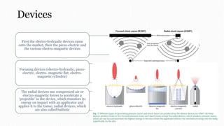
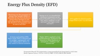
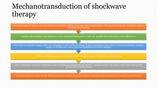
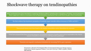
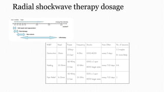
Extracorporeal Shockwave Therapy
Aditya Johan Romadhon
Introduction
ESWT is currently delivered through two primary forms: focused shockwave therapy (F-SWT) and radial shockwave therapy (R-SWT)
Shockwaves are a form of energy that has biological effects described at the cellular, tissue, and organ level
In general, a shock wave is the result of a sudden release of chemical, electrical, nuclear, or mechanical energy
Shock waves are mechanical waves, they propagate in a medium which also deforms or changes its density
The mechanical energy of shock waves was transmitted through the intact skin and concentrated on the stone without signifi cant damage of the tissue
Extracorporeal shock wave lithotripsy (SWL) is a gentle and noninvasive treatment procedure suitable for a wide range of kidney and ureteral stones
Extracorporeal shock wave therapy (ESWT) is a noninvasive form of treatment, that has been developed from ESWL (extracorporeal shock wave lithotripsy)
DOI: 10.1302/2058-
5241.5.190067
Devices
First the electro-hydraulic devices came
onto the market, then the piezo-electric and
the various electro-magnetic devices
Focusing devices (electro-hydraulic, piezo-
electric, electro- magnetic flat, electro-
magnetic cylindric)
The radial devices use compressed air or
electro-magnetic forces to accelerate a
‘projectile’ in the device, which transfers its
energy on impact with an applicator and
applies it to the tissue, radial devices, which
are also called ballistic
Physics
Typical characteristics of shock waves include a short rise time on
the order of a few nanoseconds reaching a peak pressure of up to
100 MPa or 1,000 times atmospheric pressure
Medically used shock waves are mechanical waves featuring
extremely high-pressure amplitudes in the range of 10–100 MPa
(100–1,000 bars)
The peak pressure in the focus can exceed 50 MPa for focusing
devices while the pressure peaks of approximately 15 MPa are
significantly lower for radial devices
The rise times are even more clearly different: with focusing
devices, the rise times are in the range of 5–10 ns at high
energies, with radial devices are 5–1 ms
Penetration depth of well over 10cm can be achieved with
focusing devices, whereas with radial devices the energy levels at
approximately 1.5 cm have already dropped considerably
Whereas ultrasound waves cause a smaller rise in pressure,
(perhaps 0.05 MPa) and more uniform compressions and
rarefactions of the media through which they travel
EFORT Open Rev 2020;5:584-592. DOI: 10.1302/2058-
5241.5.190067
Energy Flux Density (EFD)
Physicians use the parameter of
Energy Flux Density (EFD), to
illustrate the fact that shockwave
energy flows through an area
with perpendicular orientation to
the direction of propagation and
its unit is mJ/mm2
Rompe et al. (11) classified the
shockwave treatment on the
basis of the EFD, as low (<0.08
mJ/ mm2), medium (<0.28
mJ/mm2) and high (<0.60
mJ/mm2)
EFD applied in clinical practice
ranges from 0.001–0.4 mJ/mm2
At lower and medium EFD,
Nitric Oxide (NO) is released and
its antalgic, angiogenetic and
anti-inflammatory effects are
very useful in clinical treatment
Higher EFD is applied to treat
pseudoarthrosis, localized in the
femoral or tibial diaphysis or
metaphysis, yielding a 72%
success rate
Mechanotransduction of shockwave
therapy
In animal models it is observed, that ESWT induces free radicals and oxygen radicals, which induce the production of a number of growth factors
This is followed by activation of cell organs such as the mitochondria and the endoplasmic reticulum and the cell vesicles, which release the specific
proteins of the healing process
First the cell skeletal annexes are activated, which leads to the release of mRNA from the cell nuclei
This results in molecular changes within the cell leading to such events as a change in gene transcription (expression), or protein production, causing a
change in cell behaviour, such as increased likelihood of survival if damaged
Current understanding is that shockwaves cause mechanical deformation of cells and possible tissue destruction at the cellular level
The transmission of a shock wave or of a pressure wave leads to effects on the tissue, the transformation of the physical energy into a biological response
(mechano-transduction)
Tendon degeneration & regeneration
A tendon is a tough
band of fibrous
connective tissue,
that usually
connects muscle to
bone and is
capable of
withstanding
tension
Microscopically,
the main
components of
tendons are the
cells (tenocytes)
and the Extra
Cellular Matrix
(ECM) (collagen,
elastin, and ground
substance)
Due to overuse and
trauma, tendons
are subjected to
tendinopathies
The “injury”
generally results in
inflammation and
degeneration, or
weakening, of the
tendon, which may
eventually lead to
tendon rupture
It has been shown
more recently that
Matrix Metallo
Proteinases
(MMPs) have a
very important role
in the degradation
and remodeling of
the ECM during
the healing process
after a tendon
injury
In response to
repeated
mechanical loading
or injury, cytokines
may be released by
tenocytes and can
induce the release
of MMPs, causing
degradation of the
ECM and leading
to recurring injury
and chronic
tendinopathies
Tendon repair
In tendon repair and regeneration, there are five
growth factors that have been shown to be significantly
up-regulated and active during tendon healing:
Insulin-like Growth Factor 1 (IGF-I)
Platelet-Derived Growth Factor (PDGF)
Vascular Endothelial Growth Factor (VEGF)
Fibroblast Growth Factor (b-FGF)
Transforming Growth Factor beta (TGF-beta)
Shockwave therapy on tendinopathies
Tenocytes can respond to mechanical stimulation by increasing TGF-b1 gene expression
It has been proposed that these increased mitogenic and anabolic responses of tendon tissue can be responsible of the clinical
success of shockwave treatment in resolving tendon pathologies
These growth factors have been found to up-regulate extracellular matrix biosynthesis by tenocytes
The first evidence that shockwave promotion of tendinitis repair coincides with an increases in TGFb1 and IGF-I
Many mechanisms have been described in explaining shockwave effects, including direct stimulation of healing, neovascularisation,
direct suppressive effects on nociceptors and an hyperstimulation mechanism, that would block the gate-control mechanism
Notarnicola A, Moretti B. The biological effects of extracorporeal shock wave therapy (eswt) on
tendon tissue. Muscles Ligaments Tendons J. 2012 Jun 17;2(1):33-7. PMID: 23738271; PMCID:
PMC3666498.
Tenforde AS, Borgstrom HE, DeLuca S, et al. Best practices for extracorporeal shockwave
therapy in musculoskeletal medicine: Clinical application and training consideration. PM&R.
2022;14(5): 611-619. doi:10.1002/pmrj.12790
Clinical practice education
Radial shockwave therapy dosage
Contraindication
Absolute contraindications (all energy
treatments) : active infection (ie,
osteomyelitis), malignant tumor,
pregnancy
Relative contraindications (high energy
treatments) : brain or nerve in treatment
focus, lung or pleura in treatment focus,
significant coagulopathy, epiphyseal
plate in treatment focus
Important
considerations : cardiac
pacemakers or other
implantable devices,
current nonsteroidal
anti-inflammatory drug
use, current
anticoagulation use,
recent corticosteroid
injections
THANKS

Extracorporeal shock wave therapy.pptx

  • 1.
  • 2.
    Introduction ESWT is currentlydelivered through two primary forms: focused shockwave therapy (F-SWT) and radial shockwave therapy (R-SWT) Shockwaves are a form of energy that has biological effects described at the cellular, tissue, and organ level In general, a shock wave is the result of a sudden release of chemical, electrical, nuclear, or mechanical energy Shock waves are mechanical waves, they propagate in a medium which also deforms or changes its density The mechanical energy of shock waves was transmitted through the intact skin and concentrated on the stone without signifi cant damage of the tissue Extracorporeal shock wave lithotripsy (SWL) is a gentle and noninvasive treatment procedure suitable for a wide range of kidney and ureteral stones Extracorporeal shock wave therapy (ESWT) is a noninvasive form of treatment, that has been developed from ESWL (extracorporeal shock wave lithotripsy) DOI: 10.1302/2058- 5241.5.190067
  • 3.
    Devices First the electro-hydraulicdevices came onto the market, then the piezo-electric and the various electro-magnetic devices Focusing devices (electro-hydraulic, piezo- electric, electro- magnetic flat, electro- magnetic cylindric) The radial devices use compressed air or electro-magnetic forces to accelerate a ‘projectile’ in the device, which transfers its energy on impact with an applicator and applies it to the tissue, radial devices, which are also called ballistic
  • 4.
    Physics Typical characteristics ofshock waves include a short rise time on the order of a few nanoseconds reaching a peak pressure of up to 100 MPa or 1,000 times atmospheric pressure Medically used shock waves are mechanical waves featuring extremely high-pressure amplitudes in the range of 10–100 MPa (100–1,000 bars) The peak pressure in the focus can exceed 50 MPa for focusing devices while the pressure peaks of approximately 15 MPa are significantly lower for radial devices The rise times are even more clearly different: with focusing devices, the rise times are in the range of 5–10 ns at high energies, with radial devices are 5–1 ms Penetration depth of well over 10cm can be achieved with focusing devices, whereas with radial devices the energy levels at approximately 1.5 cm have already dropped considerably Whereas ultrasound waves cause a smaller rise in pressure, (perhaps 0.05 MPa) and more uniform compressions and rarefactions of the media through which they travel EFORT Open Rev 2020;5:584-592. DOI: 10.1302/2058- 5241.5.190067
  • 5.
    Energy Flux Density(EFD) Physicians use the parameter of Energy Flux Density (EFD), to illustrate the fact that shockwave energy flows through an area with perpendicular orientation to the direction of propagation and its unit is mJ/mm2 Rompe et al. (11) classified the shockwave treatment on the basis of the EFD, as low (<0.08 mJ/ mm2), medium (<0.28 mJ/mm2) and high (<0.60 mJ/mm2) EFD applied in clinical practice ranges from 0.001–0.4 mJ/mm2 At lower and medium EFD, Nitric Oxide (NO) is released and its antalgic, angiogenetic and anti-inflammatory effects are very useful in clinical treatment Higher EFD is applied to treat pseudoarthrosis, localized in the femoral or tibial diaphysis or metaphysis, yielding a 72% success rate
  • 6.
    Mechanotransduction of shockwave therapy Inanimal models it is observed, that ESWT induces free radicals and oxygen radicals, which induce the production of a number of growth factors This is followed by activation of cell organs such as the mitochondria and the endoplasmic reticulum and the cell vesicles, which release the specific proteins of the healing process First the cell skeletal annexes are activated, which leads to the release of mRNA from the cell nuclei This results in molecular changes within the cell leading to such events as a change in gene transcription (expression), or protein production, causing a change in cell behaviour, such as increased likelihood of survival if damaged Current understanding is that shockwaves cause mechanical deformation of cells and possible tissue destruction at the cellular level The transmission of a shock wave or of a pressure wave leads to effects on the tissue, the transformation of the physical energy into a biological response (mechano-transduction)
  • 7.
    Tendon degeneration &regeneration A tendon is a tough band of fibrous connective tissue, that usually connects muscle to bone and is capable of withstanding tension Microscopically, the main components of tendons are the cells (tenocytes) and the Extra Cellular Matrix (ECM) (collagen, elastin, and ground substance) Due to overuse and trauma, tendons are subjected to tendinopathies The “injury” generally results in inflammation and degeneration, or weakening, of the tendon, which may eventually lead to tendon rupture It has been shown more recently that Matrix Metallo Proteinases (MMPs) have a very important role in the degradation and remodeling of the ECM during the healing process after a tendon injury In response to repeated mechanical loading or injury, cytokines may be released by tenocytes and can induce the release of MMPs, causing degradation of the ECM and leading to recurring injury and chronic tendinopathies
  • 8.
    Tendon repair In tendonrepair and regeneration, there are five growth factors that have been shown to be significantly up-regulated and active during tendon healing: Insulin-like Growth Factor 1 (IGF-I) Platelet-Derived Growth Factor (PDGF) Vascular Endothelial Growth Factor (VEGF) Fibroblast Growth Factor (b-FGF) Transforming Growth Factor beta (TGF-beta)
  • 9.
    Shockwave therapy ontendinopathies Tenocytes can respond to mechanical stimulation by increasing TGF-b1 gene expression It has been proposed that these increased mitogenic and anabolic responses of tendon tissue can be responsible of the clinical success of shockwave treatment in resolving tendon pathologies These growth factors have been found to up-regulate extracellular matrix biosynthesis by tenocytes The first evidence that shockwave promotion of tendinitis repair coincides with an increases in TGFb1 and IGF-I Many mechanisms have been described in explaining shockwave effects, including direct stimulation of healing, neovascularisation, direct suppressive effects on nociceptors and an hyperstimulation mechanism, that would block the gate-control mechanism Notarnicola A, Moretti B. The biological effects of extracorporeal shock wave therapy (eswt) on tendon tissue. Muscles Ligaments Tendons J. 2012 Jun 17;2(1):33-7. PMID: 23738271; PMCID: PMC3666498.
  • 10.
    Tenforde AS, BorgstromHE, DeLuca S, et al. Best practices for extracorporeal shockwave therapy in musculoskeletal medicine: Clinical application and training consideration. PM&R. 2022;14(5): 611-619. doi:10.1002/pmrj.12790
  • 11.
  • 12.
  • 13.
  • 14.
    Contraindication Absolute contraindications (allenergy treatments) : active infection (ie, osteomyelitis), malignant tumor, pregnancy Relative contraindications (high energy treatments) : brain or nerve in treatment focus, lung or pleura in treatment focus, significant coagulopathy, epiphyseal plate in treatment focus Important considerations : cardiac pacemakers or other implantable devices, current nonsteroidal anti-inflammatory drug use, current anticoagulation use, recent corticosteroid injections
  • 15.