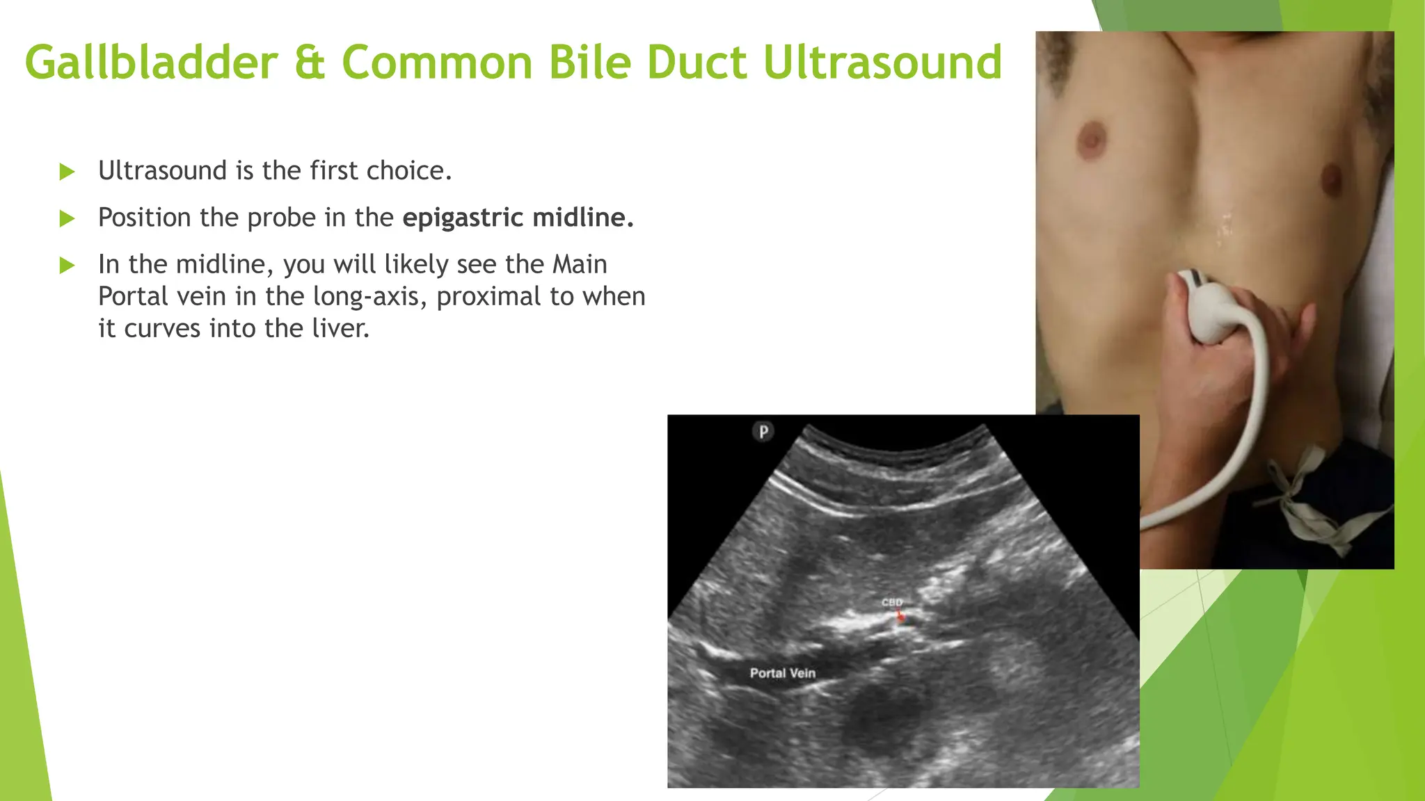
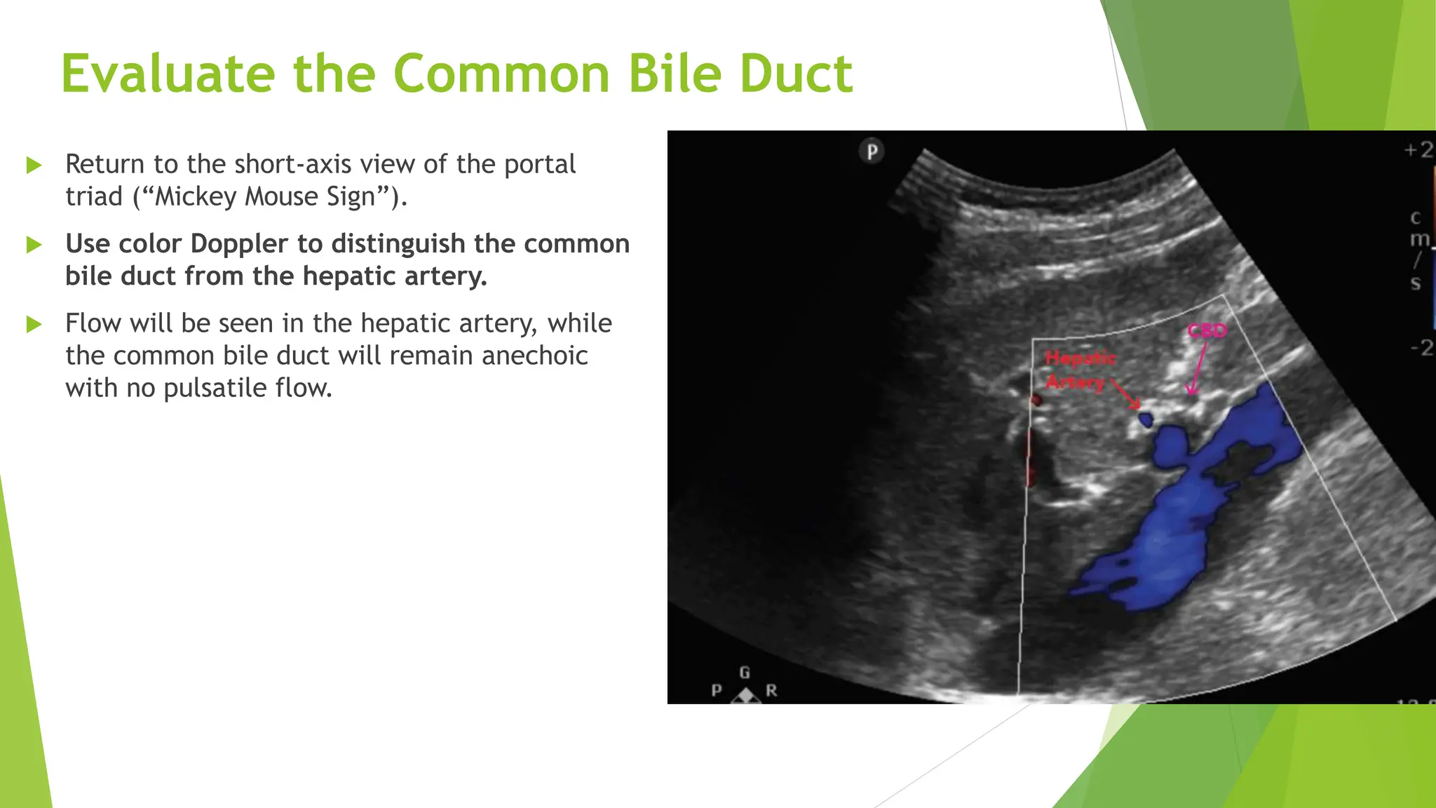
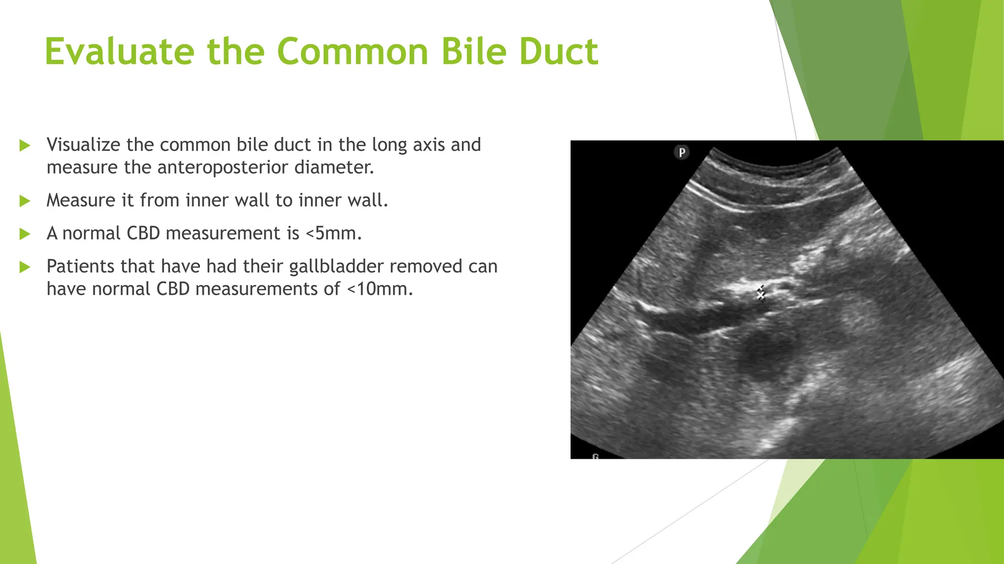
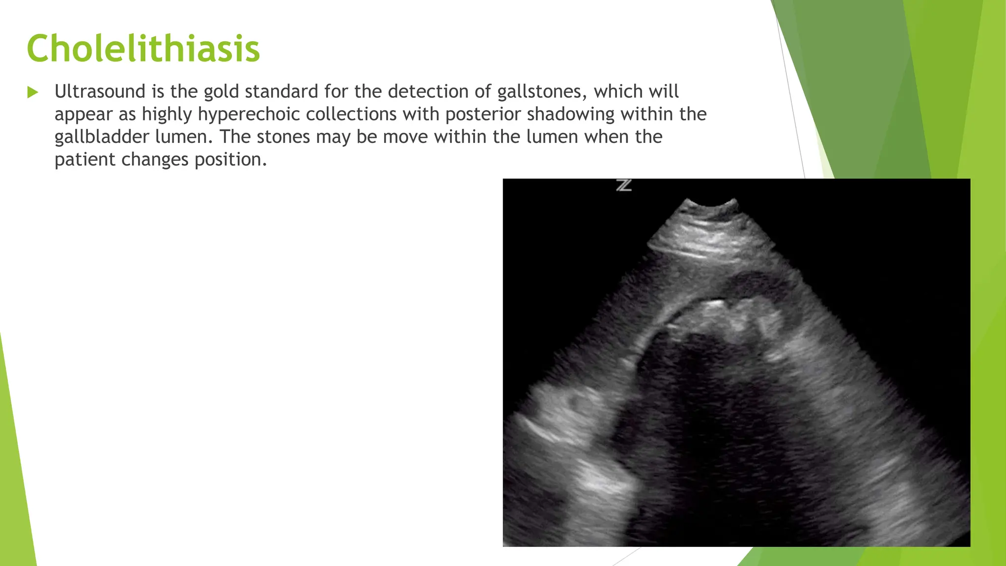
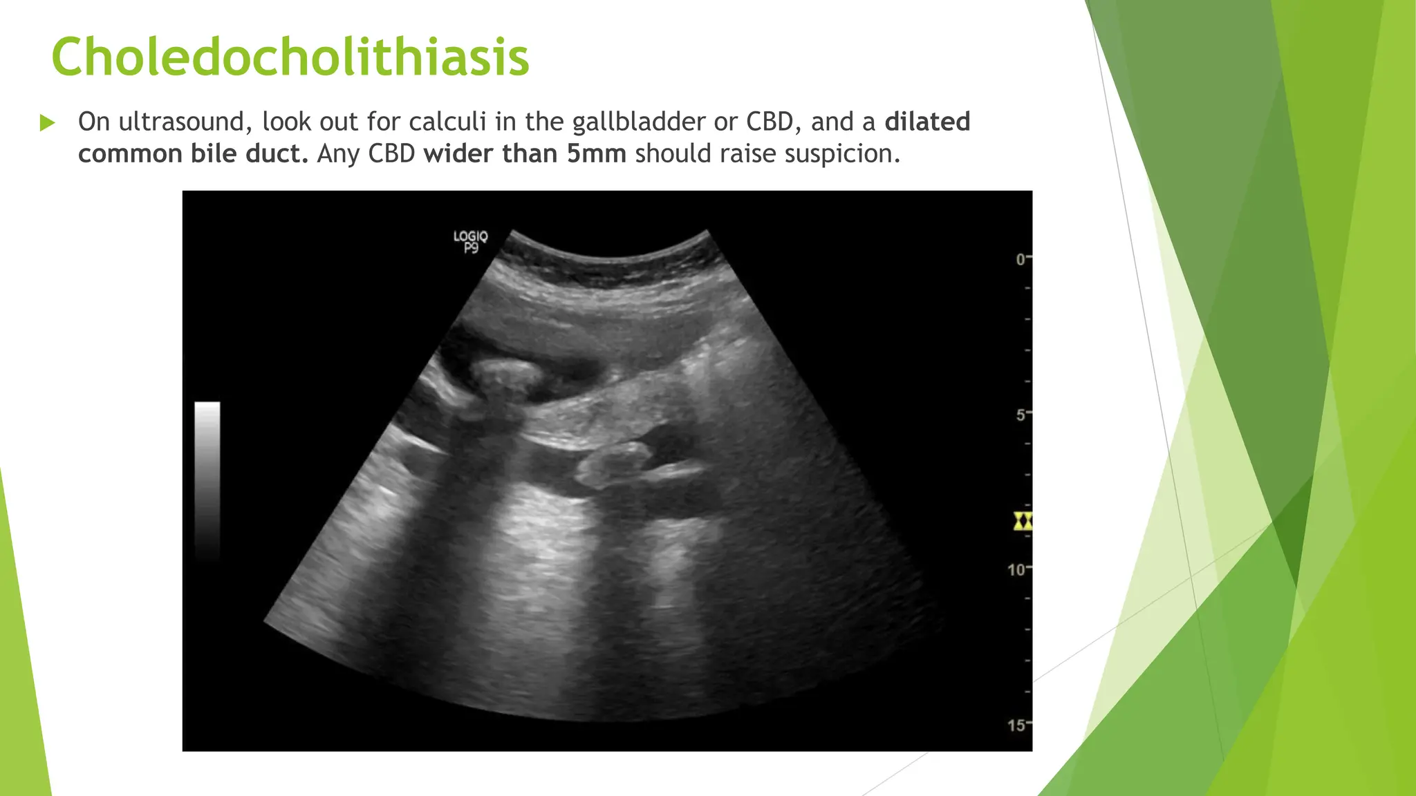
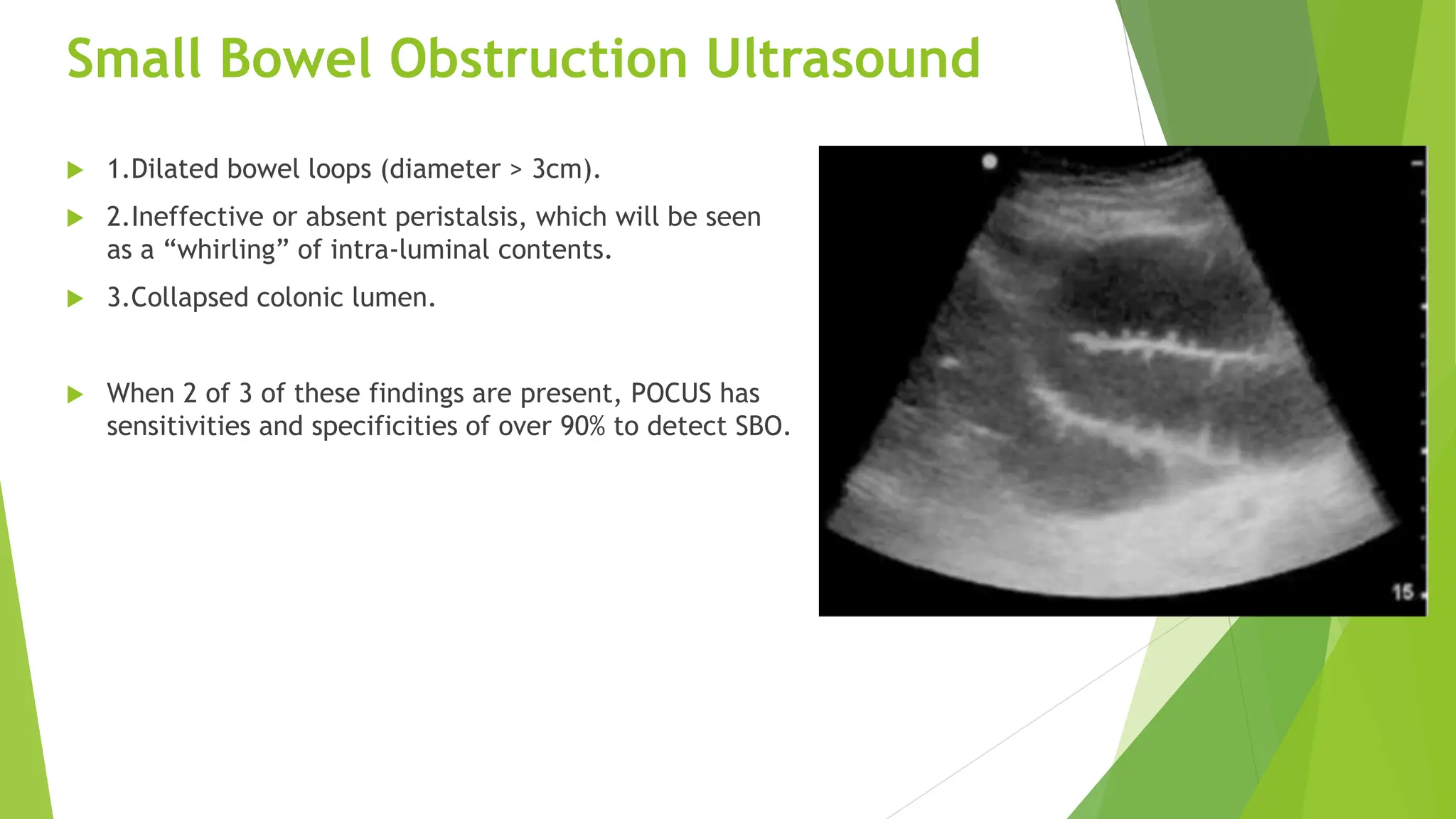
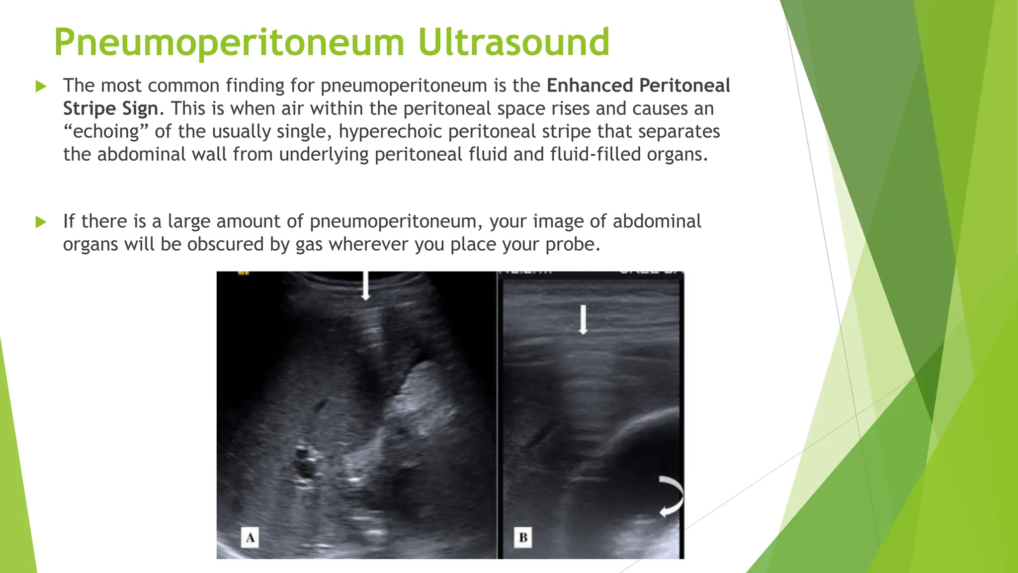
Evaluation abdominal pain
by point of care ultrasound
Tutor: Lei Man Sang
IC: Lam Ian Seng
Content
 Introduction
 What is point of care ultrasound
 Abdominal Ultrasound Preparation
 Liver ultrasound
 Gallbladder, Common Bile Duct Ultrasound
 Appendix Ultrasound
 Small Bowel Obstruction Ultrasound
 Pneumoperitoneum Ultrasound
Introduction
 Abdominal pain is the chief complaint in 5 to 10 % of emergency department visits, and
patients often require extensive evaluations, including history taking, physical examination,
blood test and imaging. The differential diagnosis is wide, ranging from benign to life-
threatening conditions.
Rapid assessment for abdominal pain
 The evaluation starts with obtaining a history and performing a physical examination.
 We simultaneously and rapidly assess if the patient may be having a life-threatening abdominal pain.
Potential indicators include shock ,presence of peritonitis, significant distress from pain, altered
mental status.
Physical examination
Point of care ultrasound
Point of care ultrasonography (POCUS)is advanced diagnostic ultrasonography that is performed by the
attending doctor as a bedside test. POCUS has been widely used in many disciplines as a rapid diagnostic tool.
POCUS has been used to aid the diagnosis of multiple medical conditions ranging from acute appendicitis,
abdominal aortic aneurysm, traumatic injury assessment.
There are several advantages of incorporating POCUS in daily clinical practice, with the major one being
integrating sonographic findings with history and clinical examination at the patient's bedside.
Point of care ultrasound
 POCUS use and implementation have expanded significantly over the past 15 years. Despite
being widely used across different medical specialization.
 In France, POCUS availability in the emergency department was as high as 71% in 2016.
 In Danish, more than 80% of the emergency departments, POCUS has been available to
emergency doctor.
 In Canada, general and emergency doctor, access to POCUS has increased from 60% access in
2013 to more than 90% access in 2019.
 In the USA, ultrasound training is now integrated into EP training.
 In China, more than half of emergency department physicians have reported having access
to POCUS, with 43% reporting using it in their clinical work.
Abdominal Ultrasound Preparation
 Patient Preparation
 Your patient should be supine with the head of the bed flat.
 Ask the patient to bend their legs at the knees to help relax
the abdominal muscles.
 Transducer: Usually curvilinear Probe.
Liver ultrasound
Liver ultrasound
 Using the lateral ultrasound approach, assess the size, parenchymal
echogenicity, and surface characteristics of the liver.
 A healthy liver should be < 16cm in this dimension.
Liver ultrasound
 Assess the liver’s echogenicity and capsular contour.
 Normally, the liver has a homogenous echogenicity similar
to the renal cortex.
 The liver should have a smooth capsular contour, similar
to that of the kidney.
This liver measures 12.6cm in the craniocaudal dimension
Acute hepatitis: Chronic hepatitis/cirrhosis: Fatty Liver Disease
↑ Brightness of portal vein
walls
↓ Brightness of portal vein walls ↓ Brightness of portal vein
walls
↓ The echogenicity of the
liver
↑ Liver echogenicity ↑ Liver echogenicity
Usually Enlarged Liver Usually Small/Atrophic Liver Usually enlarged Liver
Acute Hepatitis Ultrasound Findings
 Acute hepatitis occurs when the liver has an inflammatory reaction to an injury, whether traumatic,
infectious, drug-induced, or autoimmune.
 Hepatomegaly is the most sensitive US finding in acute hepatitis, defined as a craniocaudal length ≥
16.0cm in the midclavicular line.
 In addition, the liver may appear relatively hypoechoic, due to inflammatory fluid buildup in acute
hepatitis.
Acute Hepatitis Ultrasound Findings
Cirrhosis Ultrasound Findings
 The liver will be hyperechoic relative to the right kidney.
Liver abscess
Liver abscesses is defined as a pus-filled mass in the liver.
POCUS present a sensitivity between 85~92%
of the liver abscess.
Gallbladder & Common Bile Duct
Ultrasound
Gallbladder & Common Bile Duct Ultrasound
 Ultrasound is the first choice.
 Position the probe in the epigastric midline.
 In the midline, you will likely see the Main
Portal vein in the long-axis, proximal to when
it curves into the liver.
Gallbladder & Common Bile Duct Ultrasound
 Slide laterally along the costal margin, toward the patient’s right, until you
can clearly see the Portal Triad (Portal vein, Hepatic Artery, and Common Bile
Duct) in short axis.
Measure the anterior gallbladder wall thickness.
 Measure the anterior gallbladder wall thickness.
 The normal anterior gallbladder wall thickness is <3mm.
Evaluate the Common Bile Duct
 Return to the short-axis view of the portal
triad (“Mickey Mouse Sign”).
 Use color Doppler to distinguish the common
bile duct from the hepatic artery.
 Flow will be seen in the hepatic artery, while
the common bile duct will remain anechoic
with no pulsatile flow.
Evaluate the Common Bile Duct
 Visualize the common bile duct in the long axis and
measure the anteroposterior diameter.
 Measure it from inner wall to inner wall.
 A normal CBD measurement is <5mm.
 Patients that have had their gallbladder removed can
have normal CBD measurements of <10mm.
Cholelithiasis
 Ultrasound is the gold standard for the detection of gallstones, which will
appear as highly hyperechoic collections with posterior shadowing within the
gallbladder lumen. The stones may be move within the lumen when the
patient changes position.
Cholecystitis on Ultrasound
 A Sonographic Murphy sign, however, refers to the presence of maximal tenderness when
ultrasound probe pressure is applied over the visualized gallbladder on ultrasound. This is
a very sensitive finding for cholecystitis.
 Important secondary findings include gallstones, anterior gallbladder wall thickening
>3mm, pericholecystic fluid.
Gallstones during POCUS with positive Murphys sign has a 92%
positive for acute cholecystitis. If the US shows a normal
gallbladder wall, acute cholecystitis can be ruled out.
Choledocholithiasis
 On ultrasound, look out for calculi in the gallbladder or CBD, and a dilated
common bile duct. Any CBD wider than 5mm should raise suspicion.
Appendix Ultrasound
Appendix Ultrasound
 The appendix is located in the right lower quadrant, attached to the cecum.
However, its location can vary widely in its anatomic position.
Appendix Ultrasound
 Two ways to go about the appendix POCUS exam.
 1. Point of maximal tenderness.
 2.Systemic approach, try starting in the RUQ, trace the ascending colon
downward until reaching the cecum, and have identified the psoas major and
the iliac vessels, search slightly medial to the psoas muscle for the
appendicitis.
Appendicitis Ultrasound Findings
 Non-compressibility of the appendix (unless perforated);
 Blind ended pouch;
 Diameter of the appendix > 6 mm;
 Single wall thickness ≥ 3 mm;
 Target sign;
 Hyperechoic appendicolith with posterior shadowing.
Dilated appendix with blind ended Pouch Appendicolith in an appendix with a diameter > 6cm.
Dilated appendix with a diameter >6mm and “Target Sign”
A meta-analysis showed that POCUS for appendicitis,
has 84% sensitivity and 91% specificity, with higher
accuracy in children.
Abdominal Free Fluid Ultrasound
 Evaluating for free fluid is a common abdominal ultrasound application.
 Right Upper Quadrant (RUQ) – Morison’s Pouch.
 Left Upper Quadrant – Perisplenic Recess.
 Pelvis – Retrovesical Pouch
US has a sensitivity and specificity in the detection
of intraperitoneal free fluid of over 90%, while up to
10 mL of free fluid can be detected by POCUS.
Small Bowel Obstruction Ultrasound
 The ultrasound technique for detecting SBO is known as “mowing the lawn” .
Starting in the RLQ and using graded compression, slowly scan the abdominal
quadrants.
 1.Dilated bowel loops (diameter > 3cm).
 2.Ineffective or absent peristalsis, which will be seen
as a “whirling” of intra-luminal contents.
 3.Collapsed colonic lumen.
 When 2 of 3 of these findings are present, POCUS has
sensitivities and specificities of over 90% to detect SBO.
Small Bowel Obstruction Ultrasound
When bowel obstruction is suspected, POCUS has 90%
sensitivity and 96% specificity.
Pneumoperitoneum Ultrasound
 The most common finding for pneumoperitoneum is the Enhanced Peritoneal
Stripe Sign. This is when air within the peritoneal space rises and causes an
“echoing” of the usually single, hyperechoic peritoneal stripe that separates
the abdominal wall from underlying peritoneal fluid and fluid-filled organs.
 If there is a large amount of pneumoperitoneum, your image of abdominal
organs will be obscured by gas wherever you place your probe.
X ray had a sensitivity of 79%, specificity of 64% for
pneumoperitoneum. Ultrasound proved superior in terms of
sensitivity at 93% and at least specificity at 64%.
Reference
 Noble, V. E., & Nelson, B. (2014). Manual of Emergency and Critical Care Ultrasound. Cambridge, UK, MA: Cambridge University
Press.
 Rumack, Carol M., and Levine, Deborah. Diagnostic Ultrasound E-Book. United Kingdom, Elsevier Health Sciences, 2017.
 Arger, Peter, & Benson, Carol. Ultrasound, a practical approach to clinical problems. Thieme Publishing Group. (2008)
ISBN:3131168323.
 Point-of-care ultrasound for the diagnosis of liver abscess in a patient with HIV in the emergency department: A case report
 Normal Liver Ultrasound How To. (2020). Retrieved August 12, 2020, from https://www.ultrasoundpaedia.com
 Joshi, G., Crawford, K., Hanna, T., Herr, K., Dahiya, N., Menias, C. (2018). US of Right Upper Quadrant Pain in the Emergency
Department: Diagnosing beyond Gallbladder and Biliary Disease RadioGraphics 38(3), 766-793.
 Point-of-care ultrasound on way to the right and rapid to the diagnosis.
 Bell, D., & Sorrentino, S. (2012). Gallstones: Radiology Reference Article. Retrieved October 05, 2020
 Hartung, M., & Gaillard, F. (2010). Choledocholithiasis: Radiology Reference Article. Retrieved October 05, 2020
 Mostbeck, G., Adam, E. J., Nielsen, M. B., Claudon, M., Clevert, D., Nicolau, C., Nyhsen, C., & Owens, C. M. (2016). How to
diagnose acute appendicitis: ultrasound first. Insights into imaging, 7(2), 255–263. https://doi.org/10.1007/s13244-016-0469-6
 Unlüer EE1, Yavaşi O, Eroğlu O, et al. Ultrasonography by emergency medicine and radiology residents for the diagnosis of
small bowel obstruction. Eur J Emerg Med. 2010 Oct;17(5):260-4.)
 Tanner TN, Hall BR, Oran J. Pneumoperitoneum. Surg Clin North Am. 2018 Oct;98(5):915-932. doi: 10.1016/j.suc.2018.06.004.
PMID: 30243453.
 Indiran, V., Kumar, R. V., & Jefferson, B. (2018, May 3). Enhanced peritoneal stripe sign. Abdominal Radiology, 43, 3518-3519.
 Sonographic detection of pneumoperitoneum.
Evaluation abdominal Point of care ultrasonography (POCUS)is advanced diagnostic ultrasonography that is performed by the attending doctor as a bedside test

Evaluation abdominal Point of care ultrasonography (POCUS)is advanced diagnostic ultrasonography that is performed by the attending doctor as a bedside test

  • 1.
    Evaluation abdominal pain bypoint of care ultrasound Tutor: Lei Man Sang IC: Lam Ian Seng
  • 2.
    Content  Introduction  Whatis point of care ultrasound  Abdominal Ultrasound Preparation  Liver ultrasound  Gallbladder, Common Bile Duct Ultrasound  Appendix Ultrasound  Small Bowel Obstruction Ultrasound  Pneumoperitoneum Ultrasound
  • 3.
    Introduction  Abdominal painis the chief complaint in 5 to 10 % of emergency department visits, and patients often require extensive evaluations, including history taking, physical examination, blood test and imaging. The differential diagnosis is wide, ranging from benign to life- threatening conditions.
  • 4.
    Rapid assessment forabdominal pain  The evaluation starts with obtaining a history and performing a physical examination.  We simultaneously and rapidly assess if the patient may be having a life-threatening abdominal pain. Potential indicators include shock ,presence of peritonitis, significant distress from pain, altered mental status.
  • 5.
  • 6.
    Point of careultrasound Point of care ultrasonography (POCUS)is advanced diagnostic ultrasonography that is performed by the attending doctor as a bedside test. POCUS has been widely used in many disciplines as a rapid diagnostic tool. POCUS has been used to aid the diagnosis of multiple medical conditions ranging from acute appendicitis, abdominal aortic aneurysm, traumatic injury assessment. There are several advantages of incorporating POCUS in daily clinical practice, with the major one being integrating sonographic findings with history and clinical examination at the patient's bedside.
  • 7.
    Point of careultrasound  POCUS use and implementation have expanded significantly over the past 15 years. Despite being widely used across different medical specialization.  In France, POCUS availability in the emergency department was as high as 71% in 2016.  In Danish, more than 80% of the emergency departments, POCUS has been available to emergency doctor.  In Canada, general and emergency doctor, access to POCUS has increased from 60% access in 2013 to more than 90% access in 2019.  In the USA, ultrasound training is now integrated into EP training.  In China, more than half of emergency department physicians have reported having access to POCUS, with 43% reporting using it in their clinical work.
  • 8.
    Abdominal Ultrasound Preparation Patient Preparation  Your patient should be supine with the head of the bed flat.  Ask the patient to bend their legs at the knees to help relax the abdominal muscles.  Transducer: Usually curvilinear Probe.
  • 9.
  • 10.
    Liver ultrasound  Usingthe lateral ultrasound approach, assess the size, parenchymal echogenicity, and surface characteristics of the liver.  A healthy liver should be < 16cm in this dimension.
  • 11.
    Liver ultrasound  Assessthe liver’s echogenicity and capsular contour.  Normally, the liver has a homogenous echogenicity similar to the renal cortex.  The liver should have a smooth capsular contour, similar to that of the kidney. This liver measures 12.6cm in the craniocaudal dimension
  • 12.
    Acute hepatitis: Chronichepatitis/cirrhosis: Fatty Liver Disease ↑ Brightness of portal vein walls ↓ Brightness of portal vein walls ↓ Brightness of portal vein walls ↓ The echogenicity of the liver ↑ Liver echogenicity ↑ Liver echogenicity Usually Enlarged Liver Usually Small/Atrophic Liver Usually enlarged Liver
  • 13.
    Acute Hepatitis UltrasoundFindings  Acute hepatitis occurs when the liver has an inflammatory reaction to an injury, whether traumatic, infectious, drug-induced, or autoimmune.  Hepatomegaly is the most sensitive US finding in acute hepatitis, defined as a craniocaudal length ≥ 16.0cm in the midclavicular line.  In addition, the liver may appear relatively hypoechoic, due to inflammatory fluid buildup in acute hepatitis.
  • 14.
  • 15.
    Cirrhosis Ultrasound Findings The liver will be hyperechoic relative to the right kidney.
  • 16.
    Liver abscess Liver abscessesis defined as a pus-filled mass in the liver.
  • 17.
    POCUS present asensitivity between 85~92% of the liver abscess.
  • 18.
    Gallbladder & CommonBile Duct Ultrasound
  • 19.
    Gallbladder & CommonBile Duct Ultrasound  Ultrasound is the first choice.  Position the probe in the epigastric midline.  In the midline, you will likely see the Main Portal vein in the long-axis, proximal to when it curves into the liver.
  • 20.
    Gallbladder & CommonBile Duct Ultrasound  Slide laterally along the costal margin, toward the patient’s right, until you can clearly see the Portal Triad (Portal vein, Hepatic Artery, and Common Bile Duct) in short axis.
  • 21.
    Measure the anteriorgallbladder wall thickness.  Measure the anterior gallbladder wall thickness.  The normal anterior gallbladder wall thickness is <3mm.
  • 22.
    Evaluate the CommonBile Duct  Return to the short-axis view of the portal triad (“Mickey Mouse Sign”).  Use color Doppler to distinguish the common bile duct from the hepatic artery.  Flow will be seen in the hepatic artery, while the common bile duct will remain anechoic with no pulsatile flow.
  • 23.
    Evaluate the CommonBile Duct  Visualize the common bile duct in the long axis and measure the anteroposterior diameter.  Measure it from inner wall to inner wall.  A normal CBD measurement is <5mm.  Patients that have had their gallbladder removed can have normal CBD measurements of <10mm.
  • 24.
    Cholelithiasis  Ultrasound isthe gold standard for the detection of gallstones, which will appear as highly hyperechoic collections with posterior shadowing within the gallbladder lumen. The stones may be move within the lumen when the patient changes position.
  • 25.
    Cholecystitis on Ultrasound A Sonographic Murphy sign, however, refers to the presence of maximal tenderness when ultrasound probe pressure is applied over the visualized gallbladder on ultrasound. This is a very sensitive finding for cholecystitis.  Important secondary findings include gallstones, anterior gallbladder wall thickening >3mm, pericholecystic fluid.
  • 26.
    Gallstones during POCUSwith positive Murphys sign has a 92% positive for acute cholecystitis. If the US shows a normal gallbladder wall, acute cholecystitis can be ruled out.
  • 27.
    Choledocholithiasis  On ultrasound,look out for calculi in the gallbladder or CBD, and a dilated common bile duct. Any CBD wider than 5mm should raise suspicion.
  • 28.
  • 29.
    Appendix Ultrasound  Theappendix is located in the right lower quadrant, attached to the cecum. However, its location can vary widely in its anatomic position.
  • 30.
    Appendix Ultrasound  Twoways to go about the appendix POCUS exam.  1. Point of maximal tenderness.  2.Systemic approach, try starting in the RUQ, trace the ascending colon downward until reaching the cecum, and have identified the psoas major and the iliac vessels, search slightly medial to the psoas muscle for the appendicitis.
  • 31.
    Appendicitis Ultrasound Findings Non-compressibility of the appendix (unless perforated);  Blind ended pouch;  Diameter of the appendix > 6 mm;  Single wall thickness ≥ 3 mm;  Target sign;  Hyperechoic appendicolith with posterior shadowing. Dilated appendix with blind ended Pouch Appendicolith in an appendix with a diameter > 6cm. Dilated appendix with a diameter >6mm and “Target Sign”
  • 32.
    A meta-analysis showedthat POCUS for appendicitis, has 84% sensitivity and 91% specificity, with higher accuracy in children.
  • 33.
    Abdominal Free FluidUltrasound  Evaluating for free fluid is a common abdominal ultrasound application.  Right Upper Quadrant (RUQ) – Morison’s Pouch.  Left Upper Quadrant – Perisplenic Recess.  Pelvis – Retrovesical Pouch
  • 34.
    US has asensitivity and specificity in the detection of intraperitoneal free fluid of over 90%, while up to 10 mL of free fluid can be detected by POCUS.
  • 35.
  • 36.
    Small Bowel ObstructionUltrasound  The ultrasound technique for detecting SBO is known as “mowing the lawn” . Starting in the RLQ and using graded compression, slowly scan the abdominal quadrants.
  • 37.
     1.Dilated bowelloops (diameter > 3cm).  2.Ineffective or absent peristalsis, which will be seen as a “whirling” of intra-luminal contents.  3.Collapsed colonic lumen.  When 2 of 3 of these findings are present, POCUS has sensitivities and specificities of over 90% to detect SBO. Small Bowel Obstruction Ultrasound
  • 38.
    When bowel obstructionis suspected, POCUS has 90% sensitivity and 96% specificity.
  • 39.
    Pneumoperitoneum Ultrasound  Themost common finding for pneumoperitoneum is the Enhanced Peritoneal Stripe Sign. This is when air within the peritoneal space rises and causes an “echoing” of the usually single, hyperechoic peritoneal stripe that separates the abdominal wall from underlying peritoneal fluid and fluid-filled organs.  If there is a large amount of pneumoperitoneum, your image of abdominal organs will be obscured by gas wherever you place your probe.
  • 40.
    X ray hada sensitivity of 79%, specificity of 64% for pneumoperitoneum. Ultrasound proved superior in terms of sensitivity at 93% and at least specificity at 64%.
  • 41.
    Reference  Noble, V.E., & Nelson, B. (2014). Manual of Emergency and Critical Care Ultrasound. Cambridge, UK, MA: Cambridge University Press.  Rumack, Carol M., and Levine, Deborah. Diagnostic Ultrasound E-Book. United Kingdom, Elsevier Health Sciences, 2017.  Arger, Peter, & Benson, Carol. Ultrasound, a practical approach to clinical problems. Thieme Publishing Group. (2008) ISBN:3131168323.  Point-of-care ultrasound for the diagnosis of liver abscess in a patient with HIV in the emergency department: A case report  Normal Liver Ultrasound How To. (2020). Retrieved August 12, 2020, from https://www.ultrasoundpaedia.com  Joshi, G., Crawford, K., Hanna, T., Herr, K., Dahiya, N., Menias, C. (2018). US of Right Upper Quadrant Pain in the Emergency Department: Diagnosing beyond Gallbladder and Biliary Disease RadioGraphics 38(3), 766-793.  Point-of-care ultrasound on way to the right and rapid to the diagnosis.  Bell, D., & Sorrentino, S. (2012). Gallstones: Radiology Reference Article. Retrieved October 05, 2020  Hartung, M., & Gaillard, F. (2010). Choledocholithiasis: Radiology Reference Article. Retrieved October 05, 2020  Mostbeck, G., Adam, E. J., Nielsen, M. B., Claudon, M., Clevert, D., Nicolau, C., Nyhsen, C., & Owens, C. M. (2016). How to diagnose acute appendicitis: ultrasound first. Insights into imaging, 7(2), 255–263. https://doi.org/10.1007/s13244-016-0469-6  Unlüer EE1, Yavaşi O, Eroğlu O, et al. Ultrasonography by emergency medicine and radiology residents for the diagnosis of small bowel obstruction. Eur J Emerg Med. 2010 Oct;17(5):260-4.)  Tanner TN, Hall BR, Oran J. Pneumoperitoneum. Surg Clin North Am. 2018 Oct;98(5):915-932. doi: 10.1016/j.suc.2018.06.004. PMID: 30243453.  Indiran, V., Kumar, R. V., & Jefferson, B. (2018, May 3). Enhanced peritoneal stripe sign. Abdominal Radiology, 43, 3518-3519.  Sonographic detection of pneumoperitoneum.