Proximal gastrectomy (PG) is an alternative to total gastrectomy for early gastric cancers located in the upper third of the stomach. PG aims to preserve gastric function while achieving adequate cancer resection. PG is oncologically appropriate for cT1a/bN0 stage I gastric cancers. Lymph node dissection for PG typically involves D1 or D1+ lymphadenectomy. Various reconstruction techniques after PG aim to reduce postoperative complications like reflux esophagitis. Larger studies comparing PG to total gastrectomy find similar long-term survival but PG may better preserve nutritional parameters and quality of life. Prospective randomized trials are still needed to further establish the role of PG.
CARCINOMA ESOPHAGUS - DR ZAHID IQBAL MIR
Dr. Zahid Iqbal Mir, MBBS MS (General Surgery), DNB (General Surgery) has done his MBBS and masters in General Surgery from the prestigious Govt Medical College Jammu and DNB in General Surgery from NBEMS New Delhi. He is a passionate surgeon, earlier practising at Government Medical College, Jammu as Registrar in Department of General Surgery. Nowadays working as Senior Resident in Department of General Surgery, Government Medical College & Hospital, Sector 32, Chandigarh and a rising name in field of surgery.
He is an enthusiastic, enigmatic and dedicated teacher as well. He is not just a resolute learner, but also an awe inspiring guiding light for his juniors, which makes him the most loveable and respected senior.
Currently he is running “LOVE FOR SCALPEL” for PGMEE aspirants on most of the social platforms, which is gaining immense popularity among residents, medical graduates and undergraduates.
Minimal invasive Surgery in Management of colorectal cancerpiyushpatwa
Laparoscopic Anterior resection. After insertion of ports with patient in steep trendelenburg position Inferior mesenteric artery was identified and high ligation done with division of left colic artery and then medial to lateral dissection was done. Subsequently inferior mesenteric vein was dissected and clipped and divided. Distal dissection proceeded just behind the superior rectal artery and after identification and preservation of the hypogastric nerves, upper rectum was mobilised. Division of bowel was done at upper rectum after giving adequate distal margin and end to end anastomosis was done using circular stapler.
Colorectal anastomosis leaks are most difficult to manage for a surgeon carrying morbidity and mortality. Discussion on risk factors as well as management of anastomotic leak.
https://drdhavalmangukiya.com/
http://www.youtube.com/c/DrDhavalMangukiyaGastrosurgeonSurat
https://gastrosurgerysurat.blogspot.com/
Component separation technique for a very large abdominal wall herniaSanjiv Haribhakti
Component separation technique is an excellent technique for large ventral central defects which can allow a medial shift of approx. For More information visit at Gisurgery.info
Laparoscopic radical gastrectomy for gastric cancer management is feasible in highly complex centers with advanced laparoscopic service with comparable oncological results to open procedures with free margins, adequate lymph node count, with a low complication rate and very low recurrence rate.
MANAGEMENT OF ATRIOVENTRICULAR CONDUCTION BLOCK.pdfJim Jacob Roy
Cardiac conduction defects can occur due to various causes.
Atrioventricular conduction blocks ( AV blocks ) are classified into 3 types.
This document describes the acute management of AV block.
These lecture slides, by Dr Sidra Arshad, offer a quick overview of physiological basis of a normal electrocardiogram.
Learning objectives:
1. Define an electrocardiogram (ECG) and electrocardiography
2. Describe how dipoles generated by the heart produce the waveforms of the ECG
3. Describe the components of a normal electrocardiogram of a typical bipolar leads (limb II)
4. Differentiate between intervals and segments
5. Enlist some common indications for obtaining an ECG
Study Resources:
1. Chapter 11, Guyton and Hall Textbook of Medical Physiology, 14th edition
2. Chapter 9, Human Physiology - From Cells to Systems, Lauralee Sherwood, 9th edition
3. Chapter 29, Ganong’s Review of Medical Physiology, 26th edition
4. Electrocardiogram, StatPearls - https://www.ncbi.nlm.nih.gov/books/NBK549803/
5. ECG in Medical Practice by ABM Abdullah, 4th edition
6. ECG Basics, http://www.nataliescasebook.com/tag/e-c-g-basics
- Video recording of this lecture in English language: https://youtu.be/lK81BzxMqdo
- Video recording of this lecture in Arabic language: https://youtu.be/Ve4P0COk9OI
- Link to download the book free: https://nephrotube.blogspot.com/p/nephrotube-nephrology-books.html
- Link to NephroTube website: www.NephroTube.com
- Link to NephroTube social media accounts: https://nephrotube.blogspot.com/p/join-nephrotube-on-social-media.html
Explore natural remedies for syphilis treatment in Singapore. Discover alternative therapies, herbal remedies, and lifestyle changes that may complement conventional treatments. Learn about holistic approaches to managing syphilis symptoms and supporting overall health.
Pulmonary Thromboembolism - etilogy, types, medical- Surgical and nursing man...VarunMahajani
Disruption of blood supply to lung alveoli due to blockage of one or more pulmonary blood vessels is called as Pulmonary thromboembolism. In this presentation we will discuss its causes, types and its management in depth.
New Directions in Targeted Therapeutic Approaches for Older Adults With Mantl...i3 Health
i3 Health is pleased to make the speaker slides from this activity available for use as a non-accredited self-study or teaching resource.
This slide deck presented by Dr. Kami Maddocks, Professor-Clinical in the Division of Hematology and
Associate Division Director for Ambulatory Operations
The Ohio State University Comprehensive Cancer Center, will provide insight into new directions in targeted therapeutic approaches for older adults with mantle cell lymphoma.
STATEMENT OF NEED
Mantle cell lymphoma (MCL) is a rare, aggressive B-cell non-Hodgkin lymphoma (NHL) accounting for 5% to 7% of all lymphomas. Its prognosis ranges from indolent disease that does not require treatment for years to very aggressive disease, which is associated with poor survival (Silkenstedt et al, 2021). Typically, MCL is diagnosed at advanced stage and in older patients who cannot tolerate intensive therapy (NCCN, 2022). Although recent advances have slightly increased remission rates, recurrence and relapse remain very common, leading to a median overall survival between 3 and 6 years (LLS, 2021). Though there are several effective options, progress is still needed towards establishing an accepted frontline approach for MCL (Castellino et al, 2022). Treatment selection and management of MCL are complicated by the heterogeneity of prognosis, advanced age and comorbidities of patients, and lack of an established standard approach for treatment, making it vital that clinicians be familiar with the latest research and advances in this area. In this activity chaired by Michael Wang, MD, Professor in the Department of Lymphoma & Myeloma at MD Anderson Cancer Center, expert faculty will discuss prognostic factors informing treatment, the promising results of recent trials in new therapeutic approaches, and the implications of treatment resistance in therapeutic selection for MCL.
Target Audience
Hematology/oncology fellows, attending faculty, and other health care professionals involved in the treatment of patients with mantle cell lymphoma (MCL).
Learning Objectives
1.) Identify clinical and biological prognostic factors that can guide treatment decision making for older adults with MCL
2.) Evaluate emerging data on targeted therapeutic approaches for treatment-naive and relapsed/refractory MCL and their applicability to older adults
3.) Assess mechanisms of resistance to targeted therapies for MCL and their implications for treatment selection
The prostate is an exocrine gland of the male mammalian reproductive system
It is a walnut-sized gland that forms part of the male reproductive system and is located in front of the rectum and just below the urinary bladder
Function is to store and secrete a clear, slightly alkaline fluid that constitutes 10-30% of the volume of the seminal fluid that along with the spermatozoa, constitutes semen
A healthy human prostate measures (4cm-vertical, by 3cm-horizontal, 2cm ant-post ).
It surrounds the urethra just below the urinary bladder. It has anterior, median, posterior and two lateral lobes
It’s work is regulated by androgens which are responsible for male sex characteristics
Generalised disease of the prostate due to hormonal derangement which leads to non malignant enlargement of the gland (increase in the number of epithelial cells and stromal tissue)to cause compression of the urethra leading to symptoms (LUTS
Ozempic: Preoperative Management of Patients on GLP-1 Receptor Agonists Saeid Safari
Preoperative Management of Patients on GLP-1 Receptor Agonists like Ozempic and Semiglutide
ASA GUIDELINE
NYSORA Guideline
2 Case Reports of Gastric Ultrasound
Title: Sense of Smell
Presenter: Dr. Faiza, Assistant Professor of Physiology
Qualifications:
MBBS (Best Graduate, AIMC Lahore)
FCPS Physiology
ICMT, CHPE, DHPE (STMU)
MPH (GC University, Faisalabad)
MBA (Virtual University of Pakistan)
Learning Objectives:
Describe the primary categories of smells and the concept of odor blindness.
Explain the structure and location of the olfactory membrane and mucosa, including the types and roles of cells involved in olfaction.
Describe the pathway and mechanisms of olfactory signal transmission from the olfactory receptors to the brain.
Illustrate the biochemical cascade triggered by odorant binding to olfactory receptors, including the role of G-proteins and second messengers in generating an action potential.
Identify different types of olfactory disorders such as anosmia, hyposmia, hyperosmia, and dysosmia, including their potential causes.
Key Topics:
Olfactory Genes:
3% of the human genome accounts for olfactory genes.
400 genes for odorant receptors.
Olfactory Membrane:
Located in the superior part of the nasal cavity.
Medially: Folds downward along the superior septum.
Laterally: Folds over the superior turbinate and upper surface of the middle turbinate.
Total surface area: 5-10 square centimeters.
Olfactory Mucosa:
Olfactory Cells: Bipolar nerve cells derived from the CNS (100 million), with 4-25 olfactory cilia per cell.
Sustentacular Cells: Produce mucus and maintain ionic and molecular environment.
Basal Cells: Replace worn-out olfactory cells with an average lifespan of 1-2 months.
Bowman’s Gland: Secretes mucus.
Stimulation of Olfactory Cells:
Odorant dissolves in mucus and attaches to receptors on olfactory cilia.
Involves a cascade effect through G-proteins and second messengers, leading to depolarization and action potential generation in the olfactory nerve.
Quality of a Good Odorant:
Small (3-20 Carbon atoms), volatile, water-soluble, and lipid-soluble.
Facilitated by odorant-binding proteins in mucus.
Membrane Potential and Action Potential:
Resting membrane potential: -55mV.
Action potential frequency in the olfactory nerve increases with odorant strength.
Adaptation Towards the Sense of Smell:
Rapid adaptation within the first second, with further slow adaptation.
Psychological adaptation greater than receptor adaptation, involving feedback inhibition from the central nervous system.
Primary Sensations of Smell:
Camphoraceous, Musky, Floral, Pepperminty, Ethereal, Pungent, Putrid.
Odor Detection Threshold:
Examples: Hydrogen sulfide (0.0005 ppm), Methyl-mercaptan (0.002 ppm).
Some toxic substances are odorless at lethal concentrations.
Characteristics of Smell:
Odor blindness for single substances due to lack of appropriate receptor protein.
Behavioral and emotional influences of smell.
Transmission of Olfactory Signals:
From olfactory cells to glomeruli in the olfactory bulb, involving lateral inhibition.
Primitive, less old, and new olfactory systems with different path
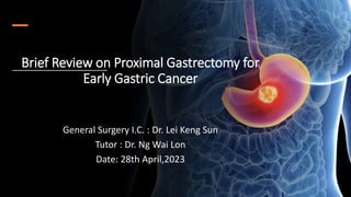
1. Brief Review on Proximal Gastrectomy for
Early Gastric Cancer
General Surgery I.C. : Dr. Lei Keng Sun
Tutor : Dr. Ng Wai Lon
Date: 28th April,2023
2. Hx of Proximal Gastrectomy
(Tetsuo Maki 1908-2006)
Tohoku University, Japan
(Tsuneo Shiratori 1922-2012)
Nara Medical University, Japan
• Tetsuo Maki published an surgical procedure, “Pylorus preserving
gastrectomy,” in 1967.
• Reduce dumping syndrome, postgastrectomy gallstone, and digestive
function disturbances after distal gastrectomy for benign ulcer.
• Tsuneo Shiratori expanded the indication for gastric cancer in 1991.
Sung HN, Woo JY Surgery for Gastric Cancer 2019
3. Hx of Proximal Gastrectomy
Over the past 30 years, the prevalence of upper third GC and EGJ
cancer has increased.
Standard surgical treatment:
Total gastrectomy with D2 lymph node dissection T2 or higher
upper third GC and GEJ cancers
TG post-gastrectomy syndrome (5–50%)
Weight loss, Dumping syndrome, and Anemia.
4. Hx of Proximal Gastrectomy
Proximal gastrectomy (PG)
Simple esophagogastrostomy after PG is the simplest and most convenient
physiological reconstruction method.
Without additional anti-reflux treatment
Several retrospective studies of esophagogastrostomy have observed
Early complications 3.1-24%
Stenosis 0-52.2 %
Reflux esophagitis 20-65.2%
Residual food 21.8%
Souya Nunobe, et la. Current status of proximal gastrectomy for gastric and esophagogastric
junctional cancer: A review . Japan, Ann Gastroenterol Surg. 2020;4:498–504
5. Hx of Proximal Gastrectomy
In recent years, anti-reflux reconstruction techniques:
Double flap technique / Double-tract reconstruction
↓Postoperative reflux esophagitis
↓postoperative weight loss and prevent anemia.
Prospective studies are underway to determine whether PG with anti-
reflux techniques improves patient-reported quality of life.
6. Aim
Reviewed available evidence for the use
of Proximal Gastrectomy (PG) for upper
third Gastric Cancer
1. Which patients are oncologically appropriated for PG?
2. Various types of reconstruction can be perfromed after PG?
3. Benefits on PG vs TG
7. UICC TNM categories and
stage grouping: Stomach
T- Primary tumour
Tis Carcinoma in situ: intraepithelial tumor without invasion of
the lamina propria, high-grade dysplasia
T1: T1a lamina propria or muscularis mucosae
T1b submucosa
T2: muscularis propria
T3: subserosa
T4: perforates serosa (visceral peritoneum)
T4a perforates serosa
T4b invades adjacent structures
N – Regional Lymph
Nodes
N1: 1 to 2 regional LNs
N2: 3 to 6 regional LNs
N3 : 7 or more regional LNs
N3a: 7 to 15 regional LNs
N3b: 16 or more regional LNs
M – Distant Metastasis
M0: No distant metastasis
M1: Distant metastasis
8. TNM categories and stage grouping
based on the 15th edition of
Japanese Classification of Gastric
Carcinoma which identical to UICC
8th edition
Japanese Gastric Cancer Treatment Guidelines 2021 (6th edition)
9. Japanese Gastric Cancer Treatment Guidelines 2021 (6th edition )
Algorithm of Standard Treatments to be Recommended in Clinical Practice
10. The standard surgery for gastric cancer
Gastrectomy
with adequate
margins
Perigastric and
extragastric LN
dissection
Consequent
gastrointestinal
reconstruction
11. Principle of Adequate Margins
KGCA = Korean Gastric Cancer Association; JGCA = Japanese Gastric Cancer Association;
CSCO = Chinese Society of Clinical Oncology; NCCN = National Comprehensive Cancer Network; ESMO = European Society for Medical
Oncology;
≥2 cm for T1 tumors (JGCA )
≥3 cm proximal margin in T2 / deeper tumors with
Borrmann type I and II tumors. (JGCA, CSCO)
A 5 cm proximal margin with Borrmann types III and IV.
(JGCA, CSCO)
5 cm for Stage IB-III gastric cancer. (KGCA, NCCN,
ESMO)
8 cm for diffuse cancer when DG, otherwise, total
gastrectomy was recommended. (ESMO)
Borramann Classification
13. Indiacations for Function-Preserving Surgery
Eom SS, A Comprehensive and Comparative Review of Global Gastric Cancer Treatment Guidelines. J Gastric Cancer. 2022 Mar;22(1):323
15. • Initially suggested by the JGCA
• Definition of D levels (Recently D1, D1+, D2)
D1: Nos 1-7.
D1+: D1 + Nos.8a, 9, 11p.
D2: D1 + Nos.8a, 9, 11p, 11d, 12a.
• The indications for different LND ranges are
heterogeneous, according to each guideline.
• In Principle:
D1 / D1+ cT1N0
D2 cN+ / ≥cT2 tumor / LN cannot be dismissed.
Lymph node dissecton
16. Oncologically appropriated patient selection
for
Proximal Gastrectomy
• Stage : Stage Ia, (cT1a /1bN0) early gastric cancer
Contraindication for ESD
• Location : Upper third of the stomach
≥ 50% of the distal gastrectomy preserved
Japanese Gastric Cancer Treatment Guidelines 2021 (6th edition )
17. Lymph node dissection
Prximal gastrectomy vs Total gastroectomy
D1 : Nos. 1, 2, 3a, 4sa, 4sb, 7
D1+: D1 + Nos. 8a, 9, 11p
D2 : D1 + Nos. 8a, 9, 11p, 11d
Japanese Gastric Cancer Treatment Guidelines 2021 (6th edition )
D1 : Nos. 1 - 7
D1+: D1 + Nos. 8a, 9, 11p
D2 : D1 + Nos. 8a, 9, 11p, 11d, 12a
For tumors invading the esophagus, Nos. 19, 20, and 110 should additionally be dissected in D2
Nos. 19 Infradiaphragmatic LNs along subphrenic artery Nos. 20 paraesophageal LNs in diaphragmatic hiatus
Nos. 110 lower thoracic para-esophageal LNs
18. Post-OP complication rate:
PG TG
10.9%
16.9%
The incidence of Los
Angeles grade C, D Severe
reflux esophagitis
Higher
:
Jan-2001 to Dec-2008
170 patients ( 64 PG, 106 TG)
19. Post-OP complication rate:
PG TG
10.9%
16.9%
The incidence of Los
Angeles grade C, D Severe
reflux esophagitis
Higher
:
Hb, Pro, vit B12 (2,3,5 ys) : Higher
Body weight (2,3,5 ys) :
loss
Albumin 3, 5ys : lower
20. Post-OP complication rate:
PG TG
10.9%
16.9%
The incidence of Los
Angeles grade C, D Severe
reflux esophagitis
Higher
:
Hb, Pro, vit B12 (2,3,5 ys) : Higher
Body weight (2,3,5 ys) :
loss
Albumin 3, 5ys : lower
5-year OS rate no significant differencec
Jan-2001 to Dec-2008
170 patients ( 64 PG, 106 TG)
22. May be for these reasons, PG is not commonly performed in Western Countries
23. Reconstruction after PG
Esophagogastric anastomosis
• Simple esophagogastrostomy
• Tube-like stomach
esophagastrostomy
• Side overlap with fundiplication by
Yamashita (SOFY)
• Double-flap technique
Reconstruction uses small
intestine
• Double-tract method
• Jejunal interposition
• Jejunal pouch interposititon
24. Simple esophagogastrostomy without
additional anti-reflux treatment, hight
incidence of postoperative reflux esophagitis
20-65%
26 studies, enroll 1439 case
Prospective case series:1
Randomized controlled trial: 1
Retrospective case series: 24
25. Yuki Hirata, et la. The role of proximal gastrectomy in gastric cancer. Chin Clin Oncol 2022;
Trend to
Reconstruction
after PG
26. Double-Tract
reconstruction
1. Roux-en Y esophagojejunostomy,
2. Gastrjejunostomy 15 cm below the
esophagojejunostomy
3. And jejunojejunostomy 25-30cm below
the gastrojejunostomy
This method maintains the continuity of the
jejunum, making it easier to perform the procedure.
27. Double-Flap Valvuloplastic Esophagogastrostomy
(a) 工-shaped seromuscular
double flap.
(b) Suturing of the
esophagus and the
gastric mucosal window.
(c) Esophagogastrostomy
covering with the double
flap.
A. The esophagogastrostomy of the
posterior wall.
B. Continous suture were used for
layer-layer suturing on the closure
of anterior wall.
C. Anastomosis was covered by
seromuscular flaps.
D. The view of completed anastomosis
with the double flaps.
Lap-PG with double-flap (Yoshihiro Saeki, 2018)
30. Comparing esophagogastrostomy (CS and DF) and DT showed that esophagogastrostomy could
significantly preserve both subcutaneous and visceral adipose tissues (P < 0.001 and P 1⁄4 0.04,
respectively).
31. Conclusion: DF is a relatively
better reconstruction method
for preserving fat mass and
preventing reflux among the
three common reconstruction
methods.
32. Postoperative QoL
Karanicolas PJ, Graham D, Gönen M, et al. Quality of life after gastrectomy for
adenocarcinoma: a prospective cohort study. Ann Surg 2013
• Enrolled 134 ( TG, DG, PG)
• PG was direct EG without specific anti-reflux procedures, more frequently reported reflux
esophagitis, nausea/vomiting, and global QoL impairment than did patients who
underwent DG or TG,
• The authors concluded that PG with direct EG should be avoided.
33. Postoperative QoL
Park JY, et al. Comparison of laparoscopic proximal gastrectomy with double-tract
reconstruction and laparoscopic total gastrectomy in terms of nutritional status or
quality of life in early gastric cancer patients. Eur J Surg Oncol 2018,
• Compared postoperative QOL between patients who underwent laparoscopic TG and
those who underwent laparoscopic PG followed by DTR in 80 GC patients, using QLQ-C30
and QLQ-STO22 administered longitudinally after surgery (every 3 months during the first
year after surgery, every 6 months for 3 years after surgery, and every 12 months for up to
5 years after surgery).
• Results showed no statistical difference in QOL scores between the two
groups
34. Postoperative QoL
• Kunisaki et al. PGSAS NEXT survey study. Ann Surg Oncol 2022;
• Enrolled 1020 (TG) + 518 (PG)
• PG (518)
EG Reconstruction (58%); (details regarding anti-reflux procedures unknown)
DTR(33%),
Jejunal interposition (6%);
Jejunal Pouch interposition (3%).
• PG patients had significantly better scores in several main outcome measures (weight loss,
dumping syndrome, necessity for additional meals, ability to work, dissatisfaction with working,
and dissatisfaction with daily life subscales; all <0.05) and generally better scores on the reflux
subscale than TG.
35. Conclusion
Proximal gastrectomy
Standard procedrue
Early gastric cancer + upper third of stomach
Proximal Gastrectomy with reconstruction may also
improve postoperative QOL.
A large-scale randomized trial comparing the long-
term survival and functional benefits after proximal
gastrectomy is required.
37. Reference:
1. Yuki Hirata, et la. The role of proximal gastrectomy in gastric cancer. Chin Clin Oncol 2022;11(5):39 |
https://dx.doi.org/10.21037/cco-22-82
2. Japanese Gastric Cancer Association(JGCA). Japanese Gastric Cancer Treatment Guidelines 2021 (6th edition) 2023
Jan;26(1):1-25. PMID: 36342574; PMCID: PMC9813208; doi: 10.1007/s10120-022-01331-8. Epub 2022 Nov 7.
3. Eom SS, Choi W, Eom BW, Park SH, Kim SJ, Kim YI, Yoon HM, Lee JY, Kim CG, Kim HK, Kook MC, Choi IJ, Kim YW, Park YI, Ryu
KW. A Comprehensive and Comparative Review of Global Gastric Cancer Treatment Guidelines. J Gastric Cancer. 2022
Mar;22(1):323.https://doi.org/10.5230/jgc.2022.22.e10
4. Wang FH, Zhang XT, Li YF, Tang L, Qu XJ, Ying JE, Zhang J, et la. The Chinese Society of Clinical Oncology (CSCO): Clinical
guidelines for the diagnosis and treatment of gastric cancer, 2021. Cancer Commun (Lond). 2021 Aug;41(8):747-795. doi:
10.1002/cac2.12193. Epub 2021 Jul 1. PMID: 34197702; PMCID: PMC8360643.
https://www.ncbi.nlm.nih.gov/pmc/articles/PMC8360643/
5. Terayama M, Ohashi M, Ida S, et la. Advantages of Function-Preserving Gastrectomy for Older Patients With Upper-Third
Early Gastric Cancer: Maintenance of Nutritional Status and Favorable Survival. J Gastric Cancer.
2023;23:e9. https://doi.org/10.5230/jgc.2023.23.e9
6. Li H, Zhang H, Zhang H, Wang Y, Wang X, Hou H; Global Health Epidemiology Reference Group. Survival of gastric cancer in
China from 2000 to 2022: A nationwide systematic review of hospital-based studies. J Glob Health. 2022 Dec 17;12:11014.
doi: 10.7189/jogh.12.11014. PMID: 36527356; PMCID:
PMC9759711.https://www.ncbi.nlm.nih.gov/pmc/articles/PMC9759711/
7. Nunobe S, Ida S. Current status of proximal gastrectomy for gastric and esophagogastric junctional cancer: A review. Ann
Gastroenterol Surg. 2020 Jun 21;4(5):498-504. doi: 10.1002/ags3.12365. PMID: 33005844; PMCID: PMC7511558.
8. Omori, T., Yamamoto, K., Yanagimoto, Y. et al. A Novel Valvuloplastic Esophagogastrostomy Technique for Laparoscopic
Transhiatal Lower Esophagectomy and Proximal Gastrectomy for Siewert Type II Esophagogastric Junction Carcinoma—the
Tri Double-Flap Hybrid Method. J Gastrointest Surg 25, 16–27 (2021). https://doi.org/10.1007/s11605-020-04547-0
9. Ri M, Nunobe S, Makuuchi R, et al. Key Factors for Maintaining Postoperative Skeletal Muscle Mass After Laparoscopic
Proximal Gastrectomy with Double-Flap Technique Reconstruction for Early Gastric Cancer. J Gastrointest Surg
2021;25:1569-72.
Editor's Notes
Japanese surgeon Tetsuo Maki published an interesting surgical procedure, “Pylorus preserving gastrectomy,” in 1967.
The intention of this procedure was to reduce dumping syndrome, postgastrectomy gallstone, and digestive function disturbances after distal gastrectomy for benign ulcer.
His colleague Tsuneo Shiratori of the Nara Medical University, Japan, expanded the indicaton for gastric cancer in 1991.
Over the past 30 years, the prevalence of upper third gastric cancer (GC) and gastroesophageal junction (GEJ) cancer has increased. Total gastrectomy with D2 lymph node dissection is the standard surgical treatment for non-early (T2 or higher) upper third and GEJ cancers, but total gastrectomy often results in post-gastrectomy syndrome (5–50%), consisting of weight loss, dumping syndrome, and anemia.
Proximal gastrectomy (PG) has the potential to avoid these postoperative problems by preserving stomach function.
However, PG has historically been discouraged by surgeons owing to the high incidence of postoperative reflux esophagitis (20–65%), anastomotic stenosis, and decreased quality of life.
In recent years, anti-reflux reconstruction techniques, such as the double flap technique and double-tract reconstruction, have been developed to be performed after PG, and evidence has emerged that these techniques not only reduce the incidence of postoperative reflux esophagitis but also decrease postoperative weight loss and prevent anemia.
Prospective studies are underway to determine whether PG with anti-reflux techniques improves patient-reported quality of life.
In this presentation, I will review available evidence for the use of proximal gastrectomy for Gastric Cance.
including oncologically appropriate patient selection for PG, and various types of reconstructions that can be performed after PG, as well as potential functional benefits of PG over TG
This UICC TNM stage grouping of Stomach
T1 consisted T1a lamina propria or musclaris mucosea; T1b is submucosa
Today presentation focal on Clinical stage 1 of upper third gastric Cancer.
CQ1 Is laparoscopic gastrectomy recommended for cStage I gastric cancer?
Laparoscopic distal gastrectomy for cStage I gastric cancer is strongly recommended as one of the standard treatments.
Laparoscopic total gastrectomy or proximal gastrectomy is weakly recommended
All surgical procedures must be conducted by a qualified surgeon in the endoscopic surgical skill qualification system of the Japanese Society of Endoscopic Surgery or a surgeon with equivalent skills or under the guidance of an instructor with equivalent skills.
And consequent gastrointestinal reconstruction.
Surgical methods should be considered to ensure safe resection margins.
The JGCA recommended a resection margin of at least 2 cm for T1 tumors.
In JGCA, CSCO guideline
D1 lymphadenectomy is indicated for cT1a tumors that do not meet the criteria for EMR/ESD, and for cT1bN0 tumors that are histologically of differentiated type and 1.5 cm or smaller in diameter.
D1+ lymphadenectomy is indicated for cT1N0 tumors other than the above.
This picture is indication for function preserving surgery
Blue one is Proximal gastrestomy
In JGCA they recommend stage Ia patient, with remnant distal stomach over 50%.
Recommond reconstruction including
Esophagogastrostomy
Jejunal interposition
Double-tract reconstration
Show it picture, review the AJCC denfinited gastric cancer or Esophageal cancer in cardiac.
A tumor that has its epicenter located >2cm from EGJ (A) or a tumor located within 2 cm of the EGJ (B)
But doss not involve the EGJ is classified as stomach cancer.
C is esophageal cancer
Your right side is showed perigastic and extragastric 16 LN station
It is Initially suggested by the JGCA
Definition of D levels (Recently D1, D1+, D2)
But the indication is according to each guideline.
In principle
D1 / D1+ cT1N0
D2 cN+ / ≥cT2 tumor / LN cannot be dismissed.
Japanese guidelines,
the indication for proximal gastrectomy is defined as early upper third gastric cancer
Stage cT1N0, for which Endoscopic submucosal dissection is not indicatied,
And in which at least half of the stomach can be preserved
Lymph node dissection in proximal gastrectomy.
Lymph node stations in blue need to be dissected in D1 dissection.
In addition, lymph node stations in orange need to be dissected in D1+ dissection
Lymph node stations in re as well in D2 dissection
The incidence of Los Angeles grade C and D reflux esophagitis was significantly higher in the TG group.
Hemoglobin level was higher and body weight loss was greater in the TG group at 2, 3, and 5 years postoperatively.
The albumin levels at 3 and 5 years were lower in the TG group.
There was no significant difference in the 5-year overall survival rates between the two groups (P=0.789).
Other one is Italian study
It showed Mortality rate was significantly higher in PG group respect to TG group (5.3 vs 1.3%; P = 0.04).
Post operative reflux esophagitis is associated with body weight loss, anastomotic stricture, and impaired QoL, and this the main reason why PG is not recommended in Western countries.
In East Asia, surgeons developed novel reconstruction techniques to prevent post-PG reflux esophagitis, such as double-tract reconstruction and the double –flap technique.
And it showed the double-tract method was good, with less stenosis and reflux;
This is simple descripted the method of esophagojejunostomy after proximal gastrectomy is double-tract reconstruction.
This technique consists of three anastomoses:
Roux-en Y esophagojejunostomy,
Gastrojejunostomy 15cm below the esophagojejunostomy,
3. And jejunojejunostomy 20cm below the gastrojejunostomy.
Double-tract reconstruction therefore adds another anastomosis (gastrojejunostomy) to the conventional Roux-en Y esophagojejunostomy.
This method maintains the continuity of the jejunum, making it easier to perform the procedure.
Another one is double flap tenique
The first step in this reconstruction is to create double-door(H-shaped) seromuscular flaps in the anterior wall of the gastric tube.
After making a mucosa window at the bottom of the flap, 3-4cm below the tip of the gastric tube, the esophageal and muco-submucosal layers of the stomach are sutured together.
Finally, the completed esophagogastrostomy is wrapped with the seromuscular flaps.
This double-flap technique can create large pseudo-fornix, with the postoperative esophagogastrostomy shape like the original cardia.
Although this reconstruction is not simple, as well as being technically demanding, a laparoscopic double-flap method has been described recently.
This study enrolled 69 patients who had undergone proximal gastrectomy for gastric cancer in our institute between 2005 and 2020.
Short-term complications, preservation of gastric remnant functions, nutritional status, and post-operative weight changes were compared.
Conclusions: The double flap technique after proximal gastrectomy was considered the most effective technique for reconstruction which leads to better bodyweight maintenance, and results in less reflux esophagitis , as well as shorter hospital stay and less PPI administration
Flowchart of the study population showed comparsion of change in body fat mass and reflux esophagitis amongreconstruction methods for PG.
Among 93 subjects, who were performed proximal gastrectomy at this institute, they excluded 17 subjects. Thus, 76 subjects were included in this study.
included 76 patients, of which 33 patients underwent esophagogastrostomy with a circular stapler (CS), 35 under double flap (DF) reconstruction, and 8 underwent double tract (DT) reconstruction.
Comparing esophagogastrostomy (CS and DF) and DT
showed that esophagogastrostomy could significantly preserve both subcutaneous and visceral adipose tissues (P < 0.001 and P 1⁄4 0.04, respectively).
However, the change in the subcutaneous and visceral adipose tissues was comparable between CS and DF.
Conclusion: DF is a relatively better reconstruction method for preserving fat mass and preventing reflux among the three common reconstruction methods.
Karanicolas PJ, Graham D, Gönen M, et al. Quality of life after gastrectomy for adenocarcinoma: a prospective cohort study. Ann Surg 2013
Enrolled 134 ( TG, DG, PG)
PG was direct EG without specific anti-reflux procedures, more frequently reported reflux esophagitis, nausea/vomiting, and global QoL impairment than did patients who underwent DG or TG,
the authors concluded that PG with direct EG should be avoided.
Park JY, et al. Comparison of laparoscopic proximal gastrectomy with double-tract reconstruction and laparoscopic total gastrectomy in terms of nutritional status or quality of life in early gastric cancer patients. Eur J Surg Oncol 2018,
Compared postoperative QOL between patients who underwent laparoscopic TG and those who underwent laparoscopic PG followed by DTR in 80 GC patients, using QLQ-C30 and QLQ-STO22 administered longitudinally after surgery (every 3 months during the first year after surgery, every 6 months for 3 years after surgery, and every 12 months for up to 5 years after surgery).
Results showed no statistical difference in QOL scores between the two groups
PG with anti-reflux reconstruction patients has generally better scores on the reflux subscale than TG.
Proximal gastrectomy may be a standard procedure for patients with early gastric cancer involving the upper third of the stomach because of its favorable outcomes.
However, with advanced stage cancer needs to be carefully debated.
To confirm this conclusion, a large-scale randomized trial comparing the long-term survival and functional benefits of reconstruction techniques after proximal gastrectomy is required.