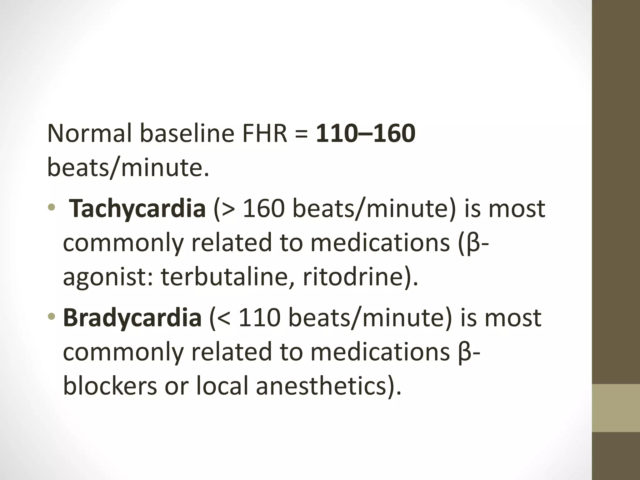
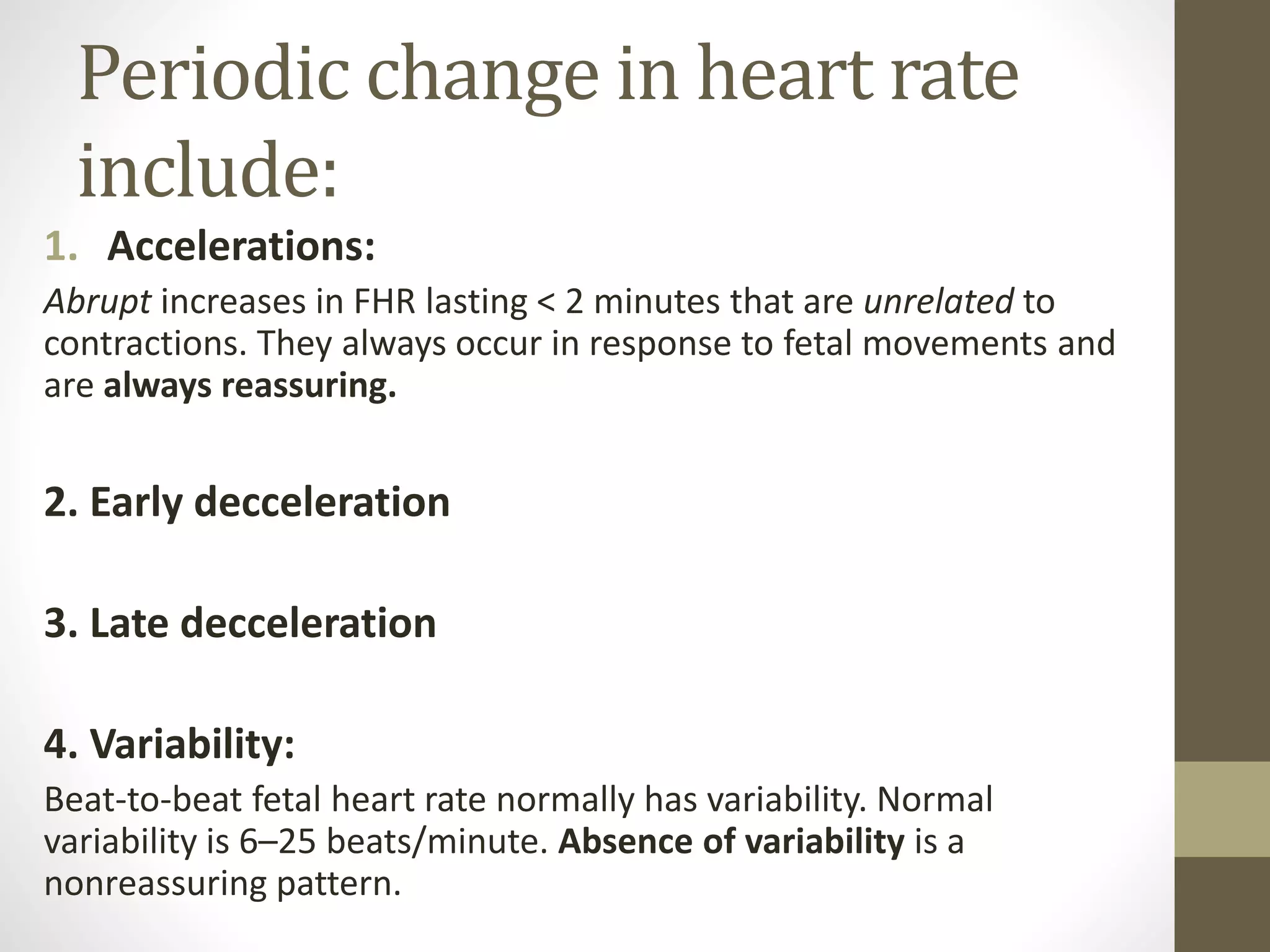
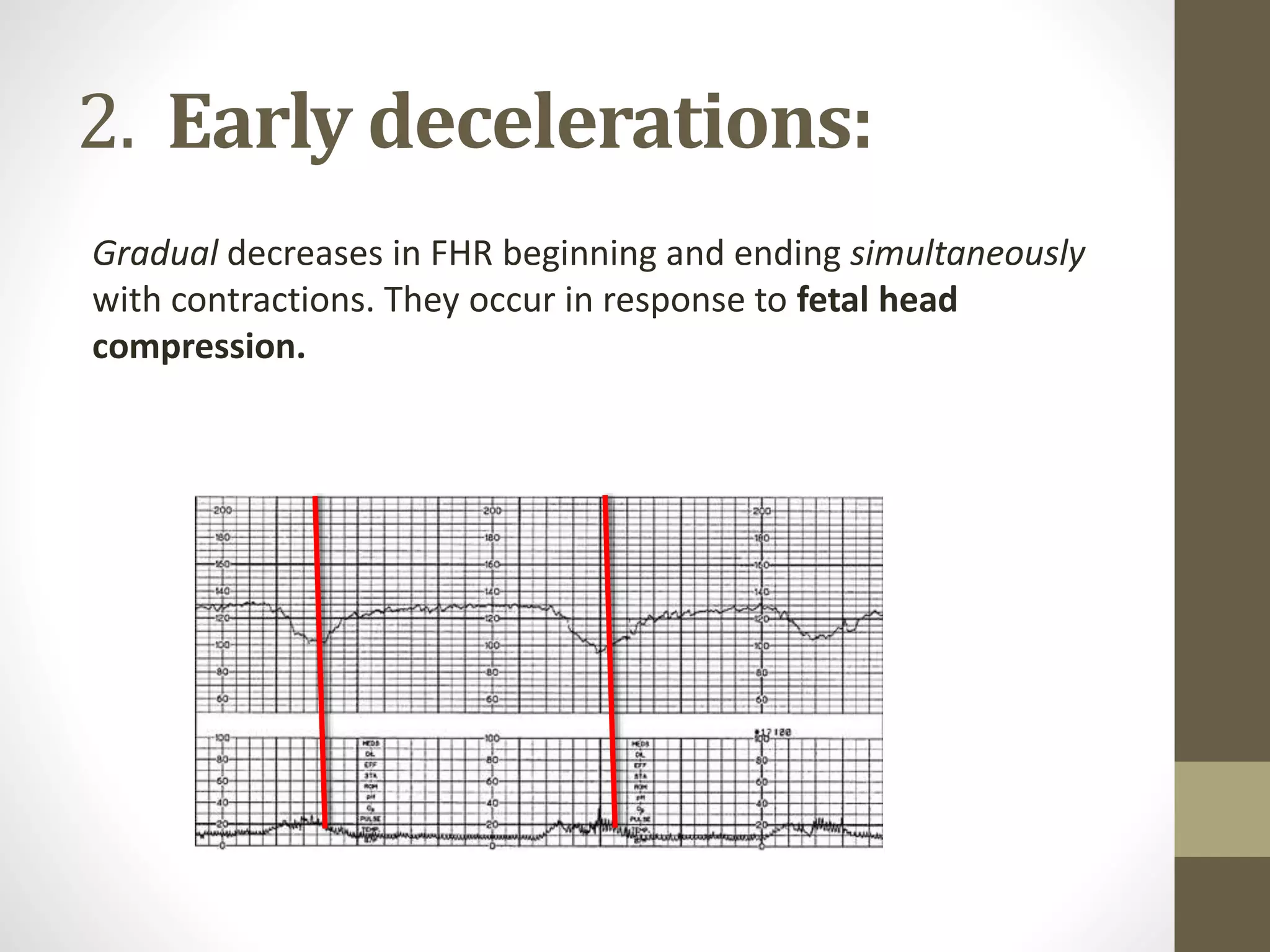
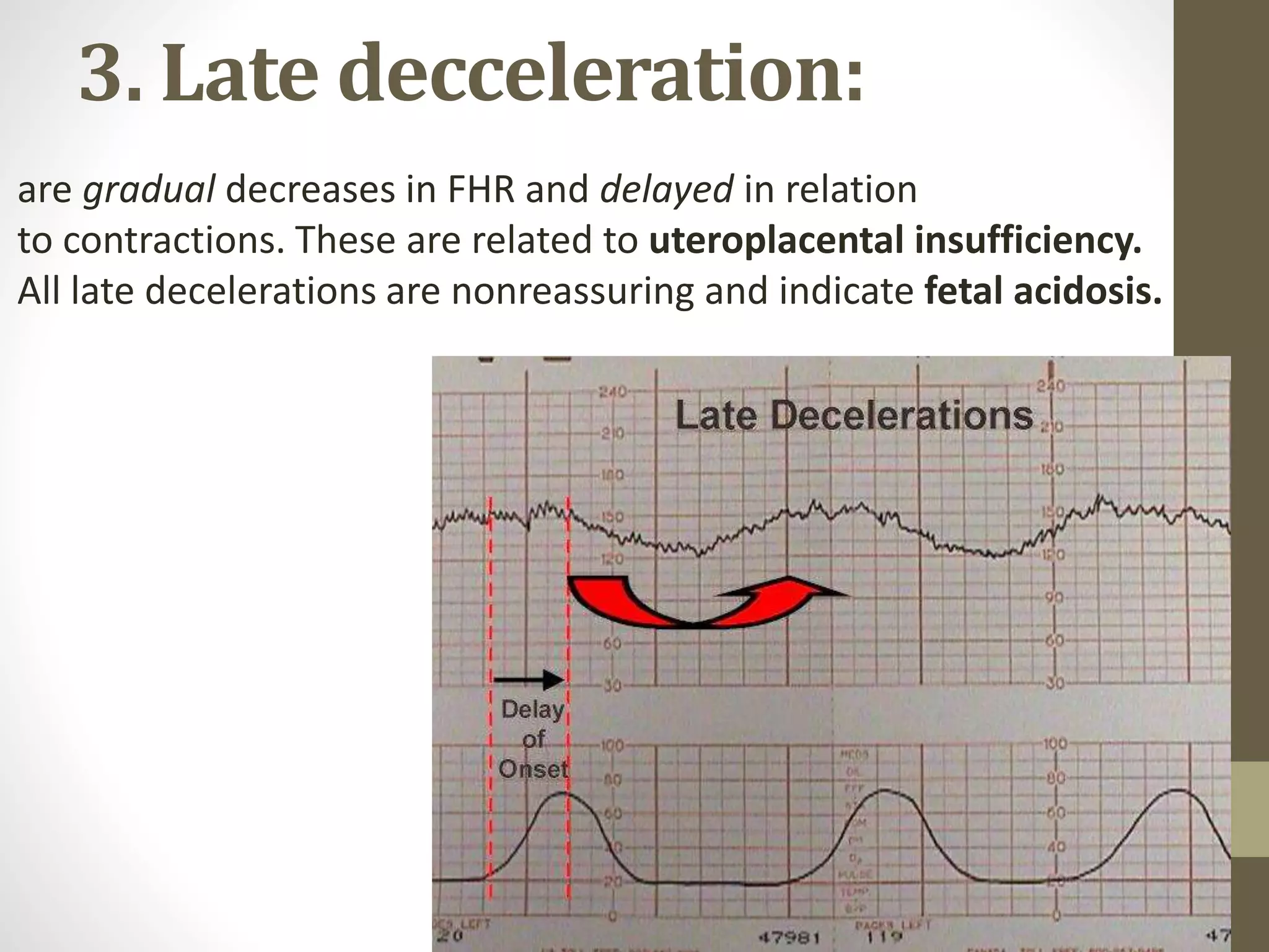
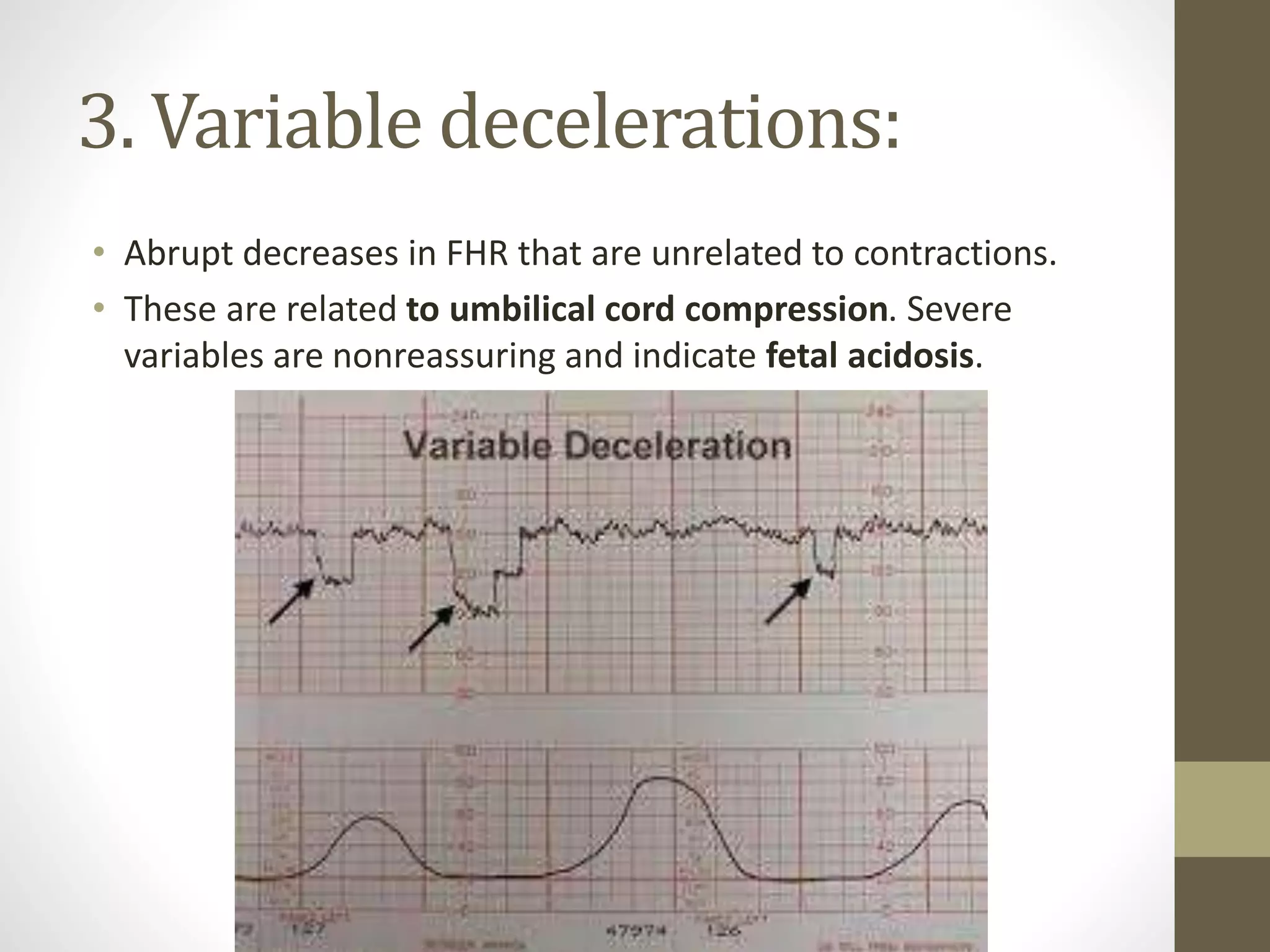
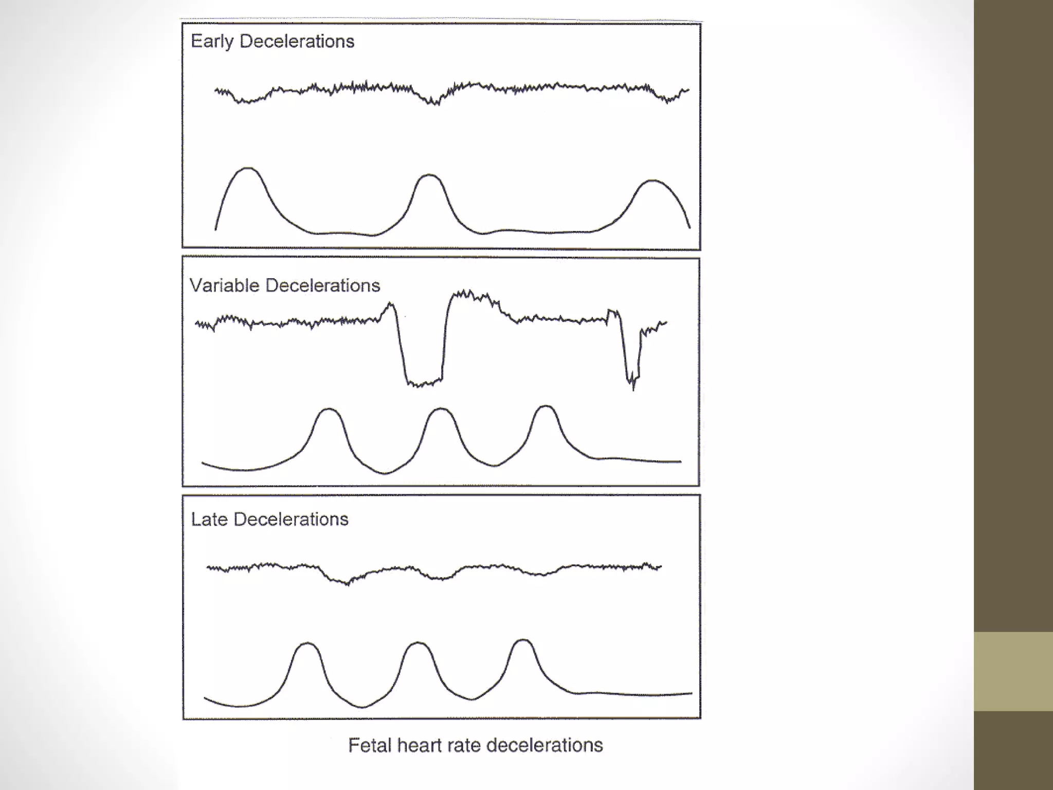
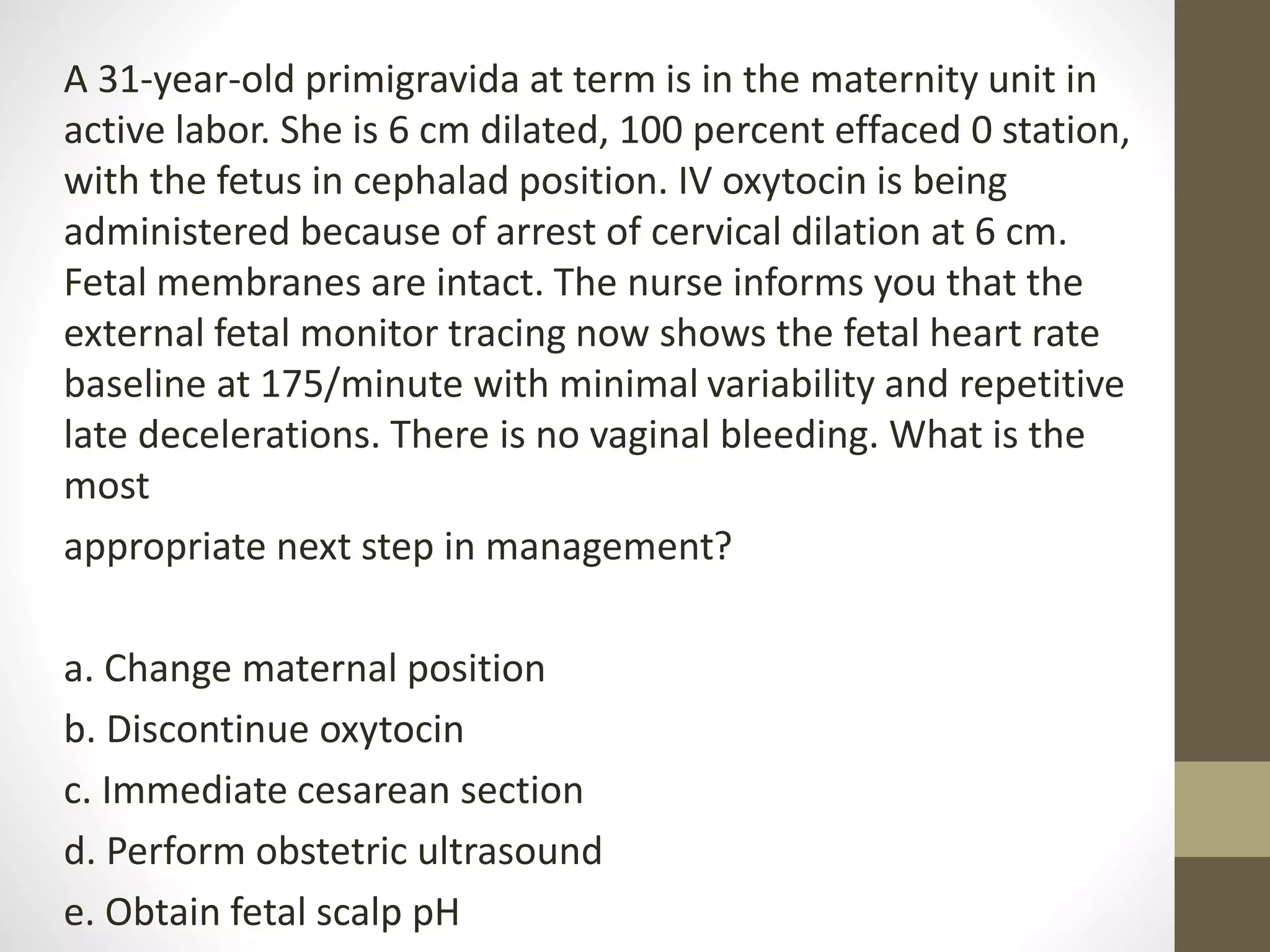
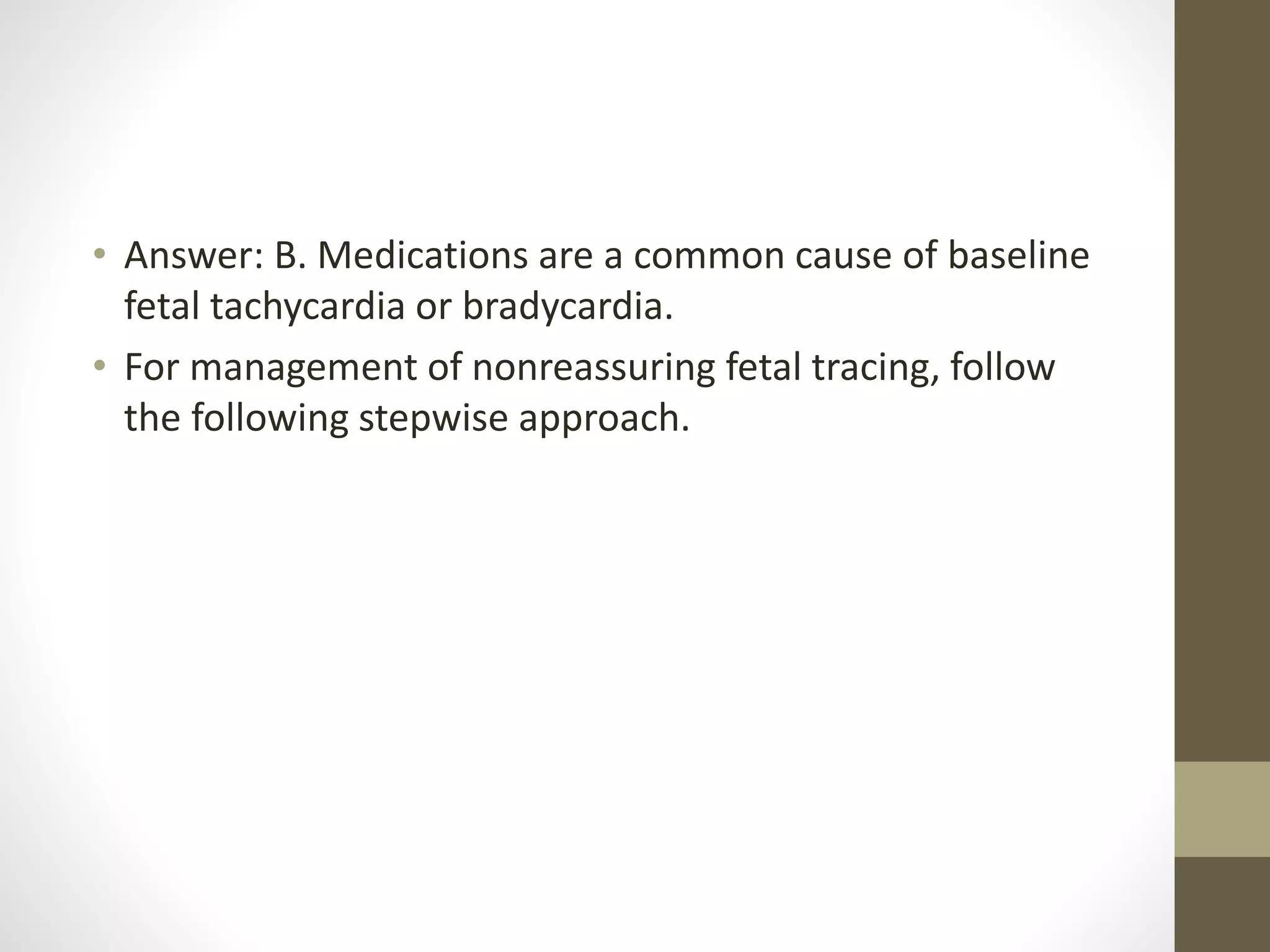
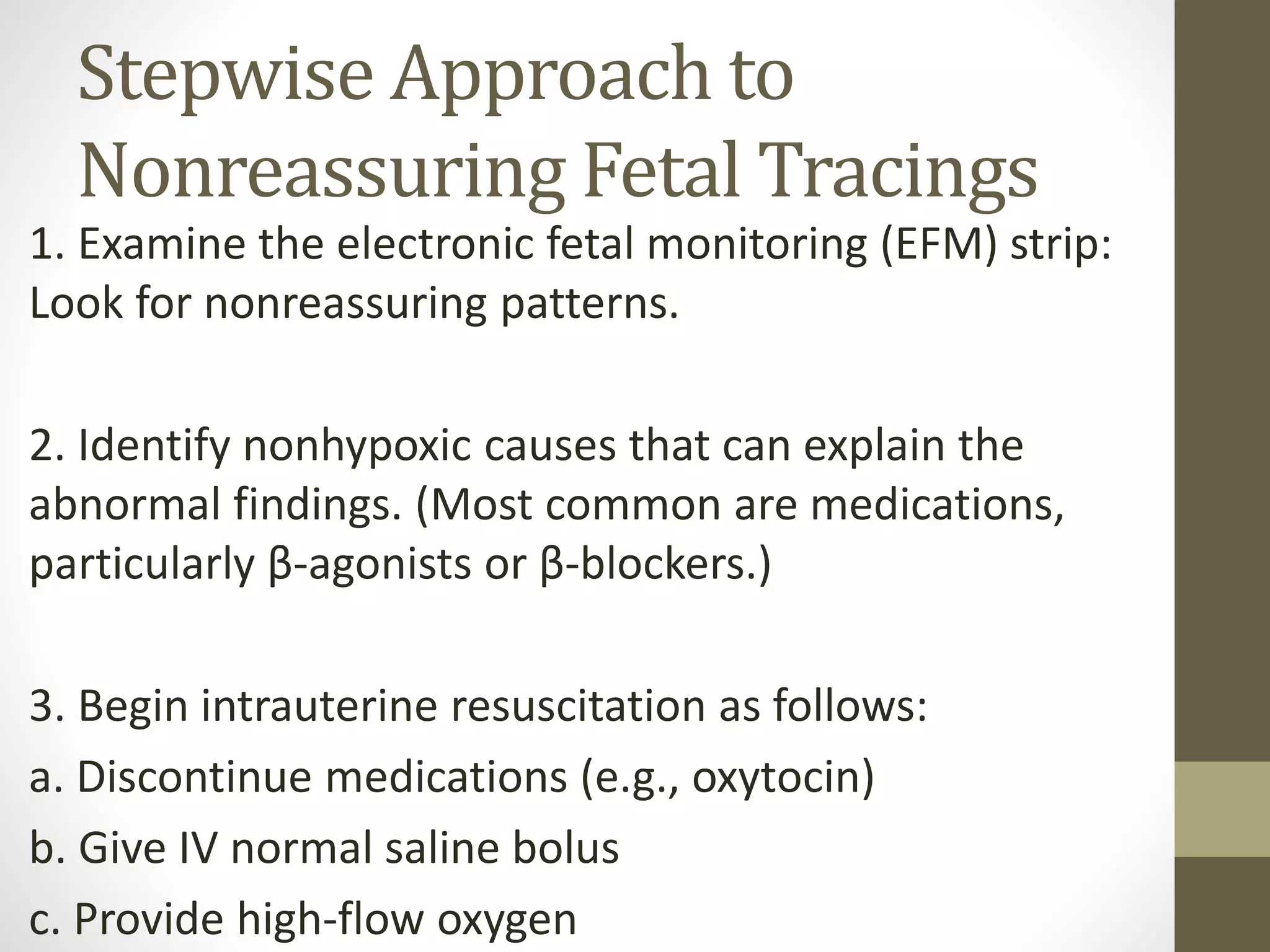
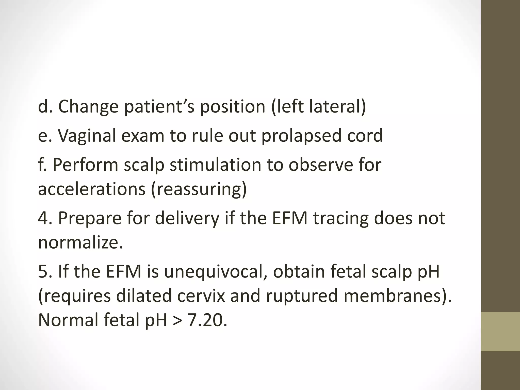
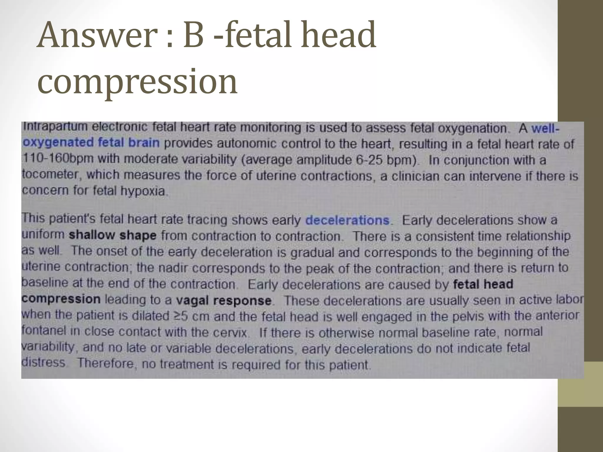
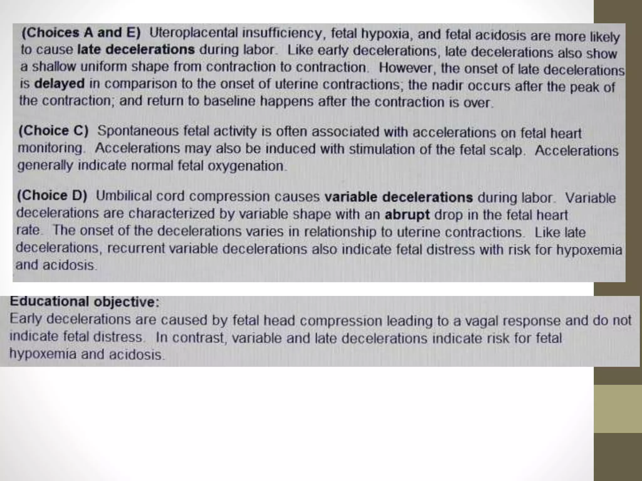
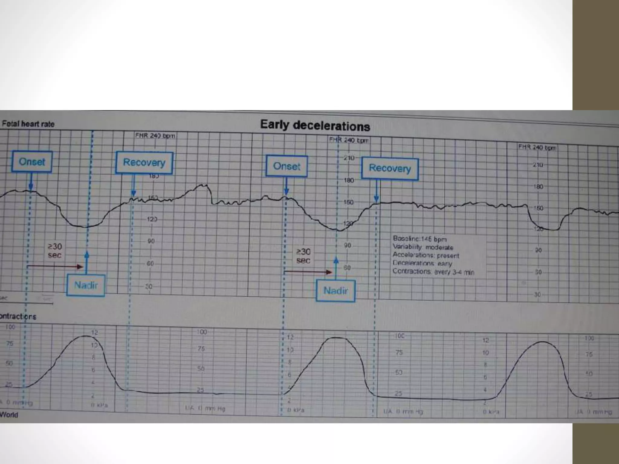
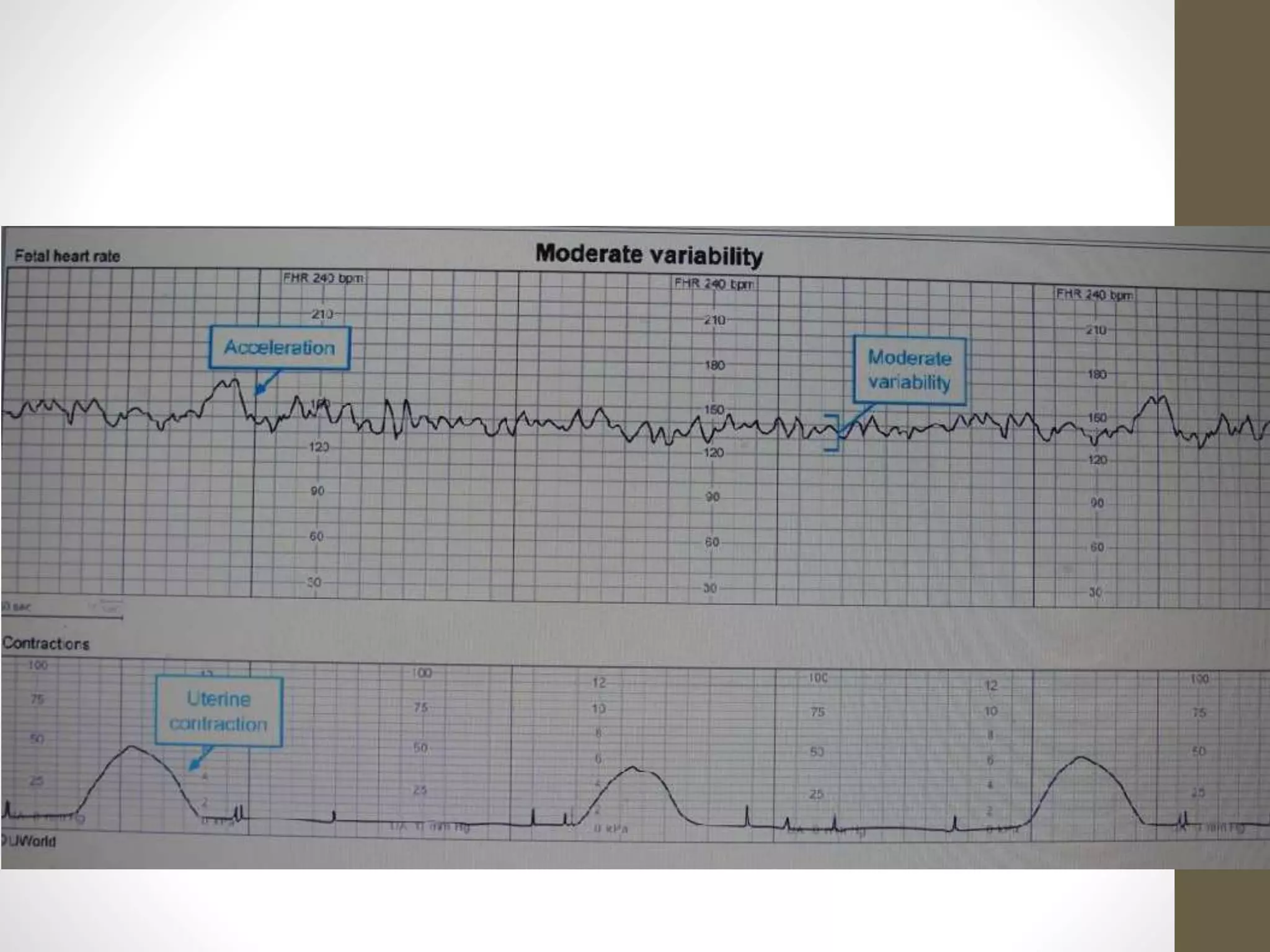
Evaluating of fetal
heart tracing
Fetal heart tracing
Baseline heart rate is the mean FHR during a 10-
minute segment of time,excluding periodic
changes. Changes in fetal heart rate and normal
periodic changes of FHR are related to the
following:
• Uterine hyperstimulation (commonly caused by
medications)
• Fetal head compression
• Umbilical cord compression
• Placental insufficiency
Normal baseline FHR = 110–160
beats/minute.
• Tachycardia (> 160 beats/minute) is most
commonly related to medications (β-
agonist: terbutaline, ritodrine).
• Bradycardia (< 110 beats/minute) is most
commonly related to medications β-
blockers or local anesthetics).
Periodic change in heart rate
include:
1. Accelerations:
Abrupt increases in FHR lasting < 2 minutes that are unrelated to
contractions. They always occur in response to fetal movements and
are always reassuring.
2. Early decceleration
3. Late decceleration
4. Variability:
Beat-to-beat fetal heart rate normally has variability. Normal
variability is 6–25 beats/minute. Absence of variability is a
nonreassuring pattern.
2. Early decelerations:
Gradual decreases in FHR beginning and ending simultaneously
with contractions. They occur in response to fetal head
compression.
3. Late decceleration:
are gradual decreases in FHR and delayed in relation
to contractions. These are related to uteroplacental insufficiency.
All late decelerations are nonreassuring and indicate fetal acidosis.
3. Variable decelerations:
• Abrupt decreases in FHR that are unrelated to contractions.
• These are related to umbilical cord compression. Severe
variables are nonreassuring and indicate fetal acidosis.
A 31-year-old primigravida at term is in the maternity unit in
active labor. She is 6 cm dilated, 100 percent effaced 0 station,
with the fetus in cephalad position. IV oxytocin is being
administered because of arrest of cervical dilation at 6 cm.
Fetal membranes are intact. The nurse informs you that the
external fetal monitor tracing now shows the fetal heart rate
baseline at 175/minute with minimal variability and repetitive
late decelerations. There is no vaginal bleeding. What is the
most
appropriate next step in management?
a. Change maternal position
b. Discontinue oxytocin
c. Immediate cesarean section
d. Perform obstetric ultrasound
e. Obtain fetal scalp pH
• Answer: B. Medications are a common cause of baseline
fetal tachycardia or bradycardia.
• For management of nonreassuring fetal tracing, follow
the following stepwise approach.
Stepwise Approach to
Nonreassuring Fetal Tracings
1. Examine the electronic fetal monitoring (EFM) strip:
Look for nonreassuring patterns.
2. Identify nonhypoxic causes that can explain the
abnormal findings. (Most common are medications,
particularly β-agonists or β-blockers.)
3. Begin intrauterine resuscitation as follows:
a. Discontinue medications (e.g., oxytocin)
b. Give IV normal saline bolus
c. Provide high-flow oxygen
d. Change patient’s position (left lateral)
e. Vaginal exam to rule out prolapsed cord
f. Perform scalp stimulation to observe for
accelerations (reassuring)
4. Prepare for delivery if the EFM tracing does not
normalize.
5. If the EFM is unequivocal, obtain fetal scalp pH
(requires dilated cervix and ruptured membranes).
Normal fetal pH > 7.20.
Answer : B -fetal head
compression
Evaluating of fetal heart tracing

Evaluating of fetal heart tracing

  • 1.
  • 2.
    Fetal heart tracing Baselineheart rate is the mean FHR during a 10- minute segment of time,excluding periodic changes. Changes in fetal heart rate and normal periodic changes of FHR are related to the following: • Uterine hyperstimulation (commonly caused by medications) • Fetal head compression • Umbilical cord compression • Placental insufficiency
  • 3.
    Normal baseline FHR= 110–160 beats/minute. • Tachycardia (> 160 beats/minute) is most commonly related to medications (β- agonist: terbutaline, ritodrine). • Bradycardia (< 110 beats/minute) is most commonly related to medications β- blockers or local anesthetics).
  • 4.
    Periodic change inheart rate include: 1. Accelerations: Abrupt increases in FHR lasting < 2 minutes that are unrelated to contractions. They always occur in response to fetal movements and are always reassuring. 2. Early decceleration 3. Late decceleration 4. Variability: Beat-to-beat fetal heart rate normally has variability. Normal variability is 6–25 beats/minute. Absence of variability is a nonreassuring pattern.
  • 5.
    2. Early decelerations: Gradualdecreases in FHR beginning and ending simultaneously with contractions. They occur in response to fetal head compression.
  • 6.
    3. Late decceleration: aregradual decreases in FHR and delayed in relation to contractions. These are related to uteroplacental insufficiency. All late decelerations are nonreassuring and indicate fetal acidosis.
  • 7.
    3. Variable decelerations: •Abrupt decreases in FHR that are unrelated to contractions. • These are related to umbilical cord compression. Severe variables are nonreassuring and indicate fetal acidosis.
  • 9.
    A 31-year-old primigravidaat term is in the maternity unit in active labor. She is 6 cm dilated, 100 percent effaced 0 station, with the fetus in cephalad position. IV oxytocin is being administered because of arrest of cervical dilation at 6 cm. Fetal membranes are intact. The nurse informs you that the external fetal monitor tracing now shows the fetal heart rate baseline at 175/minute with minimal variability and repetitive late decelerations. There is no vaginal bleeding. What is the most appropriate next step in management? a. Change maternal position b. Discontinue oxytocin c. Immediate cesarean section d. Perform obstetric ultrasound e. Obtain fetal scalp pH
  • 10.
    • Answer: B.Medications are a common cause of baseline fetal tachycardia or bradycardia. • For management of nonreassuring fetal tracing, follow the following stepwise approach.
  • 11.
    Stepwise Approach to NonreassuringFetal Tracings 1. Examine the electronic fetal monitoring (EFM) strip: Look for nonreassuring patterns. 2. Identify nonhypoxic causes that can explain the abnormal findings. (Most common are medications, particularly β-agonists or β-blockers.) 3. Begin intrauterine resuscitation as follows: a. Discontinue medications (e.g., oxytocin) b. Give IV normal saline bolus c. Provide high-flow oxygen
  • 12.
    d. Change patient’sposition (left lateral) e. Vaginal exam to rule out prolapsed cord f. Perform scalp stimulation to observe for accelerations (reassuring) 4. Prepare for delivery if the EFM tracing does not normalize. 5. If the EFM is unequivocal, obtain fetal scalp pH (requires dilated cervix and ruptured membranes). Normal fetal pH > 7.20.
  • 17.
    Answer : B-fetal head compression