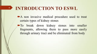
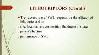
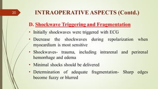
1
Extracorporeal
Shockwave Lithotripsy
(ESWL)
Presented by:
Dr. Afia Tehreem Gilanee
Resident Urology
2
OBJECTIVES
Introduction To ESWL
Working Principle of a Lithotripter
Shockwave Physics
Types of Shockwave Sources
Pre-operative, Intraoperative And Post-operative Aspects
of SWL
EAU Guidelines On SWL
Take-home Message
3
INTRODUCTION TO ESWL
A non invasive medical procedure used to treat
certain types of kidney stones
To break down kidney stones into smaller
fragments, allowing them to pass more easily
through urinary tract and be eliminated from body
4
WORKING PRINCIPLE OF A LITHOTRIPTOR
1. Energy source- creates shock waves
2. Coupling Mechanism- transfers the energy
from outside to inside the body
3. Modes (fluoroscopic, ultrasonic or both)-
identifies and positions the calculus at focus
5
LITHOTRIPTORS
Differ on the basis of
 Focal peak pressures (400-1500 bar)
 Focal dimensions (6*28mm to 50*15mm)
 Varied distances (12-17cm) between shockwave source and the target
 Generated pain
 Anaesthetic requirement
 Size
 Mobility
 Cost
 Durability
6
LITHOTRIPTORS (Contd.)
The success rate of SWL- depends on the efficacy of
lithotripter and on
o size, location, and composition (hardness) of stones
o patient’s habitus
o performance of SWL
7
SHOCKWAVE PHYSICS
Acoustic shockwaves – non harmonic
Non linear pressure characteristics
Steep rise in pressure amplitude –
compressive forces
2 basic types of shockwave sources
• Supersonic emitters
• Finite amplitude emitters
✓Piezoceramic
✓Electromagnetic
8
SUPERSONIC EMITTERS
Release energy in a confined space – producing an
expanding plasma and an acoustic shockwave
Initial compression wave travels faster than speed of
sound in water
Rapidly slows down
Analogous to thunderstorm in nature
Can successfully fragment the calculi
9
FINITE-AMPLITUDE EMITTERS
Create pulsed acoustic shockwaves
By displacing a surface activated by electrical
discharge
Piezoceramic-shockwaves after an electrical discharge
causes ceramic component to elongate- surface
displaced and acoustic pulse generated
Thousands of such components placed on concave
side of spherical surface directed towards a focus
10
11
FINITE-AMPLITUDE EMITTERS (Contd.)
Electromagnetic systems-
electric discharge to a slab,
adjacent to an insulating
foil, creates an electric
current that repulses a metal
membrane – displacing it
and generating an acoustic
pulse in an adjacent medium
12
13
14
FRAGMENTATION
Achieved by erosion and shattering
Erosion at entry and exit sites of shockwaves
Shattering –from energy absorption
Surrounding biologic tissues- resilient because neither brittle
nor focused
Acoustic energy focusing (AEF) devices- attach to the stones
External energy source- AEF device expands and cavitates-
fragmentation
15
PREOPERATIVE EVALUATION
Vital signs monitoring
Physical examination
Investigations (X-ray KUB, USG)
16
INTRAOPERATIVE ASPECTS
A.Stone Localization
➢ Proper patient positioning- pre requisite
➢Anteriorly located kidneys, medially oriented
portions of horseshoe kidney, or transplant
kidneys- best treated in prone position
➢Retrograde stents- easy identification
➢IV contrast agents- helpful in localization
17
INTRAOPERATIVE ASPECTS (Contd.)
B. Imaging
1. Flouroscopic Imaging
• Dimmed room lighting and decreased radiation exposure
• Intermittent fluoroscopy- reveals movement with respiration
2. Ultrasonic Imaging
• Eliminates radiation exposure
• Can identify radiolucent or small caculi
• Ureteral or medially located ones- difficult esp if non obstructive
• Localization is difficult in obese
18
INTRAOPERATIVE ASPECTS (Contd.)
C. Coupling
• Coupling devices- properties similar to those of
human skin
• Prevent pain, ecchymoses, hematomas, or skin
breakdown
• Water cushion coupling systems
• Coupling gel like those in USG
19
INTRAOPERATIVE ASPECTS (Contd.)
D. Shockwave Triggering and Fragmentation
• Initially shockwaves were triggered with ECG
• Decrease the shockwaves during repolarization when
myocardium is most sensitive
• Shockwaves- trauma, including intrarenal and perirenal
hemorrhage and edema
• Minimal shocks should be delivered
• Determination of adequate fragmentation- Sharp edges
become fuzzy or blurred
20
EAU GUIDELINES FOR SWL
 Routine use of internal stents before SWL does not improve stone-free
rates; however may reduce formation of steinstrasse
 Patients with a pacemaker can be treated with SWL
 Patients with implanted cardioverter defibrillators- special care;
 Starting SWL on a lower energy setting with stepwise power ramping
prevents renal injury
 Optimal shockwave frequency- 1-1.5Hz
 Repeat sessions feasible within 1 day for ureteral stones
 No standard antibiotic prophylaxis prior to SWL recommended
21
POST-OPERATIVE CARE
Fluid intake should be encouraged
Asymptomatic individuals- Follow up- serial X ray KUB and
USG
Steinstrasse or stone gravel accumulation in ureter – severe
pain or fever
PCN drainage and decompressing the collecting systems
If uneffective- retrograde endoscopic manipulations
Severe pain unresponsive to routine IV or oral medications-
rare perirenal hematomas- CT scan for staging
22
CONTRAINDICATIONS OF SWL
Pregnancy
Bleeding disorders (should be compensated 24hrs before and
48hrs after treatment)
Untreated UTIs
Severe skeletal malformations and severe obesity, (prevent
targeting of stone)
Arterial aneurysm in the vicinity of stone
Anatomical obstruction distal to the stone
23
TAKE-HOME MESSAGE
Approximately 75% of patients treated with SWL- stone free in 3
months
Large pelvic stones (>1.5cm)- stone free rates at 3 months- 75%
Vs lower calyx- 35%
Small pelvic stones (<1.5cm)- stone free rate- 90%
Vs middle calyx(75%) or lower calyx (50%)
Increasing the size of stones- stone free rates decrease (more so in
lower and middle calyces than superior calyceal and pelvic locations)
Lower calyceal stone free rates- increased with small stone burden, a
short and wide infundibulum, and a non-acute infundibulo-pelvic angle
24
25
Thank You!

ESWL (1).pdf

  • 1.
  • 2.
  • 3.
    OBJECTIVES Introduction To ESWL WorkingPrinciple of a Lithotripter Shockwave Physics Types of Shockwave Sources Pre-operative, Intraoperative And Post-operative Aspects of SWL EAU Guidelines On SWL Take-home Message 3
  • 4.
    INTRODUCTION TO ESWL Anon invasive medical procedure used to treat certain types of kidney stones To break down kidney stones into smaller fragments, allowing them to pass more easily through urinary tract and be eliminated from body 4
  • 5.
    WORKING PRINCIPLE OFA LITHOTRIPTOR 1. Energy source- creates shock waves 2. Coupling Mechanism- transfers the energy from outside to inside the body 3. Modes (fluoroscopic, ultrasonic or both)- identifies and positions the calculus at focus 5
  • 6.
    LITHOTRIPTORS Differ on thebasis of  Focal peak pressures (400-1500 bar)  Focal dimensions (6*28mm to 50*15mm)  Varied distances (12-17cm) between shockwave source and the target  Generated pain  Anaesthetic requirement  Size  Mobility  Cost  Durability 6
  • 7.
    LITHOTRIPTORS (Contd.) The successrate of SWL- depends on the efficacy of lithotripter and on o size, location, and composition (hardness) of stones o patient’s habitus o performance of SWL 7
  • 8.
    SHOCKWAVE PHYSICS Acoustic shockwaves– non harmonic Non linear pressure characteristics Steep rise in pressure amplitude – compressive forces 2 basic types of shockwave sources • Supersonic emitters • Finite amplitude emitters ✓Piezoceramic ✓Electromagnetic 8
  • 9.
    SUPERSONIC EMITTERS Release energyin a confined space – producing an expanding plasma and an acoustic shockwave Initial compression wave travels faster than speed of sound in water Rapidly slows down Analogous to thunderstorm in nature Can successfully fragment the calculi 9
  • 10.
    FINITE-AMPLITUDE EMITTERS Create pulsedacoustic shockwaves By displacing a surface activated by electrical discharge Piezoceramic-shockwaves after an electrical discharge causes ceramic component to elongate- surface displaced and acoustic pulse generated Thousands of such components placed on concave side of spherical surface directed towards a focus 10
  • 11.
  • 12.
    FINITE-AMPLITUDE EMITTERS (Contd.) Electromagneticsystems- electric discharge to a slab, adjacent to an insulating foil, creates an electric current that repulses a metal membrane – displacing it and generating an acoustic pulse in an adjacent medium 12
  • 13.
  • 14.
  • 15.
    FRAGMENTATION Achieved by erosionand shattering Erosion at entry and exit sites of shockwaves Shattering –from energy absorption Surrounding biologic tissues- resilient because neither brittle nor focused Acoustic energy focusing (AEF) devices- attach to the stones External energy source- AEF device expands and cavitates- fragmentation 15
  • 16.
    PREOPERATIVE EVALUATION Vital signsmonitoring Physical examination Investigations (X-ray KUB, USG) 16
  • 17.
    INTRAOPERATIVE ASPECTS A.Stone Localization ➢Proper patient positioning- pre requisite ➢Anteriorly located kidneys, medially oriented portions of horseshoe kidney, or transplant kidneys- best treated in prone position ➢Retrograde stents- easy identification ➢IV contrast agents- helpful in localization 17
  • 18.
    INTRAOPERATIVE ASPECTS (Contd.) B.Imaging 1. Flouroscopic Imaging • Dimmed room lighting and decreased radiation exposure • Intermittent fluoroscopy- reveals movement with respiration 2. Ultrasonic Imaging • Eliminates radiation exposure • Can identify radiolucent or small caculi • Ureteral or medially located ones- difficult esp if non obstructive • Localization is difficult in obese 18
  • 19.
    INTRAOPERATIVE ASPECTS (Contd.) C.Coupling • Coupling devices- properties similar to those of human skin • Prevent pain, ecchymoses, hematomas, or skin breakdown • Water cushion coupling systems • Coupling gel like those in USG 19
  • 20.
    INTRAOPERATIVE ASPECTS (Contd.) D.Shockwave Triggering and Fragmentation • Initially shockwaves were triggered with ECG • Decrease the shockwaves during repolarization when myocardium is most sensitive • Shockwaves- trauma, including intrarenal and perirenal hemorrhage and edema • Minimal shocks should be delivered • Determination of adequate fragmentation- Sharp edges become fuzzy or blurred 20
  • 21.
    EAU GUIDELINES FORSWL  Routine use of internal stents before SWL does not improve stone-free rates; however may reduce formation of steinstrasse  Patients with a pacemaker can be treated with SWL  Patients with implanted cardioverter defibrillators- special care;  Starting SWL on a lower energy setting with stepwise power ramping prevents renal injury  Optimal shockwave frequency- 1-1.5Hz  Repeat sessions feasible within 1 day for ureteral stones  No standard antibiotic prophylaxis prior to SWL recommended 21
  • 22.
    POST-OPERATIVE CARE Fluid intakeshould be encouraged Asymptomatic individuals- Follow up- serial X ray KUB and USG Steinstrasse or stone gravel accumulation in ureter – severe pain or fever PCN drainage and decompressing the collecting systems If uneffective- retrograde endoscopic manipulations Severe pain unresponsive to routine IV or oral medications- rare perirenal hematomas- CT scan for staging 22
  • 23.
    CONTRAINDICATIONS OF SWL Pregnancy Bleedingdisorders (should be compensated 24hrs before and 48hrs after treatment) Untreated UTIs Severe skeletal malformations and severe obesity, (prevent targeting of stone) Arterial aneurysm in the vicinity of stone Anatomical obstruction distal to the stone 23
  • 24.
    TAKE-HOME MESSAGE Approximately 75%of patients treated with SWL- stone free in 3 months Large pelvic stones (>1.5cm)- stone free rates at 3 months- 75% Vs lower calyx- 35% Small pelvic stones (<1.5cm)- stone free rate- 90% Vs middle calyx(75%) or lower calyx (50%) Increasing the size of stones- stone free rates decrease (more so in lower and middle calyces than superior calyceal and pelvic locations) Lower calyceal stone free rates- increased with small stone burden, a short and wide infundibulum, and a non-acute infundibulo-pelvic angle 24
  • 25.