SlideShare a Scribd company logo
1 of 6
Download to read offline
GDC Uttersoo
Comparative Anatomy of Brain
The brain is an organ that serves as the Centre of Nervous System in all Vertebrate animals. It is
located in the head, usually close to the sensory organs for senses such as vision.
All vertebrate brains share a common underlying form which appears most clearly during
early stages of development. In its earliest form the brain appears as three swellings at the front
end of the neural tube; these swellings eventually become the forebrain, midbrain and hindbrain
(prosencephalon, mesencephalon and rhombencephalon resp). In the initial stages of brain
development, the three areas are roughly equal in size. In many classes of vertebrates, such as
Fish and Amphibians, the three parts remain similar in size in the adult, but in Mammals the
forebrain becomes much larger than the other parts, and the midbrain becomes very small.
Functions of Brain
1. Olfactory lobes: Sense of smell.
2. Cerebral hemispheres: Seat of intelligence and memory.
3. Diencephalon: Controls the general metabolic functions of the body.
4. Optic lobes: Sense of vision.
5. Cerebellum: Co-ordinates the movements of voluntary muscles.
6. Medulla oblongata: Controls the involuntary functions of the body.
Development and differentiation of brain in Vertebrates
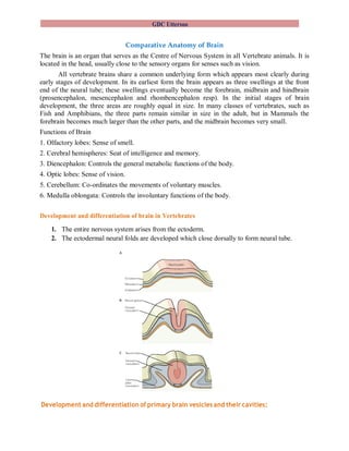
1. The entire nervous system arises from the ectoderm.
2. The ectodermal neural folds are developed which close dorsally to form neural tube.
Development and differentiation of primary brain vesicles and their cavities:
GDC Uttersoo
The vesicles of vertebrate brain:
1. Anterior part of neural tube is slightly swollen which initially differentiated in to three
primary vesicles of brain namely first prosencephalon (forebrain), second
mesencephalon (midbrain) and third rhombencephalon (hindbrain) containing cavities.
2. At later stages two secondary vesicles form in the area of the prosencephalon, 1a and
1b (see fig. below) developing into telencephalon and diencephalon respectively.
3. The diencephalon gives rise to sac like lateral outgrowths called the optic vesicles. The
optic vesicles are pushed downward by two large processes growing forward from the
anterior vesicle (the primitive cerebral hemispheres). These ultimately develop into the
retina, and other nervous parts of the eye.
4. The other secondary vesicles are developed in the rhombencephalon namely 3a and 3b
(see fig. below) developing into metencephalon and myelencephalon respectively.
5. Mesencephalon reduces to form a narrow commissure known as cerebral aqueduct (see
fig. below).
The cavities of vertebrate brain:
1. The cavities of brain are referred as ventricles. There are four ventricles in vertebrate
brain namely 1. Olfactory ventricle. 2. Lateral ventricle. 3. Third ventricle. 4. Fourth
ventricle
2. The olfactory lobes carry cavities known as olfactory ventricles arising from the
anterior part of the cerebral hemispheres. These grow forward, and soon lose olfactory
ventricles.
3. The telencephalic vesicles become the cerebral hemispheres, and their cavities become
the paired lateral ventricles (lateral telocoeles) of the adult brain.
GDC Uttersoo
4. The third ventricle lies in the diencephalon (diocoele). Later in development the lateral
walls of the diencephalon become greatly thickened to form the thalami, thus reducing
the size and changing the shape of the diocoele, which is known in adult anatomy as the
third brain ventricle.
5. The fourth ventricle lies in metencephalon. The anterior part of the fourth ventricle is
known as metacoele and the posterior part as myelocoele.
Flexures of brain:
1. As the result of unequal growth of these different parts of brain, three flexures are formed
and the embryonic brain becomes bent on itself in a somewhat zigzag fashion; the two
earliest flexures are concave ventrally and are associated with corresponding flexures of
the whole head.
2. The first flexure appears in the region of the mid-brain, and is named the ventral
cephalic flexure. By means of it the fore-brain is bent in a ventral direction around the
anterior end of the notochord and fore-gut, with the result that the floor of the fore-brain
comes to lie almost parallel with that of the hind-brain. This flexure causes the mid-brain
to become, for a time, the most prominent part of the brain, since its dorsal surface
corresponds with the convexity of the curve.
3. The second bend appears at the junction of the hind-brain and medulla spinalis. This is
termed the cervical flexure, and increases from the third to the end of the fifth week,
when the hind-brain forms nearly a right angle with the medulla spinalis; after the fifth
week erection of the head takes place and the cervical flexure diminishes and disappears.
4. The third bend is named the pontine flexure, because it is found in the region of the
future pons Varoli.
5. Both the cervical and the pontine flexures eventually straighten out, but the cephalic
flexure remains prominent throughout development.
GDC Uttersoo
Evolutionofcerebral hemispheres & cerebellum with reference to shark,frog,lizard,
pigeon & rabbit:
1. Telencephalon, called lamina terminalis and the roof of the cerebrum is called cortex or
pallium. The telencephalon then appears to be composed of two divisions, the anterior of
which is subsequently developed into the cerebral hemispheres, corpora striata, and the
olfactory lobes. The hemispheres undergo enormous enlargement in their later development
and extend dorsally and posteriorly as well as anteriorly, eventually covering the entire
diencephalon and mesencephalon under their posterior lobes.
2. Posterior part of forebrain, the diencephalon, consisting of the thalamus and
hypothalamus, representing the anterior vesicle. The median evagination in the roof of the
diencephalon develops into epiphysis.
3. The mesencephalon becomes specialized as the optic lobes, visual centers associated with
the optic nerves. The dorsal and lateral walls of the mesencephalon later increase rapidly in
thickness and become the optic lobes (copora quadrigemina) of the adult brain. . It serves as
the main pathway of the fiber tracts which connect the cerebral hemispheres with the
posterior part of the brain and the spinal cord.
4. The hindbrain became divided into anterior metencephalon and posterior myelencephalon.
The metencephalon shows ventrally and laterally an extensive ingrowth of fiber tracts giving
rise to the pons and to the cerebellar peduncles of the adult metencephalon. The roof of the
metencephalon undergoes extensive enlargement and becomes the cerebellum of the adult
brain. The ventro-lateral wall of the cerebrum becomes thick and called corpus striatum.
The ventral and lateral walls of the myelencephalon become the floor and side-walls of the
medulla of the adult brain. The medulla becomes specialized as a control center for some
autonomic and somatic pathways concerned with vital functions (such as breathing, blood
pressure, and heartbeat). The dorsal wall of myelencephalon is thin and receives rich blood
vessels thus forming posterior choroid plexus. The pons is above the medulla and also acts as
a connecting tract. The cerebellum enlarged and became a structure concerned with balance,
equilibrium, and muscular coordination.
GDC Uttersoo
Comparativeanatomyofbraininvertebrates
Brain of all vertebrates, from fish to man, is built in accordance with the same architectural plan.
However, form of brain differs in different vertebrates in accordance with the habits and
behavior of the animals.
1. Elasmobranches:
Olfactory lobes are correspondingly large.
Optic lobes and pallium are relatively moderate in size.
Saccus vasculosus, thin-walled vascular sensory organ, is attached to pituitary and
connected with cerebellum.
Pineal apparatus is well-developed.
Cerebellum is especially large due to active swimming habit.
Ruffle-like restiform bodies are present.
2. Amphibians:
Olfactory lobes are smaller than optic lobes.
Corpus striatum receives greater number of sensory fibres.
Cerebral hemispheres are more developed than in fishes.
Less-developed cerebellum.
Medulla is also small.
Pineal body is small.
3. Reptilians:
Telencephalon becomes the largest region of brain.
Olfactory lobes are larger than in amphibians.
A pair of auditory lobes is found posterior to optic lobes.
The third ventricle is reduced to a narrow cerebral aqueduct.
Cerebellum is somewhat pear-shaped and larger than in amphibians.
Modern reptiles show a great development in the basal parts of the forebrain. There are large
numbers of nervous connections between the thalamus and the hemispheres. The latter are
larger than the optic lobes, showing the increased importance of the former. The walls of the
thalamus are very thick and many of the optic nerve paths end there as well as many in the
midbrain.
4. Birds:
Brain is proportionately larger than that of a reptile.
Olfactory lobes are small.
Two cerebral hemispheres are larger, smooth and projected posteriorly over the
GDC Uttersoo
diencephalon to meet the cerebellum.
Pallium is thin but corpus striatum is greatly enlarged.
Optic lobes are conspicuously developed.
The cerebellum is greatly enlarged with several superficial folds.
5. Mammals:
Brain is proportionately larger than in other vertebrates.
Cerebral hemispheres of Prototheria, Metatheria & Eutheria are smaller & smooth, larger
& smooth, greatly enlarged & divided into lobes respectively.
The two hemispheres are jointed internally by transverse band-like fibres called corpus
callosum.
Olfactory lobes are relatively small but well defined.
Four almost solid optic lobes are present.
The mammalian brain is completely dominated by the cerebral hemispheres. The roof has
developed enormously and spread out forming the cerebral cortex which in humans is thrown
into a number of elaborate folds and almost covers the rest of the brain. The cortex is made up
millions of cells. The more folded the surface, the more cells it can contain. These cells make up
the gray matter. Their axons, which make up the tracts or pathways in the brain, form the white
matter underneath the cortex. The white matter of the spinal cord is also made up of nerve axons,
surrounding the central gray matter.
Conclusion: Discussing the evolution of brain in these vertebrate groups, it is clear that they are
originated from a common ancestral stock.

More Related Content

What's hot

Petromyzon -External features and comparative hagfish
Petromyzon -External features and comparative hagfishPetromyzon -External features and comparative hagfish
Petromyzon -External features and comparative hagfishSoniaBajaj10
 
Phylogenetic relationship of protochordates
Phylogenetic relationship of protochordatesPhylogenetic relationship of protochordates
Phylogenetic relationship of protochordatesSaira Ramzan
 
Balanoglossus- A Hemichordate
Balanoglossus- A HemichordateBalanoglossus- A Hemichordate
Balanoglossus- A HemichordatePranjal Gupta
 
Balanoglossus-Morphology structure, development , larva ,Affinities
Balanoglossus-Morphology structure, development , larva ,AffinitiesBalanoglossus-Morphology structure, development , larva ,Affinities
Balanoglossus-Morphology structure, development , larva ,AffinitiesSoniaBajaj10
 
Accessory respiratory organ in fishes
Accessory respiratory organ in fishesAccessory respiratory organ in fishes
Accessory respiratory organ in fishesAnand P P
 
Comparative Anatomy - Skeletal System
Comparative Anatomy - Skeletal SystemComparative Anatomy - Skeletal System
Comparative Anatomy - Skeletal SystemEmsi Onairpic
 
Cephalic appendages of prawn
Cephalic appendages of prawnCephalic appendages of prawn
Cephalic appendages of prawnJaleelkabdul Jaleel
 
Comparative Anatomy - Digestive System
Comparative Anatomy - Digestive SystemComparative Anatomy - Digestive System
Comparative Anatomy - Digestive SystemEmsi Onairpic
 
Comparative pro,mete,& eutheria, features of prototheria to eutheria,& af...
Comparative pro,mete,& eutheria, features of prototheria to eutheria,& af...Comparative pro,mete,& eutheria, features of prototheria to eutheria,& af...
Comparative pro,mete,& eutheria, features of prototheria to eutheria,& af...SoniaBajaj10
 
Classification of amphibia
Classification of amphibiaClassification of amphibia
Classification of amphibiaTaniya07
 
Study of Prawn (palaemon)
Study of Prawn (palaemon)Study of Prawn (palaemon)
Study of Prawn (palaemon)babusinghKirade1
 
Neurogenic and myogenic hearts
Neurogenic and myogenic heartsNeurogenic and myogenic hearts
Neurogenic and myogenic heartsMohd Asif Kanth
 
Prawn respiratory system,Types of gills,structure of gill plates,working of ...
Prawn  respiratory system,Types of gills,structure of gill plates,working of ...Prawn  respiratory system,Types of gills,structure of gill plates,working of ...
Prawn respiratory system,Types of gills,structure of gill plates,working of ...SoniaBajaj10
 

What's hot (20)

Petromyzon -External features and comparative hagfish
Petromyzon -External features and comparative hagfishPetromyzon -External features and comparative hagfish
Petromyzon -External features and comparative hagfish
 
Phylogenetic relationship of protochordates
Phylogenetic relationship of protochordatesPhylogenetic relationship of protochordates
Phylogenetic relationship of protochordates
 
Nervous systems in vertebrates: T.Y.B.Sc. Sem VI Notes
Nervous systems in vertebrates: T.Y.B.Sc. Sem VI NotesNervous systems in vertebrates: T.Y.B.Sc. Sem VI Notes
Nervous systems in vertebrates: T.Y.B.Sc. Sem VI Notes
 
Balanoglossus- A Hemichordate
Balanoglossus- A HemichordateBalanoglossus- A Hemichordate
Balanoglossus- A Hemichordate
 
Balanoglossus-Morphology structure, development , larva ,Affinities
Balanoglossus-Morphology structure, development , larva ,AffinitiesBalanoglossus-Morphology structure, development , larva ,Affinities
Balanoglossus-Morphology structure, development , larva ,Affinities
 
Aortic arches
Aortic archesAortic arches
Aortic arches
 
Accessory respiratory organ in fishes
Accessory respiratory organ in fishesAccessory respiratory organ in fishes
Accessory respiratory organ in fishes
 
Comparative Anatomy - Skeletal System
Comparative Anatomy - Skeletal SystemComparative Anatomy - Skeletal System
Comparative Anatomy - Skeletal System
 
Cephalic appendages of prawn
Cephalic appendages of prawnCephalic appendages of prawn
Cephalic appendages of prawn
 
Urino genitial system of vertebrates
Urino genitial system of vertebratesUrino genitial system of vertebrates
Urino genitial system of vertebrates
 
Origin of chordata
Origin of chordataOrigin of chordata
Origin of chordata
 
Comparative Anatomy - Digestive System
Comparative Anatomy - Digestive SystemComparative Anatomy - Digestive System
Comparative Anatomy - Digestive System
 
Comparative pro,mete,& eutheria, features of prototheria to eutheria,& af...
Comparative pro,mete,& eutheria, features of prototheria to eutheria,& af...Comparative pro,mete,& eutheria, features of prototheria to eutheria,& af...
Comparative pro,mete,& eutheria, features of prototheria to eutheria,& af...
 
Brain of vertebrates
Brain of vertebratesBrain of vertebrates
Brain of vertebrates
 
Classification of amphibia
Classification of amphibiaClassification of amphibia
Classification of amphibia
 
Study of Prawn (palaemon)
Study of Prawn (palaemon)Study of Prawn (palaemon)
Study of Prawn (palaemon)
 
Neurogenic and myogenic hearts
Neurogenic and myogenic heartsNeurogenic and myogenic hearts
Neurogenic and myogenic hearts
 
Neoteny
NeotenyNeoteny
Neoteny
 
Comparision of brain in vertebrates
Comparision of brain in vertebratesComparision of brain in vertebrates
Comparision of brain in vertebrates
 
Prawn respiratory system,Types of gills,structure of gill plates,working of ...
Prawn  respiratory system,Types of gills,structure of gill plates,working of ...Prawn  respiratory system,Types of gills,structure of gill plates,working of ...
Prawn respiratory system,Types of gills,structure of gill plates,working of ...
 

Similar to Comparative Brain Anatomy of Vertebrates

Cns development
Cns developmentCns development
Cns developmentMed Study
 
comparative_Nervous_Syst_Vertebrate_Life_Sciences_1yr_PDF.pdf
comparative_Nervous_Syst_Vertebrate_Life_Sciences_1yr_PDF.pdfcomparative_Nervous_Syst_Vertebrate_Life_Sciences_1yr_PDF.pdf
comparative_Nervous_Syst_Vertebrate_Life_Sciences_1yr_PDF.pdfMohitgupta555351
 
Development of the nervous system
Development of the nervous systemDevelopment of the nervous system
Development of the nervous systemmuhammaduzairkhan8
 
Brain strom
Brain stromBrain strom
Brain stromVamsi kumar
 
Presentation1.pptx, radiological anatomy of the brain.
Presentation1.pptx, radiological anatomy of the brain.Presentation1.pptx, radiological anatomy of the brain.
Presentation1.pptx, radiological anatomy of the brain.Abdellah Nazeer
 
Embryology development of central nervous system
Embryology   development of central nervous systemEmbryology   development of central nervous system
Embryology development of central nervous systemMBBS IMS MSU
 
Central Nervous system CNS
Central Nervous system CNSCentral Nervous system CNS
Central Nervous system CNSEduFlash
 
Overview of the anatomy of the brain and its physiology
Overview of the anatomy of the brain and its physiologyOverview of the anatomy of the brain and its physiology
Overview of the anatomy of the brain and its physiologyssuser1d880f
 
CNS PART 2.pptx
CNS PART 2.pptxCNS PART 2.pptx
CNS PART 2.pptxSanthi Dasari
 
The brain
The brainThe brain
The brainAnasAlwadi
 
Anatomy of the brain
Anatomy of the brainAnatomy of the brain
Anatomy of the brainSaika Lashari
 
BRAIN ANATOMY SHOWING THE PARTS OF THE BRAIN
BRAIN ANATOMY SHOWING THE PARTS OF THE BRAINBRAIN ANATOMY SHOWING THE PARTS OF THE BRAIN
BRAIN ANATOMY SHOWING THE PARTS OF THE BRAINJyothiK38
 
Nervous system of the frog (1)
Nervous system of the frog (1)Nervous system of the frog (1)
Nervous system of the frog (1)Timothy Uy
 

Similar to Comparative Brain Anatomy of Vertebrates (20)

Cns development
Cns developmentCns development
Cns development
 
The brain
The brainThe brain
The brain
 
Ch11 eoc
Ch11 eocCh11 eoc
Ch11 eoc
 
Cns
CnsCns
Cns
 
Human Brain
Human BrainHuman Brain
Human Brain
 
comparative_Nervous_Syst_Vertebrate_Life_Sciences_1yr_PDF.pdf
comparative_Nervous_Syst_Vertebrate_Life_Sciences_1yr_PDF.pdfcomparative_Nervous_Syst_Vertebrate_Life_Sciences_1yr_PDF.pdf
comparative_Nervous_Syst_Vertebrate_Life_Sciences_1yr_PDF.pdf
 
Development of the nervous system
Development of the nervous systemDevelopment of the nervous system
Development of the nervous system
 
Brain strom
Brain stromBrain strom
Brain strom
 
Presentation1.pptx, radiological anatomy of the brain.
Presentation1.pptx, radiological anatomy of the brain.Presentation1.pptx, radiological anatomy of the brain.
Presentation1.pptx, radiological anatomy of the brain.
 
Embryology development of central nervous system
Embryology   development of central nervous systemEmbryology   development of central nervous system
Embryology development of central nervous system
 
Central Nervous system CNS
Central Nervous system CNSCentral Nervous system CNS
Central Nervous system CNS
 
Overview of the anatomy of the brain and its physiology
Overview of the anatomy of the brain and its physiologyOverview of the anatomy of the brain and its physiology
Overview of the anatomy of the brain and its physiology
 
The human brain.pptx
The human brain.pptxThe human brain.pptx
The human brain.pptx
 
CNS PART 2.pptx
CNS PART 2.pptxCNS PART 2.pptx
CNS PART 2.pptx
 
The brain
The brainThe brain
The brain
 
Brain
BrainBrain
Brain
 
Anatomy of the brain
Anatomy of the brainAnatomy of the brain
Anatomy of the brain
 
Brain
BrainBrain
Brain
 
BRAIN ANATOMY SHOWING THE PARTS OF THE BRAIN
BRAIN ANATOMY SHOWING THE PARTS OF THE BRAINBRAIN ANATOMY SHOWING THE PARTS OF THE BRAIN
BRAIN ANATOMY SHOWING THE PARTS OF THE BRAIN
 
Nervous system of the frog (1)
Nervous system of the frog (1)Nervous system of the frog (1)
Nervous system of the frog (1)
 

Recently uploaded

Alper Gobel In Media Res Media Component
Alper Gobel In Media Res Media ComponentAlper Gobel In Media Res Media Component
Alper Gobel In Media Res Media ComponentInMediaRes1
 
Accessible design: Minimum effort, maximum impact
Accessible design: Minimum effort, maximum impactAccessible design: Minimum effort, maximum impact
Accessible design: Minimum effort, maximum impactdawncurless
 
POINT- BIOCHEMISTRY SEM 2 ENZYMES UNIT 5.pptx
POINT- BIOCHEMISTRY SEM 2 ENZYMES UNIT 5.pptxPOINT- BIOCHEMISTRY SEM 2 ENZYMES UNIT 5.pptx
POINT- BIOCHEMISTRY SEM 2 ENZYMES UNIT 5.pptxSayali Powar
 
Interactive Powerpoint_How to Master effective communication
Interactive Powerpoint_How to Master effective communicationInteractive Powerpoint_How to Master effective communication
Interactive Powerpoint_How to Master effective communicationnomboosow
 
call girls in Kamla Market (DELHI) 🔝 >༒9953330565🔝 genuine Escort Service 🔝✔️✔️
call girls in Kamla Market (DELHI) 🔝 >༒9953330565🔝 genuine Escort Service 🔝✔️✔️call girls in Kamla Market (DELHI) 🔝 >༒9953330565🔝 genuine Escort Service 🔝✔️✔️
call girls in Kamla Market (DELHI) 🔝 >༒9953330565🔝 genuine Escort Service 🔝✔️✔️9953056974 Low Rate Call Girls In Saket, Delhi NCR
 
Crayon Activity Handout For the Crayon A
Crayon Activity Handout For the Crayon ACrayon Activity Handout For the Crayon A
Crayon Activity Handout For the Crayon AUnboundStockton
 
KSHARA STURA .pptx---KSHARA KARMA THERAPY (CAUSTIC THERAPY)————IMP.OF KSHARA ...
KSHARA STURA .pptx---KSHARA KARMA THERAPY (CAUSTIC THERAPY)————IMP.OF KSHARA ...KSHARA STURA .pptx---KSHARA KARMA THERAPY (CAUSTIC THERAPY)————IMP.OF KSHARA ...
KSHARA STURA .pptx---KSHARA KARMA THERAPY (CAUSTIC THERAPY)————IMP.OF KSHARA ...M56BOOKSTORE PRODUCT/SERVICE
 
URLs and Routing in the Odoo 17 Website App
URLs and Routing in the Odoo 17 Website AppURLs and Routing in the Odoo 17 Website App
URLs and Routing in the Odoo 17 Website AppCeline George
 
Separation of Lanthanides/ Lanthanides and Actinides
Separation of Lanthanides/ Lanthanides and ActinidesSeparation of Lanthanides/ Lanthanides and Actinides
Separation of Lanthanides/ Lanthanides and ActinidesFatimaKhan178732
 
mini mental status format.docx
mini    mental       status     format.docxmini    mental       status     format.docx
mini mental status format.docxPoojaSen20
 
“Oh GOSH! Reflecting on Hackteria's Collaborative Practices in a Global Do-It...
“Oh GOSH! Reflecting on Hackteria's Collaborative Practices in a Global Do-It...“Oh GOSH! Reflecting on Hackteria's Collaborative Practices in a Global Do-It...
“Oh GOSH! Reflecting on Hackteria's Collaborative Practices in a Global Do-It...Marc Dusseiller Dusjagr
 
MENTAL STATUS EXAMINATION format.docx
MENTAL     STATUS EXAMINATION format.docxMENTAL     STATUS EXAMINATION format.docx
MENTAL STATUS EXAMINATION format.docxPoojaSen20
 
Class 11 Legal Studies Ch-1 Concept of State .pdf
Class 11 Legal Studies Ch-1 Concept of State .pdfClass 11 Legal Studies Ch-1 Concept of State .pdf
Class 11 Legal Studies Ch-1 Concept of State .pdfakmcokerachita
 
Presiding Officer Training module 2024 lok sabha elections
Presiding Officer Training module 2024 lok sabha electionsPresiding Officer Training module 2024 lok sabha elections
Presiding Officer Training module 2024 lok sabha electionsanshu789521
 
Enzyme, Pharmaceutical Aids, Miscellaneous Last Part of Chapter no 5th.pdf
Enzyme, Pharmaceutical Aids, Miscellaneous Last Part of Chapter no 5th.pdfEnzyme, Pharmaceutical Aids, Miscellaneous Last Part of Chapter no 5th.pdf
Enzyme, Pharmaceutical Aids, Miscellaneous Last Part of Chapter no 5th.pdfSumit Tiwari
 
Sanyam Choudhary Chemistry practical.pdf
Sanyam Choudhary Chemistry practical.pdfSanyam Choudhary Chemistry practical.pdf
Sanyam Choudhary Chemistry practical.pdfsanyamsingh5019
 
Organic Name Reactions for the students and aspirants of Chemistry12th.pptx
Organic Name Reactions  for the students and aspirants of Chemistry12th.pptxOrganic Name Reactions  for the students and aspirants of Chemistry12th.pptx
Organic Name Reactions for the students and aspirants of Chemistry12th.pptxVS Mahajan Coaching Centre
 
Paris 2024 Olympic Geographies - an activity
Paris 2024 Olympic Geographies - an activityParis 2024 Olympic Geographies - an activity
Paris 2024 Olympic Geographies - an activityGeoBlogs
 

Recently uploaded (20)

Alper Gobel In Media Res Media Component
Alper Gobel In Media Res Media ComponentAlper Gobel In Media Res Media Component
Alper Gobel In Media Res Media Component
 
Accessible design: Minimum effort, maximum impact
Accessible design: Minimum effort, maximum impactAccessible design: Minimum effort, maximum impact
Accessible design: Minimum effort, maximum impact
 
POINT- BIOCHEMISTRY SEM 2 ENZYMES UNIT 5.pptx
POINT- BIOCHEMISTRY SEM 2 ENZYMES UNIT 5.pptxPOINT- BIOCHEMISTRY SEM 2 ENZYMES UNIT 5.pptx
POINT- BIOCHEMISTRY SEM 2 ENZYMES UNIT 5.pptx
 
Interactive Powerpoint_How to Master effective communication
Interactive Powerpoint_How to Master effective communicationInteractive Powerpoint_How to Master effective communication
Interactive Powerpoint_How to Master effective communication
 
call girls in Kamla Market (DELHI) 🔝 >༒9953330565🔝 genuine Escort Service 🔝✔️✔️
call girls in Kamla Market (DELHI) 🔝 >༒9953330565🔝 genuine Escort Service 🔝✔️✔️call girls in Kamla Market (DELHI) 🔝 >༒9953330565🔝 genuine Escort Service 🔝✔️✔️
call girls in Kamla Market (DELHI) 🔝 >༒9953330565🔝 genuine Escort Service 🔝✔️✔️
 
Crayon Activity Handout For the Crayon A
Crayon Activity Handout For the Crayon ACrayon Activity Handout For the Crayon A
Crayon Activity Handout For the Crayon A
 
KSHARA STURA .pptx---KSHARA KARMA THERAPY (CAUSTIC THERAPY)————IMP.OF KSHARA ...
KSHARA STURA .pptx---KSHARA KARMA THERAPY (CAUSTIC THERAPY)————IMP.OF KSHARA ...KSHARA STURA .pptx---KSHARA KARMA THERAPY (CAUSTIC THERAPY)————IMP.OF KSHARA ...
KSHARA STURA .pptx---KSHARA KARMA THERAPY (CAUSTIC THERAPY)————IMP.OF KSHARA ...
 
URLs and Routing in the Odoo 17 Website App
URLs and Routing in the Odoo 17 Website AppURLs and Routing in the Odoo 17 Website App
URLs and Routing in the Odoo 17 Website App
 
Separation of Lanthanides/ Lanthanides and Actinides
Separation of Lanthanides/ Lanthanides and ActinidesSeparation of Lanthanides/ Lanthanides and Actinides
Separation of Lanthanides/ Lanthanides and Actinides
 
mini mental status format.docx
mini    mental       status     format.docxmini    mental       status     format.docx
mini mental status format.docx
 
“Oh GOSH! Reflecting on Hackteria's Collaborative Practices in a Global Do-It...
“Oh GOSH! Reflecting on Hackteria's Collaborative Practices in a Global Do-It...“Oh GOSH! Reflecting on Hackteria's Collaborative Practices in a Global Do-It...
“Oh GOSH! Reflecting on Hackteria's Collaborative Practices in a Global Do-It...
 
MENTAL STATUS EXAMINATION format.docx
MENTAL     STATUS EXAMINATION format.docxMENTAL     STATUS EXAMINATION format.docx
MENTAL STATUS EXAMINATION format.docx
 
CĂłdigo Creativo y Arte de Software | Unidad 1
CĂłdigo Creativo y Arte de Software | Unidad 1CĂłdigo Creativo y Arte de Software | Unidad 1
CĂłdigo Creativo y Arte de Software | Unidad 1
 
Class 11 Legal Studies Ch-1 Concept of State .pdf
Class 11 Legal Studies Ch-1 Concept of State .pdfClass 11 Legal Studies Ch-1 Concept of State .pdf
Class 11 Legal Studies Ch-1 Concept of State .pdf
 
Presiding Officer Training module 2024 lok sabha elections
Presiding Officer Training module 2024 lok sabha electionsPresiding Officer Training module 2024 lok sabha elections
Presiding Officer Training module 2024 lok sabha elections
 
Enzyme, Pharmaceutical Aids, Miscellaneous Last Part of Chapter no 5th.pdf
Enzyme, Pharmaceutical Aids, Miscellaneous Last Part of Chapter no 5th.pdfEnzyme, Pharmaceutical Aids, Miscellaneous Last Part of Chapter no 5th.pdf
Enzyme, Pharmaceutical Aids, Miscellaneous Last Part of Chapter no 5th.pdf
 
Model Call Girl in Tilak Nagar Delhi reach out to us at 🔝9953056974🔝
Model Call Girl in Tilak Nagar Delhi reach out to us at 🔝9953056974🔝Model Call Girl in Tilak Nagar Delhi reach out to us at 🔝9953056974🔝
Model Call Girl in Tilak Nagar Delhi reach out to us at 🔝9953056974🔝
 
Sanyam Choudhary Chemistry practical.pdf
Sanyam Choudhary Chemistry practical.pdfSanyam Choudhary Chemistry practical.pdf
Sanyam Choudhary Chemistry practical.pdf
 
Organic Name Reactions for the students and aspirants of Chemistry12th.pptx
Organic Name Reactions  for the students and aspirants of Chemistry12th.pptxOrganic Name Reactions  for the students and aspirants of Chemistry12th.pptx
Organic Name Reactions for the students and aspirants of Chemistry12th.pptx
 
Paris 2024 Olympic Geographies - an activity
Paris 2024 Olympic Geographies - an activityParis 2024 Olympic Geographies - an activity
Paris 2024 Olympic Geographies - an activity
 

Comparative Brain Anatomy of Vertebrates

  • 1. GDC Uttersoo Comparative Anatomy of Brain The brain is an organ that serves as the Centre of Nervous System in all Vertebrate animals. It is located in the head, usually close to the sensory organs for senses such as vision. All vertebrate brains share a common underlying form which appears most clearly during early stages of development. In its earliest form the brain appears as three swellings at the front end of the neural tube; these swellings eventually become the forebrain, midbrain and hindbrain (prosencephalon, mesencephalon and rhombencephalon resp). In the initial stages of brain development, the three areas are roughly equal in size. In many classes of vertebrates, such as Fish and Amphibians, the three parts remain similar in size in the adult, but in Mammals the forebrain becomes much larger than the other parts, and the midbrain becomes very small. Functions of Brain 1. Olfactory lobes: Sense of smell. 2. Cerebral hemispheres: Seat of intelligence and memory. 3. Diencephalon: Controls the general metabolic functions of the body. 4. Optic lobes: Sense of vision. 5. Cerebellum: Co-ordinates the movements of voluntary muscles. 6. Medulla oblongata: Controls the involuntary functions of the body. Development and differentiation of brain in Vertebrates 1. The entire nervous system arises from the ectoderm. 2. The ectodermal neural folds are developed which close dorsally to form neural tube. Development and differentiation of primary brain vesicles and their cavities:
  • 2. GDC Uttersoo The vesicles of vertebrate brain: 1. Anterior part of neural tube is slightly swollen which initially differentiated in to three primary vesicles of brain namely first prosencephalon (forebrain), second mesencephalon (midbrain) and third rhombencephalon (hindbrain) containing cavities. 2. At later stages two secondary vesicles form in the area of the prosencephalon, 1a and 1b (see fig. below) developing into telencephalon and diencephalon respectively. 3. The diencephalon gives rise to sac like lateral outgrowths called the optic vesicles. The optic vesicles are pushed downward by two large processes growing forward from the anterior vesicle (the primitive cerebral hemispheres). These ultimately develop into the retina, and other nervous parts of the eye. 4. The other secondary vesicles are developed in the rhombencephalon namely 3a and 3b (see fig. below) developing into metencephalon and myelencephalon respectively. 5. Mesencephalon reduces to form a narrow commissure known as cerebral aqueduct (see fig. below). The cavities of vertebrate brain: 1. The cavities of brain are referred as ventricles. There are four ventricles in vertebrate brain namely 1. Olfactory ventricle. 2. Lateral ventricle. 3. Third ventricle. 4. Fourth ventricle 2. The olfactory lobes carry cavities known as olfactory ventricles arising from the anterior part of the cerebral hemispheres. These grow forward, and soon lose olfactory ventricles. 3. The telencephalic vesicles become the cerebral hemispheres, and their cavities become the paired lateral ventricles (lateral telocoeles) of the adult brain.
  • 3. GDC Uttersoo 4. The third ventricle lies in the diencephalon (diocoele). Later in development the lateral walls of the diencephalon become greatly thickened to form the thalami, thus reducing the size and changing the shape of the diocoele, which is known in adult anatomy as the third brain ventricle. 5. The fourth ventricle lies in metencephalon. The anterior part of the fourth ventricle is known as metacoele and the posterior part as myelocoele. Flexures of brain: 1. As the result of unequal growth of these different parts of brain, three flexures are formed and the embryonic brain becomes bent on itself in a somewhat zigzag fashion; the two earliest flexures are concave ventrally and are associated with corresponding flexures of the whole head. 2. The first flexure appears in the region of the mid-brain, and is named the ventral cephalic flexure. By means of it the fore-brain is bent in a ventral direction around the anterior end of the notochord and fore-gut, with the result that the floor of the fore-brain comes to lie almost parallel with that of the hind-brain. This flexure causes the mid-brain to become, for a time, the most prominent part of the brain, since its dorsal surface corresponds with the convexity of the curve. 3. The second bend appears at the junction of the hind-brain and medulla spinalis. This is termed the cervical flexure, and increases from the third to the end of the fifth week, when the hind-brain forms nearly a right angle with the medulla spinalis; after the fifth week erection of the head takes place and the cervical flexure diminishes and disappears. 4. The third bend is named the pontine flexure, because it is found in the region of the future pons Varoli. 5. Both the cervical and the pontine flexures eventually straighten out, but the cephalic flexure remains prominent throughout development.
  • 4. GDC Uttersoo Evolutionofcerebral hemispheres & cerebellum with reference to shark,frog,lizard, pigeon & rabbit: 1. Telencephalon, called lamina terminalis and the roof of the cerebrum is called cortex or pallium. The telencephalon then appears to be composed of two divisions, the anterior of which is subsequently developed into the cerebral hemispheres, corpora striata, and the olfactory lobes. The hemispheres undergo enormous enlargement in their later development and extend dorsally and posteriorly as well as anteriorly, eventually covering the entire diencephalon and mesencephalon under their posterior lobes. 2. Posterior part of forebrain, the diencephalon, consisting of the thalamus and hypothalamus, representing the anterior vesicle. The median evagination in the roof of the diencephalon develops into epiphysis. 3. The mesencephalon becomes specialized as the optic lobes, visual centers associated with the optic nerves. The dorsal and lateral walls of the mesencephalon later increase rapidly in thickness and become the optic lobes (copora quadrigemina) of the adult brain. . It serves as the main pathway of the fiber tracts which connect the cerebral hemispheres with the posterior part of the brain and the spinal cord. 4. The hindbrain became divided into anterior metencephalon and posterior myelencephalon. The metencephalon shows ventrally and laterally an extensive ingrowth of fiber tracts giving rise to the pons and to the cerebellar peduncles of the adult metencephalon. The roof of the metencephalon undergoes extensive enlargement and becomes the cerebellum of the adult brain. The ventro-lateral wall of the cerebrum becomes thick and called corpus striatum. The ventral and lateral walls of the myelencephalon become the floor and side-walls of the medulla of the adult brain. The medulla becomes specialized as a control center for some autonomic and somatic pathways concerned with vital functions (such as breathing, blood pressure, and heartbeat). The dorsal wall of myelencephalon is thin and receives rich blood vessels thus forming posterior choroid plexus. The pons is above the medulla and also acts as a connecting tract. The cerebellum enlarged and became a structure concerned with balance, equilibrium, and muscular coordination.
  • 5. GDC Uttersoo Comparativeanatomyofbraininvertebrates Brain of all vertebrates, from fish to man, is built in accordance with the same architectural plan. However, form of brain differs in different vertebrates in accordance with the habits and behavior of the animals. 1. Elasmobranches: Olfactory lobes are correspondingly large. Optic lobes and pallium are relatively moderate in size. Saccus vasculosus, thin-walled vascular sensory organ, is attached to pituitary and connected with cerebellum. Pineal apparatus is well-developed. Cerebellum is especially large due to active swimming habit. Ruffle-like restiform bodies are present. 2. Amphibians: Olfactory lobes are smaller than optic lobes. Corpus striatum receives greater number of sensory fibres. Cerebral hemispheres are more developed than in fishes. Less-developed cerebellum. Medulla is also small. Pineal body is small. 3. Reptilians: Telencephalon becomes the largest region of brain. Olfactory lobes are larger than in amphibians. A pair of auditory lobes is found posterior to optic lobes. The third ventricle is reduced to a narrow cerebral aqueduct. Cerebellum is somewhat pear-shaped and larger than in amphibians. Modern reptiles show a great development in the basal parts of the forebrain. There are large numbers of nervous connections between the thalamus and the hemispheres. The latter are larger than the optic lobes, showing the increased importance of the former. The walls of the thalamus are very thick and many of the optic nerve paths end there as well as many in the midbrain. 4. Birds: Brain is proportionately larger than that of a reptile. Olfactory lobes are small. Two cerebral hemispheres are larger, smooth and projected posteriorly over the
  • 6. GDC Uttersoo diencephalon to meet the cerebellum. Pallium is thin but corpus striatum is greatly enlarged. Optic lobes are conspicuously developed. The cerebellum is greatly enlarged with several superficial folds. 5. Mammals: Brain is proportionately larger than in other vertebrates. Cerebral hemispheres of Prototheria, Metatheria & Eutheria are smaller & smooth, larger & smooth, greatly enlarged & divided into lobes respectively. The two hemispheres are jointed internally by transverse band-like fibres called corpus callosum. Olfactory lobes are relatively small but well defined. Four almost solid optic lobes are present. The mammalian brain is completely dominated by the cerebral hemispheres. The roof has developed enormously and spread out forming the cerebral cortex which in humans is thrown into a number of elaborate folds and almost covers the rest of the brain. The cortex is made up millions of cells. The more folded the surface, the more cells it can contain. These cells make up the gray matter. Their axons, which make up the tracts or pathways in the brain, form the white matter underneath the cortex. The white matter of the spinal cord is also made up of nerve axons, surrounding the central gray matter. Conclusion: Discussing the evolution of brain in these vertebrate groups, it is clear that they are originated from a common ancestral stock.