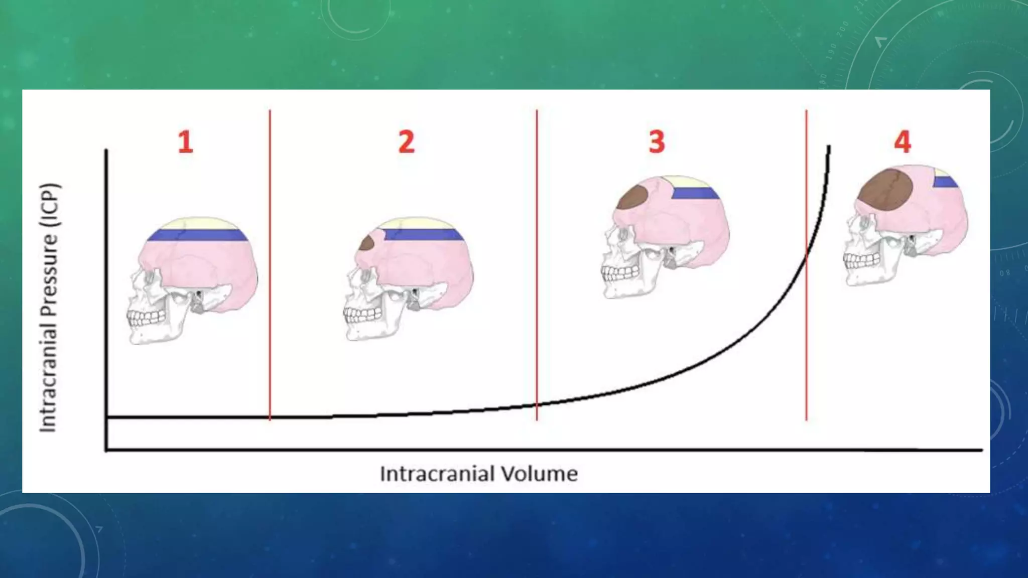
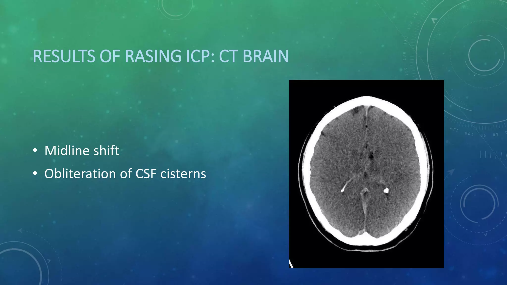
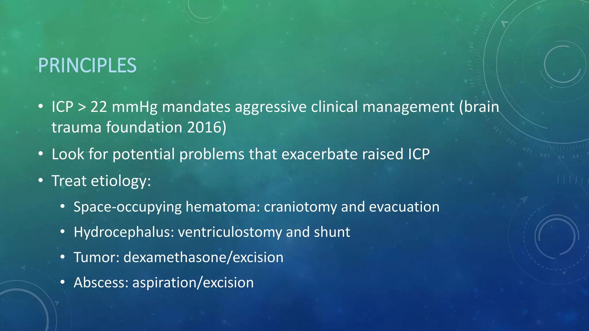
ESSENTIALS
OF
INTRACRANIAL PRESSURE
FACEBOOK: HAPPY FRIDAY KNIGHT
MARCH, 12TH, 2020
OUTLINES
• Production and flow of CSF
• Physiology of ICP and Pathophysiology of increased ICP
• ICP waveform and interpretation
• ICP monitoring and technology
• Treatment of elevated ICP
PRODUCTION AND FLOW OF
CEREBROSPINAL FLUID (CSF)
PRODUCTION AND FLOW OF CSF
• CSF: predominately produced by choroid plexuses
• Absorption at cranial and spinal arachnoid villi (granulations)
• Volume: 125 – 150 ml: 25 ml in ventricles, 125 ml in subarachnoid
spaces
• Active and delicately controlled secretion + normal absorption =
ICP stable
Sakka L, Coll G, Cazal J. Anatomy and physiology of cerebrospinal
fluid. European annals of otorhinolaryngology, head and neck
diseases. 2011;128:309-316.
• Variation
• Adult humans normally form CSF 0.35 ml/min  400 - 600 ml/day
• CSF dynamics affect brain metabolism
• CSF formation fluctuates in disease
• Hypersecretion: choroid plexus papillomas
• Hyposecretion: NPH, AD
PRODUCTION AND FLOW OF CSF
PHYSIOLOGY OF CSF: PRESSURE
• CSF Pressure: 10 – 15 mmHg in adult that varies with
• Systolic pulse wave
• Respiratoy cycle
• Abdominal pressure
• Jugular venous pressure
• State of arousal
• Physical activity
• posture
COMPOSITION OF CSF
• 99% water
• Na, Cl, Mg: higher concentration than those in plasma
• K, Ca: lower concentration than those in plasma
• CSF cell count: < 5cells/mL, no PMN, no RBC
• Loop diuretics and carbonic anhydrase: reduce CSF secretion and
turnover (3-5 times daily)
PHYSIOLOGY OF INTRACRANIAL PRESSURE (ICP)
AND PATHOPHYSIOLOGY OF INCREASED ICP
PHYSIOLOGY OF ICP
• ICP = CSF pressure
• dynamic
• Then ICP is reflective of
• CSF formation
• Volume storage or compliance
• CSF absorption
MONROE-KELLIE DOCTRINE
• Vother: volume of any abnormal component  hematoma, tumor
• Cranium is
• Nonexpandable
• incompressible
VOLUME
• Brain parenchyma: 1400 ml
• Cerebral blood volume: 150 ml
• CSF volume: 150 ml
Reilly P. Management of intracranial pressure and cerebral perfusion. Head injury. London, 1997.
CEREBRAL BLOOD FLOW
• CPP = MAP - ICP
• Pressure autoregulation: MAP 50-150 mmHg is autoregulated to
maintain constant CBF
PATHOPHYSIOLOGY
• A change in components at intracranial space
RESULTS OF RASING ICP
• CSF can be displaced from ventricles and subarachnoid spaces and
exit intracranial compartment via foramen magnum
• Intravenous blood: displaced via IJVs
• Arterial vasoconstriction: diffuse cerebral ischemia
• Cerebral herniation
RESULTS OF RASING ICP: CT BRAIN
• Midline shift
• Obliteration of CSF cisterns
PATHOPHYSIOLOGY: CSF
• Acute obstruction of CSF flow: acute hydrocephalus
• Hypersecretion of CSF: structurally or functionally alteration of
choroid plexus secretion
• Choroid plexus papillomas and hyperplasia
• Malabsorption of CSF: cribiform plate obstruction
• Obstruction of venous outflow
PATHOPHYSIOLOGY: BLOOD
• Arterial system
• Vasodilatation: hypercapnia, drug reaction
• Venous system
• Obstruction of venous outflow: thrombosis, inappropriate position
of head and neck, pneumothorax
PATHOPHYSIOLOGY: BRAIN EDEMA
• Vasogenic
• Cytotoxic
• Interstitial
• osmotic
PATHOPHYSIOLOGY: MASS OCCUPYING LESION
• Tumor
• Hemorrhage
• Infection: abscess
CLINICAL PRESENTATION OF RAISED ICP
• Quite unreliable
• pupil asymmetry
• Unilateral abnormal posture
• Signs of brain herniation: Cushing’s triad
• Hypertension
• Bradycardia
• Respiratory irregularity
ICP WAVEFORM AND INTERPRETATION
ICP WAVEFORM
• Gold standard: interventricular monitoring through
ventriculostomy
• Other technologies: devices inserted into
• Epidural space
• Subdural space
• Subarachnoid space
• Brain parenchyma
• 3 characteristic peak of decreasing height + correlate with arterial
waveform:
• P1: percussion wave  arterial pulse
• P2: tidal wave  cerebral compliance
• P3: dicrotic wave  aortic valve closure
ICP WAVEFORM: NORMAL
https://derangedphysiology.com/main/required-reading/neurology-and-neurosurgery/Chapter%201.0.5/interpretation-
intracranial-pressure-waveforms
https://derangedphysiology.com/main/required-reading/neurology-and-neurosurgery/Chapter%201.0.5/interpretation-
intracranial-pressure-waveforms
Cardiac cycle
• This reflects changes in intrathoracic pressure with respiration.
• This respiratory variation in ICP diminishes and eventually disappears altogether as
intracranial pressure increases.
ICP INTERPRETATION: RISING ICP
• Increasing all amplitudes
• Waveform morphology
doesn’t change
ICP INTERPRETATION: DECREASING CEREBRAL
PREFUSION
• Decrease amplitude of P1
• Due to P1=reflective of
arterial pulse in choroid
plexus
• Vasospasm if no raised ICP
ICP INTERPRETATION: DECREASING CEREBRAL
COMPLIANCE
• Prominent P2
• Increase in cerebral
bulk: edema
• Vasospasm if no
raised ICP
ICP INTERPRETATION: INCREASING ICP
• Flattening of ICP
breathing variation
ICP INTERPRETATION: LUNDBERG WAVES
• Pathological waveforms defined by Dr. Nils Lundberg
• Not frequently seen but if so, in patients with resistant to
therapeutic interventions, is the sign of worse outcome
• Designated as
• A: plateau wave
• B: pressure pulse
• C: Traube-Hering arterial waves
LUNDBERG “A” WAVES
• Plateau waves
• ICP elevation to 50 mmHg
for 2 – 20 minutes then
abrupt fall to baseline
• Intact cerebral blood flow
autoregulation
• But impending
uncontrollable ICP
LUNDBERG “A” WAVES
• 4 phases
1. Drift: decrease CPP  vasodilatation
2. Plateau: vasodilatation  increase ICP
3. Ischemic response phase: decrease CPP  cerebral ischemia 
brainstem vasomotor trigger  cushing response
4. Resolution: cushing response  restore CPP
LUNDBERRG “B” WAVES
• ICP elevation 10 – 20 mmHg
lasting 30 sec – 2 minutes
• Depends on periodic breathing
(PaCO2), increased ICP,
decrease cerebral compliance
• Unclear value in clinical
practice
LUNDBERG “C” WAVES
• Similar to B waves but
more rapid sinusoidal
fluctuation
• Seen in normal ICP
waveform
• Suggestive but not
pathognomonic of
increased ICP
ICP MONITORING: METHODS AND TECHNOLOGIES
• Anatomical locations of device insertion
• Standard ICP monitoring: external ventricular drain
• Indications
• Points of insertion
• Insertion technique
• Complications
• Other technologies
ANATOMICAL LOCATIONS OF DEVICE INSERTION
• Intraventricular – gold standard
• Intraparenchymal
• Subarachnoid
• Subdural
• epidural
https://www.openpr.com/news/476693/global-intracranial-
pressure-icp-monitoring-devices-market-raumedic-medtronic-
vittamed-spiegelberg-sophysa-ltd.html
EXTERNAL VENTRICULAR DRAIN
• A catheter placed into one of the ventricles through burr hole
• Gold standard
• Cost effective
• Most invasive
• Can also be used for CSF drainage and administration of medicine
intrathecally
Winn, H. Richard. Youmans and Winn’s Neurological Surgery. Phildelphia: Elsevier, 2017.
ICP MONITORING: INDICATIONS
• Traumatic brain injury with GCS ≤ 8
• Subarachnoid hemorrhage
• Intracranial tumor
• Intracranial hemorrhage
• Stroke
• Hydrocephalus
• CNS infection
• Fulminant hepatic failure
ICP MONITORING: POINTS OF INSERTION
• Anterior access
• Kocher: frontal (coronal) – 2
• Paine
• Tubbs - 6
• Kaufman - 5
• Posterior access
• Keen: posterior parietal - 1
• Frazier: occipital-parietal - 4
• Dandy: occipital - 3
KOCHER’S POINT
• 3 cm from midline
• 1 cm anterior to coronal
suture
• Point needle to nasion
and tragus
• Most common point
https://link.springer.com/referenceworkentry/10.1007%2F978-
3-642-00418-6_544
PAINE’S POINT
• 2.5 cm superior to supraorbital
margin
• Anterior edge of temporalis muscle
• Used in aneurysmal surgery via an
interhemispheric approach
TUBBS’ POINT
• Transorbital access to frontal
horn
• Just medial to midpapilary
point to roof of orbit
KAUFMAN’S POINT
• 4 cm superior to nasion
• 3 cm lateral to midline
• Cosmetic concern but rapidly
drained
KEEN’S POINT
• 3 FB superior and posterior to
unfold pinna
• Placement in trigone
• Point for VP shunt
FRAZIER’S POINT
• Occipital-parietal
• 6 cm above inion
• 3-4 cm from midline
• Point for VP shunt
DANDY’S POINT
• Occipital bur hole
• 3 cm above 2 cm lateral to inion
• Infants: correspond with lambdoid
suture in the midpupillary line
• Higher risk for visual impairment
INSERTION TECHNIQUE
• Right (non-dominant) side is preferred unless contraindication
• Kocher’s point: Incision is made in sagittal plane
• Elevate periosteum
• Apply self-retaining retractor
• Make a bur hole 1 – 2 cm anterior to coronal suture
• Control bleeding by bone wax and gelfoam
• Durotomy by blade No.11 and cauterize dural edge by bipolar
INSERTION TECHNIQUE
• Insert ventriculostomy needle perpendicular to brain surface (point to
nasion and tragus), not more than 7 cm depth
• Feel popping sensation and remove stylet
• Make a tunnel for the catheter
• Insert catheter
• Measure opened pressure, collect CSF, and measure closed pressure
• Fix catheter, hemostasis, and close the skin
COMPLICATIONS
• Intracranial and tract hemorrhage
• Infection: Ventriculitis
• Technical failure
• Over-drainage: transtentorial herniation
• Kinks and blockage by air, blood, and debris
• Inadvertent vascular injury.
• Pneumocephalus and pneumoventriculi.
• CSF leak.
• Accidental fracture
• Accidental pull
• Obstruction by choroid plexus, blood clot or proteinous plug
TECHNOLOGIES OF ICP MONITORING
• Fiberoptic intracranial pressure monitor
• Miniature strain gauge
• Spiegelberg parenchymal transducer
• Hummingbird synergy
• Telemetric ICP monitoring
TREATMENT OF INTRACRANIAL HYPERTENSION
PRINCIPLES
• ICP > 22 mmHg mandates aggressive clinical management (brain
trauma foundation 2016)
• Look for potential problems that exacerbate raised ICP
• Treat etiology:
• Space-occupying hematoma: craniotomy and evacuation
• Hydrocephalus: ventriculostomy and shunt
• Tumor: dexamethasone/excision
• Abscess: aspiration/excision
Czosnyka M, Pickard JD, Steiner LA. Principles of intracranial pressure
monitoring and treatment in Handbook of Clinical Neurology. Elsevier,
2017
FIRST LEVEL: PREVENTION
• Head elevation: up to 30o
• Maintain SBP:
• Age 50 – 69 keep SBP ≥ 100 mmHg, other keep SBP ≥ 110 mmHg
• Normocarbia (PaCO2 35 – 40 mmHg): no role of prophylactiv
hyperventilation
• Normothermia (36 – 37.5oC)
• Light sedation
• CT brain and check for surgical treatment options if ICP > 20 mmHg
• Avoid hyperglycemia
SECOND LEVEL: MORE INVASIVE
• CSF drainage in hydrocephalus
• Hyperosmolar treatment if evidence of herniation shown
• 20% mannitol 0.25 – 1 g/kg
• Hypertonic saline: keep Na < 155 meq/L
• Steroid in vasogenic edema
• Mild hyperventilation (PaCO2 30 – 34 mmHg): too long will cause
global ischemia
THIRD LEVEL: CONTROVERSY
• Decompressive craniectomy
• Consider deeper hypothermia: 33 – 34oC
• Consider barbiturate coma to maintain CPP
CONCLUSION
REFERENCES
Winn, H. Richard. Youmans and Winn’s Neurological Surgery. Phildelphia: Elsevier, 2017.
Greenberg MS. Handbook of Neurosurgery. 8th ed. New York: Thieme, 2016.
American college of surgeon committee on trauma. ATLS student course manual. 10th ed. Chicago: American
college of surgeon, 2018.
Sakka L, Coll G, Cazal J. Anatomy and physiology of cerebrospinal fluid. European annals of
otorhinolaryngology, head and neck diseases. 2011;128:309-316.
Tumani H, Huss A, and Buchhuber F. The cerebrospinal fluid and barriers – anatomic and physiologic
considerations in Handbook of clinical neurology. Vol3 (3rd series), 2018.
Kirkness CJ et al. Intracranial pressure waveform analysis: clinical and research implications. Journal of neuroscience
nursing.2000:32(5):271-277.
REFERENCES
Kasprowicz M, Lalou DA, Czosnyka M, Garnett M, Czosnyka Z.
Intracranial pressure, its components and cerebrospinal fluid pressure–volume compensation. Acta Neurol
Scand 2016: 134: 168–180.
Czosnyka M. Increased intracranial pressure: what to do and when?. www.ccmjournal.org. 2013:41(2):688-
689.
Czosnyka M, Pickard JD, Steiner LA. Principles of intracranial pressure monitoring and treatment in
Handbook of Clinical Neurology. Elsevier, 2017.
Karimy JK et al. Cerebrospinal fluid hypersecretion in pediatric hydrocephalus. Neurosurg focus.
2016:41(5):1-11.
Johanson CE et al. Multiplicity of cerebrospinal fluid functions: new challenges in health and disease.
Cerebrospinal fluid research. 2008:5(10):1-32.
https://derangedphysiology.com/main/required-reading/neurology-and-neurosurgery/Chapter%201.0.5/interpretation-
intracranial-pressure-waveforms
REFERENCES
https://www.researchgate.net/figure/Lundberg-waves-Lundberg-A-waves-are-ICP-increases-for-5-to-
10-minutes-They-reflect_fig2_311525129
Raboel PH et al. Intracranial pressure monitoring: invasive versus non-invasive methods – a review.
Critical care research and practice. 2012:1-14.
https://www.ncbi.nlm.nih.gov/books/NBK545317/
Mortazavi MM et al. The ventricular system of the brain: a comprehensive review of its history,
anatomy, histology, embryology, and surgical considerations. Childs Nerv Syst. 2013:
Wilson WC et al. Trauma: critical care vol 2. New York: Informa, 2007.
Morone PJ et al. Craniometrics and Ventricular Access: A Review of Kocher’s, Kaufman’s, Paine’s, Menovksy’s,
Tubbs’, Keen’s, Frazier’s, Dandy’s, and Sanchez’s Points. Operative neurosurgery.2019:0:1-9.
REFERENCES
Carney N et al. Guidelines for the Management of Severe Traumatic Brain
Injury, Fourth Edition. 2016
Reilly P. Management of intracranial pressure and cerebral perfusion.
Head injury. London, 1997.
Yoon SY, Kwak Y, Park J. Adjustable Ghajar Guide Technique for Accurate
Placement of Ventricular Catheters: A Pilot Study. J Korean Neurosurg Soc.
2017 Sep;60(5):604-609.

essentials of intracranial pressure (ICP)

  • 1.
  • 2.
    OUTLINES • Production andflow of CSF • Physiology of ICP and Pathophysiology of increased ICP • ICP waveform and interpretation • ICP monitoring and technology • Treatment of elevated ICP
  • 3.
    PRODUCTION AND FLOWOF CEREBROSPINAL FLUID (CSF)
  • 4.
    PRODUCTION AND FLOWOF CSF • CSF: predominately produced by choroid plexuses • Absorption at cranial and spinal arachnoid villi (granulations) • Volume: 125 – 150 ml: 25 ml in ventricles, 125 ml in subarachnoid spaces • Active and delicately controlled secretion + normal absorption = ICP stable
  • 6.
    Sakka L, CollG, Cazal J. Anatomy and physiology of cerebrospinal fluid. European annals of otorhinolaryngology, head and neck diseases. 2011;128:309-316.
  • 7.
    • Variation • Adulthumans normally form CSF 0.35 ml/min  400 - 600 ml/day • CSF dynamics affect brain metabolism • CSF formation fluctuates in disease • Hypersecretion: choroid plexus papillomas • Hyposecretion: NPH, AD PRODUCTION AND FLOW OF CSF
  • 8.
    PHYSIOLOGY OF CSF:PRESSURE • CSF Pressure: 10 – 15 mmHg in adult that varies with • Systolic pulse wave • Respiratoy cycle • Abdominal pressure • Jugular venous pressure • State of arousal • Physical activity • posture
  • 9.
    COMPOSITION OF CSF •99% water • Na, Cl, Mg: higher concentration than those in plasma • K, Ca: lower concentration than those in plasma • CSF cell count: < 5cells/mL, no PMN, no RBC • Loop diuretics and carbonic anhydrase: reduce CSF secretion and turnover (3-5 times daily)
  • 10.
    PHYSIOLOGY OF INTRACRANIALPRESSURE (ICP) AND PATHOPHYSIOLOGY OF INCREASED ICP
  • 11.
    PHYSIOLOGY OF ICP •ICP = CSF pressure • dynamic • Then ICP is reflective of • CSF formation • Volume storage or compliance • CSF absorption
  • 12.
    MONROE-KELLIE DOCTRINE • Vother:volume of any abnormal component  hematoma, tumor • Cranium is • Nonexpandable • incompressible
  • 13.
    VOLUME • Brain parenchyma:1400 ml • Cerebral blood volume: 150 ml • CSF volume: 150 ml
  • 14.
    Reilly P. Managementof intracranial pressure and cerebral perfusion. Head injury. London, 1997.
  • 15.
    CEREBRAL BLOOD FLOW •CPP = MAP - ICP • Pressure autoregulation: MAP 50-150 mmHg is autoregulated to maintain constant CBF
  • 17.
    PATHOPHYSIOLOGY • A changein components at intracranial space
  • 18.
    RESULTS OF RASINGICP • CSF can be displaced from ventricles and subarachnoid spaces and exit intracranial compartment via foramen magnum • Intravenous blood: displaced via IJVs • Arterial vasoconstriction: diffuse cerebral ischemia • Cerebral herniation
  • 20.
    RESULTS OF RASINGICP: CT BRAIN • Midline shift • Obliteration of CSF cisterns
  • 21.
    PATHOPHYSIOLOGY: CSF • Acuteobstruction of CSF flow: acute hydrocephalus • Hypersecretion of CSF: structurally or functionally alteration of choroid plexus secretion • Choroid plexus papillomas and hyperplasia • Malabsorption of CSF: cribiform plate obstruction • Obstruction of venous outflow
  • 22.
    PATHOPHYSIOLOGY: BLOOD • Arterialsystem • Vasodilatation: hypercapnia, drug reaction • Venous system • Obstruction of venous outflow: thrombosis, inappropriate position of head and neck, pneumothorax
  • 23.
    PATHOPHYSIOLOGY: BRAIN EDEMA •Vasogenic • Cytotoxic • Interstitial • osmotic
  • 24.
    PATHOPHYSIOLOGY: MASS OCCUPYINGLESION • Tumor • Hemorrhage • Infection: abscess
  • 25.
    CLINICAL PRESENTATION OFRAISED ICP • Quite unreliable • pupil asymmetry • Unilateral abnormal posture • Signs of brain herniation: Cushing’s triad • Hypertension • Bradycardia • Respiratory irregularity
  • 26.
    ICP WAVEFORM ANDINTERPRETATION
  • 27.
    ICP WAVEFORM • Goldstandard: interventricular monitoring through ventriculostomy • Other technologies: devices inserted into • Epidural space • Subdural space • Subarachnoid space • Brain parenchyma
  • 28.
    • 3 characteristicpeak of decreasing height + correlate with arterial waveform: • P1: percussion wave  arterial pulse • P2: tidal wave  cerebral compliance • P3: dicrotic wave  aortic valve closure ICP WAVEFORM: NORMAL
  • 29.
  • 30.
    https://derangedphysiology.com/main/required-reading/neurology-and-neurosurgery/Chapter%201.0.5/interpretation- intracranial-pressure-waveforms Cardiac cycle • Thisreflects changes in intrathoracic pressure with respiration. • This respiratory variation in ICP diminishes and eventually disappears altogether as intracranial pressure increases.
  • 31.
    ICP INTERPRETATION: RISINGICP • Increasing all amplitudes • Waveform morphology doesn’t change
  • 32.
    ICP INTERPRETATION: DECREASINGCEREBRAL PREFUSION • Decrease amplitude of P1 • Due to P1=reflective of arterial pulse in choroid plexus • Vasospasm if no raised ICP
  • 33.
    ICP INTERPRETATION: DECREASINGCEREBRAL COMPLIANCE • Prominent P2 • Increase in cerebral bulk: edema • Vasospasm if no raised ICP
  • 34.
    ICP INTERPRETATION: INCREASINGICP • Flattening of ICP breathing variation
  • 35.
    ICP INTERPRETATION: LUNDBERGWAVES • Pathological waveforms defined by Dr. Nils Lundberg • Not frequently seen but if so, in patients with resistant to therapeutic interventions, is the sign of worse outcome • Designated as • A: plateau wave • B: pressure pulse • C: Traube-Hering arterial waves
  • 36.
    LUNDBERG “A” WAVES •Plateau waves • ICP elevation to 50 mmHg for 2 – 20 minutes then abrupt fall to baseline • Intact cerebral blood flow autoregulation • But impending uncontrollable ICP
  • 37.
    LUNDBERG “A” WAVES •4 phases 1. Drift: decrease CPP  vasodilatation 2. Plateau: vasodilatation  increase ICP 3. Ischemic response phase: decrease CPP  cerebral ischemia  brainstem vasomotor trigger  cushing response 4. Resolution: cushing response  restore CPP
  • 38.
    LUNDBERRG “B” WAVES •ICP elevation 10 – 20 mmHg lasting 30 sec – 2 minutes • Depends on periodic breathing (PaCO2), increased ICP, decrease cerebral compliance • Unclear value in clinical practice
  • 39.
    LUNDBERG “C” WAVES •Similar to B waves but more rapid sinusoidal fluctuation • Seen in normal ICP waveform • Suggestive but not pathognomonic of increased ICP
  • 40.
    ICP MONITORING: METHODSAND TECHNOLOGIES
  • 41.
    ICP MONITORING: METHODSAND TECHNOLOGIES • Anatomical locations of device insertion • Standard ICP monitoring: external ventricular drain • Indications • Points of insertion • Insertion technique • Complications • Other technologies
  • 42.
    ANATOMICAL LOCATIONS OFDEVICE INSERTION • Intraventricular – gold standard • Intraparenchymal • Subarachnoid • Subdural • epidural https://www.openpr.com/news/476693/global-intracranial- pressure-icp-monitoring-devices-market-raumedic-medtronic- vittamed-spiegelberg-sophysa-ltd.html
  • 43.
    EXTERNAL VENTRICULAR DRAIN •A catheter placed into one of the ventricles through burr hole • Gold standard • Cost effective • Most invasive • Can also be used for CSF drainage and administration of medicine intrathecally
  • 44.
    Winn, H. Richard.Youmans and Winn’s Neurological Surgery. Phildelphia: Elsevier, 2017.
  • 45.
    ICP MONITORING: INDICATIONS •Traumatic brain injury with GCS ≤ 8 • Subarachnoid hemorrhage • Intracranial tumor • Intracranial hemorrhage • Stroke • Hydrocephalus • CNS infection • Fulminant hepatic failure
  • 46.
    ICP MONITORING: POINTSOF INSERTION • Anterior access • Kocher: frontal (coronal) – 2 • Paine • Tubbs - 6 • Kaufman - 5 • Posterior access • Keen: posterior parietal - 1 • Frazier: occipital-parietal - 4 • Dandy: occipital - 3
  • 47.
    KOCHER’S POINT • 3cm from midline • 1 cm anterior to coronal suture • Point needle to nasion and tragus • Most common point https://link.springer.com/referenceworkentry/10.1007%2F978- 3-642-00418-6_544
  • 48.
    PAINE’S POINT • 2.5cm superior to supraorbital margin • Anterior edge of temporalis muscle • Used in aneurysmal surgery via an interhemispheric approach
  • 49.
    TUBBS’ POINT • Transorbitalaccess to frontal horn • Just medial to midpapilary point to roof of orbit
  • 50.
    KAUFMAN’S POINT • 4cm superior to nasion • 3 cm lateral to midline • Cosmetic concern but rapidly drained
  • 51.
    KEEN’S POINT • 3FB superior and posterior to unfold pinna • Placement in trigone • Point for VP shunt
  • 52.
    FRAZIER’S POINT • Occipital-parietal •6 cm above inion • 3-4 cm from midline • Point for VP shunt
  • 53.
    DANDY’S POINT • Occipitalbur hole • 3 cm above 2 cm lateral to inion • Infants: correspond with lambdoid suture in the midpupillary line • Higher risk for visual impairment
  • 54.
    INSERTION TECHNIQUE • Right(non-dominant) side is preferred unless contraindication • Kocher’s point: Incision is made in sagittal plane • Elevate periosteum • Apply self-retaining retractor • Make a bur hole 1 – 2 cm anterior to coronal suture • Control bleeding by bone wax and gelfoam • Durotomy by blade No.11 and cauterize dural edge by bipolar
  • 55.
    INSERTION TECHNIQUE • Insertventriculostomy needle perpendicular to brain surface (point to nasion and tragus), not more than 7 cm depth • Feel popping sensation and remove stylet • Make a tunnel for the catheter • Insert catheter • Measure opened pressure, collect CSF, and measure closed pressure • Fix catheter, hemostasis, and close the skin
  • 56.
    COMPLICATIONS • Intracranial andtract hemorrhage • Infection: Ventriculitis • Technical failure • Over-drainage: transtentorial herniation • Kinks and blockage by air, blood, and debris • Inadvertent vascular injury. • Pneumocephalus and pneumoventriculi. • CSF leak. • Accidental fracture • Accidental pull • Obstruction by choroid plexus, blood clot or proteinous plug
  • 57.
    TECHNOLOGIES OF ICPMONITORING • Fiberoptic intracranial pressure monitor • Miniature strain gauge • Spiegelberg parenchymal transducer • Hummingbird synergy • Telemetric ICP monitoring
  • 58.
  • 59.
    PRINCIPLES • ICP >22 mmHg mandates aggressive clinical management (brain trauma foundation 2016) • Look for potential problems that exacerbate raised ICP • Treat etiology: • Space-occupying hematoma: craniotomy and evacuation • Hydrocephalus: ventriculostomy and shunt • Tumor: dexamethasone/excision • Abscess: aspiration/excision
  • 60.
    Czosnyka M, PickardJD, Steiner LA. Principles of intracranial pressure monitoring and treatment in Handbook of Clinical Neurology. Elsevier, 2017
  • 61.
    FIRST LEVEL: PREVENTION •Head elevation: up to 30o • Maintain SBP: • Age 50 – 69 keep SBP ≥ 100 mmHg, other keep SBP ≥ 110 mmHg • Normocarbia (PaCO2 35 – 40 mmHg): no role of prophylactiv hyperventilation • Normothermia (36 – 37.5oC) • Light sedation • CT brain and check for surgical treatment options if ICP > 20 mmHg • Avoid hyperglycemia
  • 62.
    SECOND LEVEL: MOREINVASIVE • CSF drainage in hydrocephalus • Hyperosmolar treatment if evidence of herniation shown • 20% mannitol 0.25 – 1 g/kg • Hypertonic saline: keep Na < 155 meq/L • Steroid in vasogenic edema • Mild hyperventilation (PaCO2 30 – 34 mmHg): too long will cause global ischemia
  • 63.
    THIRD LEVEL: CONTROVERSY •Decompressive craniectomy • Consider deeper hypothermia: 33 – 34oC • Consider barbiturate coma to maintain CPP
  • 65.
  • 66.
    REFERENCES Winn, H. Richard.Youmans and Winn’s Neurological Surgery. Phildelphia: Elsevier, 2017. Greenberg MS. Handbook of Neurosurgery. 8th ed. New York: Thieme, 2016. American college of surgeon committee on trauma. ATLS student course manual. 10th ed. Chicago: American college of surgeon, 2018. Sakka L, Coll G, Cazal J. Anatomy and physiology of cerebrospinal fluid. European annals of otorhinolaryngology, head and neck diseases. 2011;128:309-316. Tumani H, Huss A, and Buchhuber F. The cerebrospinal fluid and barriers – anatomic and physiologic considerations in Handbook of clinical neurology. Vol3 (3rd series), 2018. Kirkness CJ et al. Intracranial pressure waveform analysis: clinical and research implications. Journal of neuroscience nursing.2000:32(5):271-277.
  • 67.
    REFERENCES Kasprowicz M, LalouDA, Czosnyka M, Garnett M, Czosnyka Z. Intracranial pressure, its components and cerebrospinal fluid pressure–volume compensation. Acta Neurol Scand 2016: 134: 168–180. Czosnyka M. Increased intracranial pressure: what to do and when?. www.ccmjournal.org. 2013:41(2):688- 689. Czosnyka M, Pickard JD, Steiner LA. Principles of intracranial pressure monitoring and treatment in Handbook of Clinical Neurology. Elsevier, 2017. Karimy JK et al. Cerebrospinal fluid hypersecretion in pediatric hydrocephalus. Neurosurg focus. 2016:41(5):1-11. Johanson CE et al. Multiplicity of cerebrospinal fluid functions: new challenges in health and disease. Cerebrospinal fluid research. 2008:5(10):1-32. https://derangedphysiology.com/main/required-reading/neurology-and-neurosurgery/Chapter%201.0.5/interpretation- intracranial-pressure-waveforms
  • 68.
    REFERENCES https://www.researchgate.net/figure/Lundberg-waves-Lundberg-A-waves-are-ICP-increases-for-5-to- 10-minutes-They-reflect_fig2_311525129 Raboel PH etal. Intracranial pressure monitoring: invasive versus non-invasive methods – a review. Critical care research and practice. 2012:1-14. https://www.ncbi.nlm.nih.gov/books/NBK545317/ Mortazavi MM et al. The ventricular system of the brain: a comprehensive review of its history, anatomy, histology, embryology, and surgical considerations. Childs Nerv Syst. 2013: Wilson WC et al. Trauma: critical care vol 2. New York: Informa, 2007. Morone PJ et al. Craniometrics and Ventricular Access: A Review of Kocher’s, Kaufman’s, Paine’s, Menovksy’s, Tubbs’, Keen’s, Frazier’s, Dandy’s, and Sanchez’s Points. Operative neurosurgery.2019:0:1-9.
  • 69.
    REFERENCES Carney N etal. Guidelines for the Management of Severe Traumatic Brain Injury, Fourth Edition. 2016 Reilly P. Management of intracranial pressure and cerebral perfusion. Head injury. London, 1997. Yoon SY, Kwak Y, Park J. Adjustable Ghajar Guide Technique for Accurate Placement of Ventricular Catheters: A Pilot Study. J Korean Neurosurg Soc. 2017 Sep;60(5):604-609.

Editor's Notes

  • #9 Hypersecretion: ทำลาย choroid plexus สามารถลด ICP ได้ เพราฉะนั้น การรู้ว่า choroid plexus ทำงานเยอะหรือน้อย จะบอกได้ว่าจะรักษายังไง
  • #32 พีคต้องไล่จากสูงสุดไปต่ำสุด