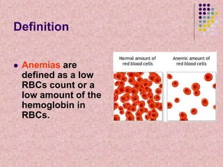
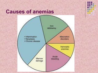
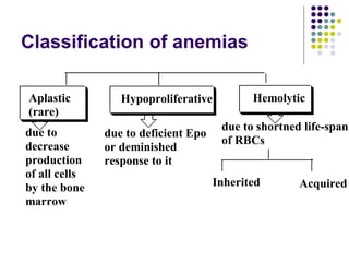
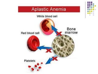
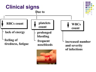
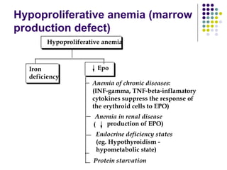
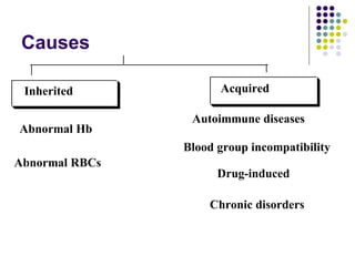
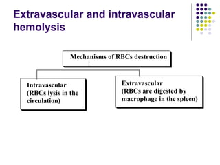
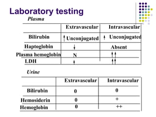
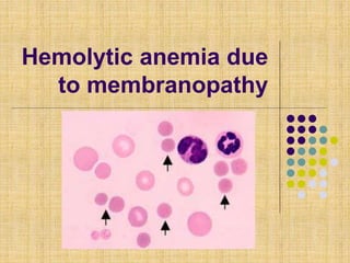
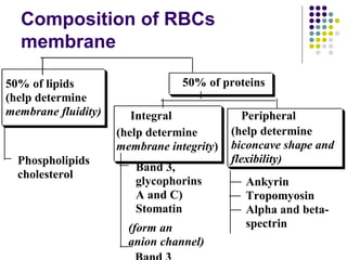
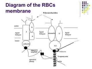
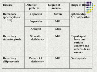
Anemias
Definition
 Anemias are
defined as a low
RBCs count or a
low amount of the
hemoglobin in
RBCs.
Causes of anemias
Production of blood cells
Pluripotential
stem cell
GEMML (cell giving rise to Granulocyte, Erythroid,
Monocyte, Megakaryocyte, Lymphoid cells)
Site:
bone marrow
Time: 7 days
Differentiation,
proliferation
Lymphoid stem cells (L)
IL-2
T-cells
IL-15
NK-cells
Il-7
B-cells,
plasma cells
GEMM
ME
(megakaryocyte
erythroid)
GM
(granulocytes,
monocytes)
eosinophil
M-CSF
MEGM
(granulocyte
monocyte)
(megakaryocyte
erythroid)
IL-5
Basophil/
mast cells
IL-3
Monocyte/
macrophage
G-CSF
Neutrophils
BFU-E
EPO
Megakaryoblast
Plateletes
basophilic
normoblast
CFU-E
(colony forming unit
erythroid)
(burst-forming unit
erythroid)
EPO
can undergo
4-5 cell divisions
that give
16-32 mature RBCs
polychromic
normoblast
ortochromic
normoblast
Reticulocytes
circulation
(maturation to)
Erythrocytes
Maturation of reticulocytes to
RBCs in blood circulation (1-2
days)
 Reticulocytes still
contain ribosomes
and elements of
endoplasmic
reticulum.
 The RNA of the
ribosome can be
detected by cresyl
blue.
Reticulocyte count
 A typical normal range
is 0.5-1.5 %.
 Increase level of
reticulocytes as known
as reticulocytosis
Regulation of Erythropoiesis
 Growth factor:
 GM-CSF granylocyte-macrophage colony
stimulating factor
 Cytokines:
 IL-3, IL-4, IL-9, IL-11 – stimulate erythroid
stem cell proliferation.
 IL-2 inhibits erythropoiesis
Erythropoietin (EPO)
Structure of EPO
 Is a glycoprotein growth
factor (165 amino
acids) with high
carbohydrates content
which increase the
molecule’s stability in
the circulation
Erythropoietin (Epo)
 Site of synthesis:
Peritubular
lining cells of
the renal cortex
Liver
Ito cells
EPO production
predominates
in the adult
EPO production
predominates in the
fetal and perinatal period
Factors which stimulate Epo
production:
 Hypoxia of different genesis
 Increase hemolyzates level (products of
RBCs destruction)
 Anterior pituitary hormones: LH, FSH,
ACTH, TSH, GH
 Androgens
Factors which decrease Epo
production
 Estrogens decrease synthesis of Epo
 This is why in females RBCs count is less
then in males.
Factors affecting EPO
production
Stem cells BFU-E CFU-E Erythroid
precursor
Red-cell mass
EPO
production
Oxygen
sensor
EPO
Atmospheric
oxygen
Blood volume
Hb concentration
Oxygen affinity
Cardiopulmanary
functionRenal blood
flow
Renal oxygen
consumption
Regulation of the EPO production
by blood oxygenation (feed-back)
O2
Oxygen
sensor
protein (heme-containing)
changes the
conformation
to Deoxy-form
HIF
(hypoxia inducible factor-1)
O2
Hydroxylation
of HIF
EPO gene transcription
EPO production
Proteosomal
degradation
EPO production
Oxygen
sensor
protein (heme-containing)
changes the
conformation
to Oxy-form
Effect of Epo
 1. Epo induces proliferation and maturation
of erythroid progenitor cells (CFU-E and
BFU-E have receptors for EPO)
 2. Epo increases release of reticulocytes
from the bone marrow
 3. Epo stimulates absorption of hemoglobin
by suppressing hepcidin.
Role of hepcidin
 Is peptide hormone
produced by the liver.
 Increase iron storage
in cells
 Prevents enterocytes
from secreting iron
into the hepatic portal
system
 Antifungal and
antibacterial activities
Classification of anemias
Aplastic
(rare)
Hypoproliferative Hemolytic
Inherited Acquired
due to
decrease
production
of all cells
by the bone
marrow
due to deficient Epo
or deminished
response to it
due to shortned life-span
of RBCs
Causes:
 Causes of aplastic anemia usually bind
with damage of the stem cells in the bone
marrow:
 Exposure to toxic substances
 Cancer therapy
 Autoimmune disease
 Viral infection
Clinical signs
Due to
RBCs count platelets
count
WBCs
count
lack of energy
feeling of
tiredness, fatigue
prolonged
bleeding
frequent
nosebleeds
increased number
and severity
of infections
Hypoproliferative anemia (marrow
production defect)
Iron
deficiency
Epo
Anemia of chronic diseases:
(INF-gamma, TNF-beta-inflamatory
cytokines suppress the response of
the erythroid cells to EPO)
Anemia in renal disease
production of EPO)(
Endocrine deficiency states
(eg. Hypothyroidism -
hypometabolic state)
Protein starvation
Hypoproliferative anemia
Treatment of hypoproliferative
anemia
 1. Transfusions
 2. Epo therapy
Hemolytic anemia
 Definition. Hemolytic anemias are defined as
decreased red cell mass resulting from an
increase rate of erythrocytes destruction.
 Normal red cells have a lifespan 100-120
days.
 At the end of this lifespan, both normal and
damaged cells are ‘recognized’ by
macrophages in the spleen and removed
from the circulation
 In hemolytic anemia,
red cell lifespan is
shortened.
 The bone marrow is
not able to produce
new RBCs quickly
to replace destroyed
RBCs
Causes
Inherited
Abnormal Hb
Abnormal RBCs
Acquired
Autoimmune diseases
Blood group incompatibility
Drug-induced
Chronic disorders
Extravascular and intravascular
hemolysis
Mechanisms of RBCs destruction
Extravascular
(RBCs are digested by
macrophage in the spleen)
Intravascular
(RBCs lysis in the
circulation)
Laboratory testing
Bilirubin
Plasma
Extravascular Intravascular
Unconjugated Unconjugated
Haptoglobin Absent
Plasma hemoglobin N
LDH
Bilirubin
Extravascular Intravascular
Hemosiderin
Hemoglobin
0 0
0 +
0 ++
Urine
Hemolytic anemia due
to membranopathy
Composition of RBCs
membrane
50% of lipids
(help determine
membrane fluidity)
Phospholipids
cholesterol
50% of proteins
Integral Peripheral
(help determine
membrane integrity)
Band 3,
glycophorins
A and C)
Stomatin
(help determine
biconcave shape and
flexibility)
Ankyrin
Tropomyosin
Alpha and beta-
spectrin(form an
anion channel)
Diagram of the RBCs
membrane
3
A
A
stomatin C
4.1
ankyrin
actin
spectrin
(alpha)
spectrin
(beta)
outer
inner
lipid
bilayer
lipid
bilayer
lipid
bilayer
Polysaccharides
Tropomyosin
Adductin
Disease Defect of
proteins
Degree of
anemia
Shape of RBCs
Hereditary
spherocytosis
(HS)
α-spectrin Severe Spherocytes
Are not flexible
β-spectrin Mild
Ankyrin Mild
Hereditary
stomatocytosis
Stomatin
deficiency
Mild Cup-shaped
have one
surface
concave and
other side as
convex
Hereditary
elliptocytosis
Protein 4.1
deficiency
Mild Ovalocytosis
Clinical presentation:
 Extravascular Hemolysis in the spleen.
Plasma hemoglobin is slightly raised.
 Splenomegaly
 UCB-emia and risk for jaundice and pigment
stones
 Reticulocytosis (5-20%).
Osmotic fragility test is positive
 When RBCs are exposed to a hypotonic
saline solution (<0.9%) only few normal
RBCs are hemolyzed.
 But 50% of spherocytes lyses under this
conditions.
 ↓ ability of RBCs to extrude sodium →
retention of water → osmotic injury

Anaemia, Erythropoiesis

  • 1.
  • 2.
    Definition  Anemias are definedas a low RBCs count or a low amount of the hemoglobin in RBCs.
  • 3.
  • 4.
    Production of bloodcells Pluripotential stem cell GEMML (cell giving rise to Granulocyte, Erythroid, Monocyte, Megakaryocyte, Lymphoid cells) Site: bone marrow Time: 7 days Differentiation, proliferation Lymphoid stem cells (L) IL-2 T-cells IL-15 NK-cells Il-7 B-cells, plasma cells GEMM ME (megakaryocyte erythroid) GM (granulocytes, monocytes)
  • 5.
    eosinophil M-CSF MEGM (granulocyte monocyte) (megakaryocyte erythroid) IL-5 Basophil/ mast cells IL-3 Monocyte/ macrophage G-CSF Neutrophils BFU-E EPO Megakaryoblast Plateletes basophilic normoblast CFU-E (colony formingunit erythroid) (burst-forming unit erythroid) EPO can undergo 4-5 cell divisions that give 16-32 mature RBCs polychromic normoblast ortochromic normoblast Reticulocytes circulation (maturation to) Erythrocytes
  • 6.
    Maturation of reticulocytesto RBCs in blood circulation (1-2 days)  Reticulocytes still contain ribosomes and elements of endoplasmic reticulum.  The RNA of the ribosome can be detected by cresyl blue.
  • 7.
    Reticulocyte count  Atypical normal range is 0.5-1.5 %.  Increase level of reticulocytes as known as reticulocytosis
  • 8.
    Regulation of Erythropoiesis Growth factor:  GM-CSF granylocyte-macrophage colony stimulating factor  Cytokines:  IL-3, IL-4, IL-9, IL-11 – stimulate erythroid stem cell proliferation.  IL-2 inhibits erythropoiesis
  • 9.
  • 10.
    Structure of EPO Is a glycoprotein growth factor (165 amino acids) with high carbohydrates content which increase the molecule’s stability in the circulation
  • 11.
    Erythropoietin (Epo)  Siteof synthesis: Peritubular lining cells of the renal cortex Liver Ito cells EPO production predominates in the adult EPO production predominates in the fetal and perinatal period
  • 12.
    Factors which stimulateEpo production:  Hypoxia of different genesis  Increase hemolyzates level (products of RBCs destruction)  Anterior pituitary hormones: LH, FSH, ACTH, TSH, GH  Androgens
  • 13.
    Factors which decreaseEpo production  Estrogens decrease synthesis of Epo  This is why in females RBCs count is less then in males.
  • 14.
    Factors affecting EPO production Stemcells BFU-E CFU-E Erythroid precursor Red-cell mass EPO production Oxygen sensor EPO Atmospheric oxygen Blood volume Hb concentration Oxygen affinity Cardiopulmanary functionRenal blood flow Renal oxygen consumption
  • 15.
    Regulation of theEPO production by blood oxygenation (feed-back) O2 Oxygen sensor protein (heme-containing) changes the conformation to Deoxy-form HIF (hypoxia inducible factor-1) O2 Hydroxylation of HIF EPO gene transcription EPO production Proteosomal degradation EPO production Oxygen sensor protein (heme-containing) changes the conformation to Oxy-form
  • 16.
    Effect of Epo 1. Epo induces proliferation and maturation of erythroid progenitor cells (CFU-E and BFU-E have receptors for EPO)  2. Epo increases release of reticulocytes from the bone marrow  3. Epo stimulates absorption of hemoglobin by suppressing hepcidin.
  • 17.
    Role of hepcidin Is peptide hormone produced by the liver.  Increase iron storage in cells  Prevents enterocytes from secreting iron into the hepatic portal system  Antifungal and antibacterial activities
  • 18.
    Classification of anemias Aplastic (rare) HypoproliferativeHemolytic Inherited Acquired due to decrease production of all cells by the bone marrow due to deficient Epo or deminished response to it due to shortned life-span of RBCs
  • 20.
    Causes:  Causes ofaplastic anemia usually bind with damage of the stem cells in the bone marrow:  Exposure to toxic substances  Cancer therapy  Autoimmune disease  Viral infection
  • 21.
    Clinical signs Due to RBCscount platelets count WBCs count lack of energy feeling of tiredness, fatigue prolonged bleeding frequent nosebleeds increased number and severity of infections
  • 22.
    Hypoproliferative anemia (marrow productiondefect) Iron deficiency Epo Anemia of chronic diseases: (INF-gamma, TNF-beta-inflamatory cytokines suppress the response of the erythroid cells to EPO) Anemia in renal disease production of EPO)( Endocrine deficiency states (eg. Hypothyroidism - hypometabolic state) Protein starvation Hypoproliferative anemia
  • 23.
    Treatment of hypoproliferative anemia 1. Transfusions  2. Epo therapy
  • 24.
  • 25.
     Definition. Hemolyticanemias are defined as decreased red cell mass resulting from an increase rate of erythrocytes destruction.  Normal red cells have a lifespan 100-120 days.  At the end of this lifespan, both normal and damaged cells are ‘recognized’ by macrophages in the spleen and removed from the circulation
  • 26.
     In hemolyticanemia, red cell lifespan is shortened.  The bone marrow is not able to produce new RBCs quickly to replace destroyed RBCs
  • 27.
    Causes Inherited Abnormal Hb Abnormal RBCs Acquired Autoimmunediseases Blood group incompatibility Drug-induced Chronic disorders
  • 28.
    Extravascular and intravascular hemolysis Mechanismsof RBCs destruction Extravascular (RBCs are digested by macrophage in the spleen) Intravascular (RBCs lysis in the circulation)
  • 29.
    Laboratory testing Bilirubin Plasma Extravascular Intravascular UnconjugatedUnconjugated Haptoglobin Absent Plasma hemoglobin N LDH Bilirubin Extravascular Intravascular Hemosiderin Hemoglobin 0 0 0 + 0 ++ Urine
  • 30.
  • 31.
    Composition of RBCs membrane 50%of lipids (help determine membrane fluidity) Phospholipids cholesterol 50% of proteins Integral Peripheral (help determine membrane integrity) Band 3, glycophorins A and C) Stomatin (help determine biconcave shape and flexibility) Ankyrin Tropomyosin Alpha and beta- spectrin(form an anion channel)
  • 32.
    Diagram of theRBCs membrane 3 A A stomatin C 4.1 ankyrin actin spectrin (alpha) spectrin (beta) outer inner lipid bilayer lipid bilayer lipid bilayer Polysaccharides Tropomyosin Adductin
  • 33.
    Disease Defect of proteins Degreeof anemia Shape of RBCs Hereditary spherocytosis (HS) α-spectrin Severe Spherocytes Are not flexible β-spectrin Mild Ankyrin Mild Hereditary stomatocytosis Stomatin deficiency Mild Cup-shaped have one surface concave and other side as convex Hereditary elliptocytosis Protein 4.1 deficiency Mild Ovalocytosis
  • 34.
    Clinical presentation:  ExtravascularHemolysis in the spleen. Plasma hemoglobin is slightly raised.  Splenomegaly  UCB-emia and risk for jaundice and pigment stones  Reticulocytosis (5-20%).
  • 35.
    Osmotic fragility testis positive  When RBCs are exposed to a hypotonic saline solution (<0.9%) only few normal RBCs are hemolyzed.  But 50% of spherocytes lyses under this conditions.  ↓ ability of RBCs to extrude sodium → retention of water → osmotic injury