This study evaluated a new minimally invasive technique for inserting an equine stented pulmonary valve through a right ventricular approach without using cardiopulmonary bypass. The valve consisted of an equine jugular vein sutured into a self-expanding nitinol stent with a sinus portion. In seven lambs, the valve was successfully inserted through a right minithoracotomy and released in the pulmonary position using a flexible hydraulic delivery system. At follow-up of up to six months, echocardiography showed the valves were well-positioned and functioning properly. Histological analysis demonstrated endothelialization of the valves. This new approach may provide an alternative to existing surgical and percutaneous pulmonary valve replacement methods.
Cardiac Interventions in Pediatric Cardiology: The FutureApollo Hospitals
Pediatric Cardiac interventions have come a long way from the initial intervention in the 1950's. Balloon angioplasty
has been accepted as the procedure of choice in several congenital anomalies. Apart from balloon angioplasties/
valvuloplasties, Atrial Septal Defect, Ventricular Septal Defect (muscular) device closure have been FDA approved
with adequate world wide clinical experience and long-term follow-up. In addition, newer procedures are under
clinical trial for perimembranous VSD device closure in the catheterization lab; per operative closed heart procedure
in the operation theatre or as a hybrid procedure. Palliative procedures like flow restriction to lungs with devices to
equate with surgical pulmonary artery banding; stenting of the patent ductus arteriosus in duct dependent cyanotic
heart disease in the newborn or a combination of these form transcatheter Norwood stage I in the cath lab.
Experience and technology will also help make transcatheter Fontan operation possible and that does not seem too far. The emphasis in pediatric cardiac interventions shall always remain that the decision, procedure, and management of their complications is a joint effort of the surgeon and the interventionalist.
A heart coping with a dysfunctional prosthetic valveescardio
A heart coping with a dysfunctional prosthetic valve (at least once in every few beats…)
http://www.escardio.org/communities/Working-Groups/valvular/Pages/welcome.aspx
Evaluation of prosthetic valve function and clinical utility.Ramachandra Barik
Many of the prosthesis-related complications can be prevented or their impact minimized through optimal prosthesis selection in the individual patient and careful medical management and follow-up after implantation.
Successful Valve in Ring Abolishing the Stenosis and Regurgitation with Robus...Premier Publishers
Transcatheter mitral valve-in ring implantation (TMViRI), is a novel alternative treatment strategy and promising technique for patients at high risk of repeat open-heart surgery. In this report we demonstrate a case of 61 years old male with multiple co morbidities who underwent mitral valve repair long time ago who successfully treated and dramatically improved through trans-septal approach, under trans oesophageal echocardiography and fluoroscopic guidance in Hybrid catheterization laboratory.
Arterial bleeding during EUS-guided pseudocyst drainage stopped by placement...Enrique Moreno Gonzalez
Hemorrhagic complications during EUS-guided pseudocyst drainage can occur, because the vessels on the internal wall of the pseudocyst might be compressed by the fluid and thus not visible on color Doppler or even power Doppler EUS.
ECMO CPR
ECMO in Cardiac arrest has increased exponentially in the past 10 years, on the back of, up until very recently, non-randomised, predominantly retrospective studies.
What is the efficacy?
Appropriate patient selection?
Cost effectiveness and model of delivery of ECPR?
Finally is ECMO really the intervention or just optimising the chain of survival?
PHP Albania User Group - What is this about?andrixh
Charter presentation for PHP Albania User Group. What is the group about? What is planned in the short and long term? How to contribute help build a better developer community.
1. PEDIATRIC AND CONGENITAL HEART DISEASE
Original Studies
Minimally Invasive Insertion of an Equine Stented
Pulmonary Valve with a Built-in Sinus Portion in a
Sheep Model
Ali Dodge-Khatami,1* MD, PhD, Stefan Hallhagen,2
MD, Kuno Limacher,3
MSC,
Bjo¨ rn So¨ derberg,2
MD, and Rolf Jenni,4
MD, MSEE
Objectives: This study evaluated the feasibility of inserting a new equine stented-valve
with a sinus portion in a lamb survival model, through a minimally invasive thoracot-
omy with right ventricular access without cardiopulmonary bypass. Background:
Extant surgical or percutaneous methods for inserting biological valves in the right
outflow tract have drawbacks and limitations. Methods: A decellularized equine valved
jugular vein, sutured to a self-expanding stent with a sinus portion, was placed
through a minimal right thoracotomy using a newly developed flexible hydraulic release
device in seven lambs. The approach through the right ventricle into the pulmonary
valve position is achieved on a beating heart. Results: The stented valves were cor-
rectly positioned in the right outflow tract, were competent up to 6 months as con-
firmed by angiography and echocardiography, and were well-tolerated by the animals,
with endothelialization of the valve demonstrated at 6 months. Conclusions: The newly
developed hydraulic release system allowed for safe and reliable insertion of an equine
stented-valve with a sinus portion, through a right transventricular approach on a
beating heart, in a sheep survival model. VC 2011 Wiley Periodicals, Inc.
Key words: animal model; biomaterials; heart valve (transapical, percutaneous); off-
pump surgery; pulmonary valve
INTRODUCTION
The importance of a correctly functioning pulmonary
valve is increasingly recognized. The extent to which
various degrees of pulmonary valve stenosis or insuffi-
ciency can or should be tolerated is still subject to
debate, but increasing quantitative and qualitative diag-
nostic measurements are being established better to
determine the timing for pulmonary valve replacement
(PVR). Current established possibilities for PVR
1
Divison of Pediatric Cardiac Surgery, University Heart Center,
Hamburg, Germany
2
Divisons of Pediatric Cardiac Surgery and Pediatric Cardiol-
ogy, Queen Silvia Children’s Hospital, Gothenburg, Sweden
3
Carag AG, Baar, Switzerland
4
Division of Cardiology, University Hospital, Zurich, Switzer-
land
Conflict of interest: The coauthors (A.D.K., S.H., B.S., and R.J.) are
research consultants to Carag AG Company who manufactured the
valve. However, we have no further financial relationship with the
company, had full control and freedom of investigation with regards
to the design of the study, the methods used, as well as the outcome
measurements, data analysis, and the writing of the final manu-
script.
Grant sponsor: Carag AG (Baar, Switzerland)
*Correspondence to: Ali Dodge-Khatami, MD, PhD, Chief of Pediat-
ric Cardiac Surgery, Program Head for Congenital Heart Disease,
University Heart Center Hamburg-Eppendorf, Martinistrasse 52,
20246 Hamburg, Germany. E-mail: a.dodge-khatami@uke.de
Received 2 March 2011; Revision accepted 9 August 2011
DOI 10.1002/ccd.23354
Published online 8 December 2011 in Wiley Online Library
(wileyonlinelibrary.com)
VC 2011 Wiley Periodicals, Inc.
Catheterization and Cardiovascular Interventions 79:654–658 (2012)
2. include either surgical reoperation with cardiopulmo-
nary bypass [1], or percutaneous PVR under fluoro-
scopic guidance [2]. In animal experimental models,
research is underway to investigate off-pump insertion
of a functional pulmonary valve, using a combination
of minimal invasive surgical technique and percutane-
ous release systems [3–6], which have also been
reported in scant case reports in humans [7,8]. Inde-
pendently of the mode of insertion, the extant biologi-
cal valvar implants all have a limited life span, so that
the grail in the search of the ideal pulmonary valve
replacement remains to be found. Furthermore, percuta-
neous valve insertion has inherent limitations due to
the size of the device, the vessels and the patients, ren-
dering new solutions to approach mandatory to expand
the indications for an ever-growing patient population
in need of a competent pulmonary valve.
Attempting to overcome the drawbacks inherent to
the current systems, we describe the first time implan-
tation of a decellularized biological valve made from
an equine jugular vein, sutured into a self-expanding
nitinol stent with a sinus portion using a hydraulic
stent-delivery and release system, and describe our ex-
perience with regards to minimally invasive surgical
insertion of this prosthesis in a sheep survival model.
MATERIAL AND METHODS
A new equine stented pulmonary valve, with a built-
in sinus portion, and a hydraulic mechanism release de-
vice to allow transventricular valve release were newly
developed. The valve consisted of an equine jugular
vein, freshly harvested from the slaughterhouse. After
removal of surrounding tissue, the valve areas were
identified and the vein was cut in pieces containing
working valves. The grafts were decellularized and
sutured with running sutures into a self-expanding niti-
nol stent.
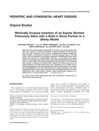
The self-expanding stent is laser-cut out of a nitinol
tube, with a sinus portion fitting to the natural shape of
the valve area. Two sizes of stents were used: the di-
ameter of the stent at the tubular part was 22 and 25
mm, respectively, and the sinus portion had a maxi-
mum diameter of 25 and 27.5 mm, respectively. The
length of the stent in its expanded shape was 29 mm.
Thanks to the sinus portion of the stent, the valve is
able to work in its natural manner to reduce or elimi-
nate the so-called ‘‘hammock effect.’’ The stent has
eyes at the proximal end to allow for a controlled
release (Fig. 1).
The delivery instrument consists of a stent chamber
with an outer diameter of 25 F and a very flexible
delivery catheter with a diameter of 6 F. The stent con-
taining the valve is crimped down to the necessary di-
ameter and loaded into the stent chamber prior to
delivery. A silicone tip is fixed to the distal end of the
delivery instrument to ease insertion of the instrument
through the myocardium, and reduce the risk of injury
to the vessels (Fig. 2). At the distal end of the instru-
ment, a luer lock connection allows safe and leak-proof
hydraulic connection of a standard 5 ml syringe. The
stent release is carried out by loading hydraulic pres-
sure on the stent chamber using sterile saline. During
release, the instrument can be locked into position,
which eases positioning. The instrument can be deliv-
ered over a 0.03500
guide wire.
Between November 2009 and August 2010, seven
Gotlandsfar lambs weighing 34–56 kg underwent a
right minithoracotomy for surgical insertion of the
stented equine valves, through direct right ventricular
Fig. 1. Stented equine graft displaying the sinus configuration of the stent and the tricuspid leaflets.
Equine Stented Pulmonary Valve Insertion 655
Catheterization and Cardiovascular Interventions DOI 10.1002/ccd.
Published on behalf of The Society for Cardiovascular Angiography and Interventions (SCAI).
3. access. Institutional Review Board approval was
granted for the survival study in sheep. After shaving
and prepping of the right thorax, the lambs were placed
on their left side. Antibiotic prophylaxis was given
with Cloxacillin 6 mg/kg prior to skin incision and one
dose afterwards. Intravenous heparin was administered
with a dose of 100 U/kg. A short right lateral thoracot-
omy incision was made and the fourth intercostal space
was entered. After pericardial opening, the lateral as-
pect and apex of the right ventricle were accessed, and
purse strings were placed for insertion of the angio-
catheter and delivery instrument, respectively. After
visualization of the right outflow tract and the pulmo-
nary valve by dye contrast injection through the angio-
catheter, a suitably sized prosthesis was selected, and
mounted onto the introducer. Following the placement
of a guidewire (Amplatz Super Stiff, Boston Scientific
Corp., Natick, MA) through the purse string into the
pulmonary artery, the introducer and prosthesis were
inserted into the right outflow tract, and the valve
released under angiographic visualization (Fig. 3). The
introducer was removed and the purse string tied. After
insertion of a chest drain, the chest was closed in
layers, and the animals rapidly extubated. Post-opera-
tive pain management was given with local administra-
tion of Lidocaine by use of the Painbuster pain relief
system (IFlow Corp., Lake Forrest, CA) for three days.
While the first animal was planned for an acute experi-
ment, six sheep were scheduled for sacrifice at 6
months.
RESULTS
The off-bypass procedure was well tolerated by all
animals, except for one, which suffered ventricular fi-
brillation and expired after stent deployment, despite
angiographically apparent correct positioning of the
valve. Good access to the right ventricle was achieved
through the right minithoracotomy, and positioning the
introducer device to the right outflow tract through the
purse-string was relatively straightforward. After angio-
graphic measurements of the pulmonary annulus (range
16.5–24 mm), the sinus of Valsalva portion (range 23–
31 mm), and of the pulmonary artery at the sinotubular
junction (range 20–26 mm), the stented grafts (22–24
mm) were successfully and smoothly released through
the native pulmonary valve. Correct position and func-
tioning of the valve was assessed by intraoperative an-
giography and confirmed by transesophageal echocardi-
ography. Procedure times ranged between 85 and 130
min. One animal suffered from ventricular fibrillation
after stent release and expired, and another died from
respiratory insufficiency from pneumothorax on post-
operative day 1 due to injury by another animal. At
last follow-up, transesophageal echocardiography dem-
onstrated good position of the stented valve, minimal
Fig. 3. Implantation sequence in the right outflow tract with correct positioning of the
equine stented valve through the native pulmonary valve.
Fig. 2. The hydraulic delivery device.
656 Dodge-Khatami et al.
Catheterization and Cardiovascular Interventions DOI 10.1002/ccd.
Published on behalf of The Society for Cardiovascular Angiography and Interventions (SCAI).
4. to mild central pulmonary insufficiency, and peak gra-
dients of 3–16 mm Hg over the right outflow tract.
Histological analysis of the explanted grafts was per-
formed in one sheep after 1 month (unplanned death
from pneumonia), in another after 3 months (unplanned
death from bacteremia, heart failure with pericardial
effusion and tamponnade), and in two animals as
planned at 6 months. Macroscopically, pliable free-
moving transparent non-calcified leaflets were observed
in all four animals. At 3 months in one animal, no
reendothelialization on the decellularized graft was
observed. Bacterial colonies together with findings
from pathology confirmed a postoperative bacterial
infection leading to sepsis, with valve leaflets sur-
rounded by fibrin. Histological examination at 6
months showed a layer of endothelial cells on the sur-
face of the valve (Fig. 4). Scant lymph follicles in one
graft wall indicated a mild unspecific immunogenic
reaction. Light vascularization of the graft wall was
observed.
DISCUSSION
Surgical pulmonary valve replacement through
repeat sternotomy using cardiopulmonary bypass is a
standardized procedure with excellent results [1], but is
considered a relatively invasive procedure. Percutane-
ous approaches for PVR may reduce the invasiveness,
are also widely accepted, but currently still have lim-
ited application due to prosthesis-vascular access mis-
match. Common to both approaches is the biological
fate and reduced longevity of the extant prostheses
available, be they of human or animal origin, rendering
any measure at best palliative. More recently, research
and clinical attempts are striving for transventricular
access to the right outflow, using a combination of
minimally invasive surgical techniques and percutane-
ous technology [3–8].
Advantages of the newly designed stent with a sinus
portion include the creation of vortex flow, which is
very important to promote active closure of the valve,
also avoiding the so called ‘‘hammock effect’’ [9].
Vortex flow reduces wear and shear stress, and there-
fore increases the durability of the valve cusps [10,11].
In contrast, in extant stents without a sinus portion, the
valve closes only passively because of backflow. The
direct apical approach to the right ventricle allows for
insertion of stented valves with a larger diameter than
is otherwise possible with the current percutaneous sys-
tems. Stent release with a hydraulic mechanism allows
for a very flexible and narrow shaft of the instrument.
The design permits easily following of the guide wire
to the desired position. The release mechanism can be
controlled with one hand, keeping the other free to
secure the position of the device during release. The
stented valves have been echocardiographically docu-
mented to function correctly up to 6 months, with his-
tological evidence of vascularization of the graft wall,
and endothelialization of the valve.
CONCLUSIONS
In an experimental sheep survival model, we have
demonstrated the feasibility and ease of insertion of a
new stented equine valve with a sinus portion, using a
new hydraulic device release system through a mini
right thoracotomy, on a beating heart. This approach
may become a complement to the percutaneous trans-
catheter one, either in much younger patients with
small vascular access, or on the contrary in very large
patients with much wider outflow tracts than the extant
percutaneous valves. With direct access to the right
outflow, problems pertaining to these two extremes in
patient vessel or outflow versus device mismatch
potentially allow a broader patient population to benefit
from this type of therapy. Insertion through a small
incision avoiding repeat sternotomy, as well of the fea-
sibility to insert the prosthesis without the use of car-
diopulmonary bypass make this a more attractive and
less invasive procedure. Mid to long-term functional
and histological results are required for further evalua-
tion of this new valve.
ACKNOWLEDGEMENTS
The authors thank Monika Hilbe, MD, Institute of
Veterinary Pathology, Vetsuisse Faculty Zurich,
Fig. 4. Histology of an explanted competent valve at 6
months, with pliable noncalcified leaflets (*), and an endothe-
lial layer (arrows).
Equine Stented Pulmonary Valve Insertion 657
Catheterization and Cardiovascular Interventions DOI 10.1002/ccd.
Published on behalf of The Society for Cardiovascular Angiography and Interventions (SCAI).
5. Zurich, Switzerland, for conducting the histological
investigation.
Disclosures and Freedom of Investigation
This work was supported by Carag AG (Baar, Swit-
zerland). The nitinol stent was designed by the authors,
and the valves were fabricated by Carag prior to inser-
tion. The authors are research consultants to Carag AG
but have no further financial relationship with the com-
pany. They had full control and freedom of investiga-
tion with regards to the design of the study, the meth-
ods used, as well as the outcome measurements, data
analysis, and writing of the final manuscript.
REFERENCES
1. Valsangiacomo-Bu¨chel ER, Dave HH, Kellenberger CJ, Dodge-
Khatami A, Preˆtre R, Berger F, Bauersfeld U. Remodeling of
the right ventricle after early pulmonary valve replacement
in children with repaired tetralogy of Fallot: Assessment by car-
diovascular magnetic resonance. Eur Heart J 2005;26:2721–
2727.
2. Bonhoeffer P, Boudjemline Y, Qureshi SA, Le Bidois J, Iserin
L, Acar P, Merckx J, Kachaner J, Sidi D. Percutaneous insertion
of the pulmonary valve. J Am Coll Cardiol 2002;39:1664–1669.
3. Meng G-W, Zhou J-Y, Tang Y, Ye Z-K, Zhang Y, Liu G-M,
Hu S-S. Off-pump pulmonary valve implantation of a valved
stent with an anchoring mechanism. Ann Thorac Surg 2009;87:
597–602.
4. Huber CH, Hurni M, Tsang V, von Segesser LK. Valved stents
for transapical pulmonary valve replacement. J Thorac Cardio-
vasc Surg 2009;137:914–918.
5. Attmann T, Jahnke T, Quaden R, Boening A, Muller-Hulsbeck S,
Cremer J, Lutter G. Advances in experimental percutaneous pul-
monary valve replacement. Ann Thorac Surg 2005;80:969–975.
6. Godart F, Bouzguenda I, Juthier F, Wautot F, Prat A, Rey C,
Corseaux D, Ung A, Jude B, Vincentelli A. Experimental off-
pump transventricular pulmonary valve replacement using a
self-expandable valved stent: A new approach for pulmonary
incompetence after repaired tetralogy of Fallot? J Thorac Cardi-
ovasc Surg 2009;137:1141–1145.
7. Ferrari E, Sulzer C, Rizzo E, von Segesser LK. Transcatheter
stent-valve implantation in a stenotic pulmonary conduit via a sub-
xyphoidian access. Eur J Cardiothorac Surg 2009;36:595–597.
8. Berdat PA, Carrel T. Off-pump pulmonary valve replacement
with a new Shelhigh injectable stented pulmonary valve.
J Thorac Cardiovasc Surg 2006;131:1192–1193.
9. Nordmeyer J, Coats L, Lurz, Lee T-Y, Derrick G, Rees P, Cullen
S, Taylor AM, Khambadkone S, Bonhoeffer P. Percutaneous pul-
monary valve-in-valve implantation: A successful treatment con-
cept for early device failure. Eur Heart J 2008;29:810–815.
10. Katayama S, Utemani N, Sugiura S, Hisada T. The sinus of
Valsalva relieves abnormal stress on aortic valve leaflets by
facilitating smooth closure. J Thorac Cardiovasc Surg 2008;136:
1528–1535.
11. Grande-Allen KJ, Cochran RP, Reinhall PG, Kunzelman KS. Re-
creation of sinuses is important for sparing the aortic valve: A fi-
nite element study. J Thorac Cardiovasc Surg 2000;119:753–763.
658 Dodge-Khatami et al.
Catheterization and Cardiovascular Interventions DOI 10.1002/ccd.
Published on behalf of The Society for Cardiovascular Angiography and Interventions (SCAI).