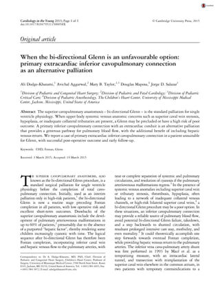
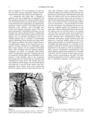
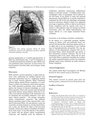
Anaesthetic Management of A Rare Case of Hemianomalous Pulmonary Venous Conne...Apollo Hospitals
We present a case report on anaesthetic management of a case of hemianomalous pulmonary venous connection with VSD, ASD for total intracardiac repair. A balanced anaesthetic technique was used with oxygen, isoflurane, fentanyl, midazolam, vecuronium. Patient was successfully operated under cardiopulmonary bypass with hypothermia.
Total Situs Inversus and D- Transposition of Great Arteries Managed in 2 Surg...semualkaira
A case report of a pediatric patient in whom multiple congenital
cardiac malformations coexist is presented below. The presentation of D-transposition of the great arteries (TGA) associated with
total situs inversus and pulmonary artery atresia presented a challenge in surgical management, however, it could be carried out
successfully using the Rastelli technique.
Drs. Potter and Richardson's CMC Pediatric X-Ray Mastery October CasesSean M. Fox
Drs. Potter and Richardson are interested in education and Pediatric Emergency Medicine. Follow along with the EMGuideWire.com team and Dr. Michael Gibbs as they post these educational, self-guided radiology slides on Pediatric Emergency Medicine Radiology Topics including:
• Scoliosis
• Pneumothorax
• Parapneumonic Effusion
• Cardiomegaly
• Vaping associated lung injury
2. Fontan completion.5
To our knowledge, we report the
first successful primary extracardiac inferior cavopul-
monary connection with satisfactory early follow-up.
A 3-month-old male infant after a Norwood I
operation with Sano modification for hypoplastic left
heart syndrome presented for routine pre-Glenn evalua-
tion. Ventricular function was normal with no semilunar
or atrioventricular valve regurgitation. At catheterisation,
pulmonary vascular resistance (2.94 Wood Units) and
pulmonary artery pressures (18/13/15 mmHg) were
normal, with adequate pulmonary arteries. The veno-
gram demonstrated a thrombosed innominate vein and
multiple veno–venous collaterals between a smaller right
superior caval vein, a left remnant superior caval vein, the
azygous vein, an accessory hemiazygous, and para-
vertebral plexuses (Fig 1). Attempts at interventional
catheter improvement of the situation were unsuccessful.
The surgery was deferred until nearly 6 months of age to
encourage further superior caval vein growth, but intra-
operative findings confirmed the unfavourable upper
body venous anatomy for the planned bi-directional
Glenn. After consulting with the referring paediatric
cardiologist and family, we proceeded with primary
inferior cavopulmonary connection. Through redo med-
ian sternotomy, arterial inflow was accomplished via the
previous 3.5-mm GoreTex graft to the innominate
artery, along with a low inferior caval vein and right atrial
cannulation for venous return. The superior caval vein
was mobilised, and the azygous vein was doubly ligated
and transected, so that the future inferior caval vein–
pulmonary artery conduit could travel unobstructed
behind the superior caval vein–right atrial junction and
avoid right pulmonary venous compression. Cardio-
pulmonary bypass was initiated and the procedure was
performed on a beating heart. After takedown of the Sano
shunt, the inferior caval vein was transected from the
common atrium and the cardiac side was oversewn. A
tube of bovine photofixed xenopericardium was created –
with 14-mm diameter based on intra-operative inferior
caval vein measurement – and anastomosed to the infer-
ior caval vein opening. The pulmonary arteries were iso-
lated, the underside opened, and anastomosed to the
extracardiac conduit, working first between the aorta and
the superior caval vein and then lateral to the superior
caval vein (Fig 2). An intra-cardiac line was placed and
tunnelled to the skin for common atrial pressure mon-
itoring. Weaning from cardiopulmonary bypass was
uneventful, with systemic oxygen saturations in the low
eighties on a fractional inspired oxygen of 50%, pul-
monary artery pressures via a femoral venous line of 14–
18 mmHg, and atrial pressures of 4–6 mmHg. In the
paediatric cardiac ICU, low-dose intravenous heparin was
transitioned to oral Aspirin as with our standard post-
Glenn protocol. Post-operative haemodynamics were
stable, allowing planned early extubation, an uneventful
3-day ICU stay with removal of chest drains, and dis-
charge to home on post-operative day eight. The infant
maintained saturations in the low eighties on room air,
had no abdominal symptoms, and was feeding and
Figure 1.
Pre-operative antero-posterior angiogram showing a small stenotic
superior caval vein and multiple systemic veno–venous collaterals.
Figure 2.
Artist’s illustration of the inferior cavopulmonary connection and
the free lie posterior to the superior caval vein–common atrial
junction.
2 Cardiology in the Young 2015
3. growing appropriately at 5 months post-operatively. A
follow-up catheterisation (Fig 3) and CT-angiogram at
4 months post-operatively disclosed an open inferior caval
vein–pulmonary artery connection, no veno–venous col-
lateral systems in the lower body, and no hepatic venous
congestion.
Discussion
With anatomic concerns pertaining to upper body sys-
temic veins, performing the standard superior cavo-
pulmonary anastomosis may be a challenging forward
step in single ventricle palliation. When considering
optimal staging towards Fontan completion, it is para-
mount to protect the pulmonary arteries from hyperten-
sion while promoting pulmonary artery growth, and
shorten the duration of shunted physiology yet avoid
Glenn takedown. Faced with a patient with sub-optimal
upper body systemic venous anatomy, likely due to vas-
cular access-induced thrombosis of the innominate vein,
we attempted and successfully performed a new alter-
native surgical operation – the primary extracardiac
inferior cavopulmonary connection. This palliation
allowed saturations equal to those expected after a stan-
dard bi-directional Glenn, the post-operative course was
uneventful, of similar duration to a bi-directional Glenn,
and, to date, has allowed the infant to feed and grow
normally, without any evidence of morbidity. If a hepatic
factor entity exists,2
it may flow directly to the pulmon-
ary arteries with the primary inferior cavopulmonary
connection, and hopefully avoid the formation of
troublesome pulmonary arteriovenous malformations
leading to increased cyanosis.3
A bubble-contrast echo-
cardiographic study or angiography will be planned in
the near future of our patient to study this potential
phenomenon. Longer follow-up is certainly warranted to
understand the fate of this new physiology and perhaps
extend its indications: whether it leads to an unplanned
inter-stage re-operation, will be an ideal set-up for future
Fontan completion with the superior caval vein,5
or
results in a final palliative stage not needing any further
intervention, given the similar haemodynamics and
oxygen delivery to a very largely fenestrated Fontan
circulation.
Limitations to the technique and future considerations
In the absence of a right-sided pumping chamber,
creating an inferior caval vein–pulmonary artery con-
nection with laminar flow, which goes against gravity, in
an infant who is not yet ambulating or even crawling
may be haemodynamically sub-optimal. The true sig-
nificance of this phenomenon compared with the bi-
directional Glenn, which theoretically uses gravity to
flow downwards is however unknown. The use of an
interposition graft of any nature, which will not grow,
may render future re-operations inevitable due to poten-
tial conduit outgrowth (patient-conduit size mismatch),
although many centres uniformly use small conduits at
Fontan completion.
Acknowledgements
The authors thank Kyle Cunningham and Michael
Schenk for their superb surgical illustration.
Financial Support
This research received no specific grant from any
funding agency, commercial, or not-for-profit sectors.
Conflicts of Interest
None.
References
1. Pridjian AK, Mendelsohn AM, Lupinetti FM, et al. Usefulness of
the bidirectional Glenn procedure as staged reconstruction for the
functional single ventricle. Am J Cardiol 1993; 71: 959–962.
2. Bernstein HS, Brook MM, Silverman NH, Bristow J. Development
of pulmonary arteriovenous fistulae in children after
cavopulmonary shunt. Circulation 1995; 92 (Suppl): II309–II314.
3. Knight WB, Mee RBB. A cure for pulmonary arteriovenous fistulas?
Ann Thorac Surg 1995; 59: 999–1001.
4. Iyer GK, Van Arsdell GS, Dicke FP, McCrindle BW, Coles JG,
Williams WG. Are bilateral superior vena cavae a risk factor for
single ventricle palliation? Ann Thorac Surg 2000; 70: 711–716.
5. Macé L, Dervanian P, Losay J, et al. Bidirectional inferior vena cava-
pulmonary artery shunt. Ann Thorac Surg 1997; 63: 1321–1325.
Figure 3.
Post-operative early injection angiogram showing the inferior
cavopulmonary connection with widely patent anastomoses compatible
with generous pulmonary artery flow.
Dodge-Khatami et al: When the bi-directional Glenn is an unfavourable option 3