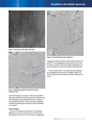
1. Supplement to Sponsored by Boston Scientific Corporation
April 2013
Versatility in
the Embolic
Spectrum
Making successful outcomes possible
even in challenging cases.
2. 2 Supplement to Endovascular Today April 2013
Versatility in the Embolic Spectrum
By J. Timothy Riley, MD, St. Joseph’s Hospital
Embolization of Challenging
Type II Endoleaks
CASE 1
An 84-year-old man underwent endovascular aortic
repair and was monitored for an enlarging aneurysm sac
and a type II endoleak.
As a result of the location of the stent graft and the
endoleak, an endovascular approach via the lumbar ves-
sels was selected. Using a 5-F, 0.038-inch inner diameter
selective diagnostic catheter, the origin of the feeding
vessel was identified with a contrast injection. Because
of the small diameter and the significant tortuosity of
the lumbar vessel, the 5-F catheter was advanced only
slightly beyond the origin of the vessel.
A 0.021-inch inner lumen diameter microcatheter
(Renegade® STC Microcatheter) and a torquable 0.016-inch
INTRODUCTION
Despite significant advances in the devices used in the endovascular repair of abdominal aortic aneurysms, the
occurrence of clinically relevant type II endoleaks remains relatively common. Although there are a variety of ways
to successfully treat type II endoleaks, one of the most frequently used techniques involves accessing the feeding
vessels via an endovascular approach. As a result of the significant tortuosity, the navigation of catheters and wires
to these aneurysms can be difficult, and it typically requires the clinician to use smaller, more precise microemboli-
zation devices (0.018-inch), which are often less occlusive than larger 0.035-inch embolization devices. Historically,
this requirement has made it challenging to achieve the desired level of hemostasis because large aneurysmal
volumes typically need a large quantity of highly thrombogenic devices to achieve stasis. As a result of these con-
flicting priorities, very few embolization platforms are available that satisfy the need for precise delivery and high
thrombogenicity.
One of the most important advances in the field of embolization has been the development of long detachable
coils that contain significant amounts of thrombogenic fibers. These devices allow delivery of large volumes of embol-
ic materials quickly and accurately. As a result, many of the procedures that are traditionally very time consuming
and challenging have become common practice for experienced interventionists.
The following cases demonstrate the utility of the Interlock™ Fibered IDC™ Occlusion System in patients with chal-
lenging type II endoleaks.
Figure 1. The origin of the feeding vessel was identified with
a contrast injection.
Figure 2. Selective contrast injection revealed a large
aneurysm.
Results from case studies are not necessarily predictive of results in other cases. Results in other cases may vary.
3. April 2013 Supplement to Endovascular Today 3
Versatility in the Embolic Spectrum
microwire (Fathom® – 16 Steerable Guidewire) were
passed through the diagnostic catheter into the distal
vasculature. After significant catheter and wire manipula-
tion, access to the sac was achieved, and a contrast injec-
tion revealed a large aneurysmal void that was causing
the endoleak.
After positioning the microcatheter within the aneu-
rysm and removing the guidewire, the process of emboli-
zing both the sac and the feeding vasculature began. The
embolization procedure was completed using a number
of Interlock™ – 18 Fibered Platinum Coils, each with a
wide diameter and a long length to ensure adequate wall
apposition. Using a total of seven coils, 210 cm of coil
was rapidly deployed, and hemostasis was achieved.
CASE 2
Approximately 6 months after endograft
implantation, a CT scan revealed the contin-
ued expansion of a large aneurysm sac.
Using a radial access site, a 120-cm, 5-F
hydrophilic catheter was used to select the
left internal iliac artery. After cannulation
of the internal iliac artery with the 5-F cath-
eter, a lumbar vessel was selected using a
150-cm Renegade® STC Microcatheter and
a Fathom® – 16 Guidewire. Selective angio
graphy revealed that the large aneurysm sac
was being fed by two lumbar vessels.
Once positioned appropriately, the
microcatheter was used to deliver embol-
ic coils in an attempt to halt flow into
the enlarging aneurysm sac. To begin the
coil embolization, a number of 14-mm
Interlock™–18 Fibered Platinum Coils were
packed into the wide-diameter aneurysm
sac. In an effort to fill the aneurysm with a dense net-
work of coils, varying diameters were selected. After
the embolization of the sac itself, the same microcath-
eter was used to deliver additional long coils into the
feeding vessel. In total, 22 coils were used, and com-
plete stasis was achieved.
CASE 3
A 76-year-old man with an enlarging aneurysm and
type II endoleak was treated via the inferior mesenteric
artery (IMA).
Interrogation of the superior mesenteric artery was done
using a standard technique from the right femoral artery.
Using the 5-F, 0.038-inch catheter, the superior mesenteric
artery was imaged, and the middle colic artery was identi-
fied. The 5-F catheter was used to cannulate the origin of
the middle colic artery, and a small-lumen microcatheter
was then used to further access the distal vasculature.
Using a small-lumen 0.021-inch inner lumen micro-
catheter (Renegade® STC Microcatheter) and a torqueable
microguidewire (Fathom® – 16 Steerable Guidewire),
navigation through the mesenteric arch and into the
ascending branch of the left colic artery was gradually
achieved. Due to the tortuous path of the vasculature
and the relative lack of proximal support, a guidewire
with significant rail support was required to ensure ade-
quate push through the vasculature.
After significant manipulation of the microcatheter and
microwire, access to the ostium of the IMA was achieved.
Figure 3. Coil embolization of the aneurysm sac.
Figure 5. Twenty-two coils were
used, and complete stasis was
achieved.
Figure 4. Selective angiography
revealed that a large aneurysm sac
was being fed by two lumbar vessels.
4. 4 Supplement to Endovascular Today April 2013
Versatility in the Embolic Spectrum
After removal of the microwire, a contrast injection revealed
a large, patent IMA feeding the aneurysm sac.
After further advancement of the microcatheter,
numerous detachable coils (Interlock™ – 18 Fibered IDC™
Occlusion System) were deployed in an attempt to fully
embolize the vessels feeding the endoleak. In total, nine
long, fibered detachable coils were used, and complete
stasis was achieved.
DISCUSSION
Although improvements have been made to commer-
cially available endovascular aortic repair devices over the
past decade, the endovascular repair of type II endoleaks
remains a relatively common procedure. As the cases pre-
sented illustrate, significant technical skills with small-lumen
delivery systems and embolic platforms are often required
due to the challenging nature of the vasculature that is
involved. In addition, although the vasculature that must
be traversed is often challenging due to the small diam-
eter of the vessels and the tortuosity of the path, the area
that must ultimately be embolized is often quite large and
requires a significant amount of embolic material to achieve
adequate stasis. Embolic systems that offer long coil lengths
and a dense network of thrombogenic fibers can be valu-
able tools in these procedures. n
J. Timothy Riley, MD, is a Vascular Surgeon at St. Joseph’s
Hospital Health Center in Syracuse, New York. All cases per-
formed in collaboration with Interventional Radiology. He has
disclosed that he has no financial interests related to this article.
Figure 6. Interrogation of the superior mesenteric artery
was done using a standard technique from the right femoral
artery.
Figure 7. Navigation through the mesenteric arch was
achieved with a microcatheter and microguidewire.
Figure 9. Nine long, fibered detachable coils were used, and
complete stasis was achieved.
Figure 8. Contrast injection revealed a large, patent IMA
feeding the aneurysm sac.
5. 6 Supplement to Endovascular Today April 2013
Versatility in the Embolic Spectrum
By Peter Waybill, MD, and Jerron L. Fischer, MD, Harrisburg Medical Center
Percutaneous Management of a
Large Renal AVM
INTRODUCTION
A 30-year-old man with no significant medical history experienced blunt trauma to the right flank while playing
flag football. Subsequently, he developed gross hematuria and visited the emergency room. The imaging workup
revealed a large right renal vascular lesion (Figure 1). At the patient’s initial evaluation by the urology department,
it was suggested that the lesion might be a congenital arteriovenous malformation (AVM), which can be managed
surgically, but would likely require a nephrectomy. The other option was to pursue a percutaneous nephron-sparing
intervention.
PROCEDURE DESCRIPTION
Abdominal aortography was performed to identify
the location and number of renal arteries (Figure 2).
This revealed single renal arteries bilaterally. A diagnostic
catheter was exchanged for a 5-F, RC-1 catheter, which
was advanced into the right renal artery. Right renal
arteriography was performed, and the images showed a
massive right renal AVM arising from a dilated branch of
a lower pole right renal artery.
The 5-F catheter was then exchanged for a 6-F Ansel
sheath. Through the sheath, a 5-F Cobra 2 catheter was
used to select the lower pole right renal artery branch,
which supplied the AVM. There was a very small vessel
supplying a small segment of normal renal parenchyma
arising at the origin of the AVM (Figure 3). We decided
to start coil embolization in this small branch vessel and
then allow the coil to prolapse into the large vessel sup-
plying the AVM. This allowed the coil to be anchored
and was a good starting point for our coil nest by ensur-
ing that the coil would not migrate through the AVM
to the venous outflow. As an additional safety measure to
prevent the coil from migrating, an Interlock™ Coil was cho-
sen for the first coil. The coil was deployed in the appropri-
ate position, without prolapse into the outflow vein.
A total of three Interlock™ 0.035-inch Coils were
placed: first, a 15-mm X 40-cm coil, followed by two
12-mm X 20-cm coils. Two pushable vortex-shaped coils
were then used as fillers. Follow-up arteriography showed
no flow within the AVM. Flow was maintained to 80% of
the right renal parenchyma (Figure 4).
Figure 1. The imaging workup revealed a large right renal
vascular lesion.
Figure 2. Abdominal aortography was performed to identify
the location and number of renal arteries.
Results from case studies are not necessarily predictive of results in other cases. Results in other cases may vary.
6. April 2013 Supplement to Endovascular Today 7
Versatility in the Embolic Spectrum
DISCUSSION
AVMs refer to a congenital type of malformation
and are found more often in women. Renal AVMs are
very rare. There are two types of congenital renal AVMs
described in literature: the cirsoid type, which is more
common, and the cavernous type. The cirsoid type con-
sists of multiple small, dilated arteriovenous communica-
tions with corkscrew appearances and tends to be adja-
cent to the collecting system. Patients present with gross
hematuria. Cavernous AVMs have a single dilated vessel.
We suspect that our patient had a cavernous AVM.
In comparison, arteriovenous fistulas (AVFs) are
acquired and typically have a single feeding artery and
a single draining vein. Acquired AVFs are the most
common type of renal arteriovenous communication,
and represent 75% to 80% of renal vascular anomalies.
These are usually a result of trauma, surgery, biopsy,
tumors, or erosion of aneurysms into a vein. Renal
AVMs and AVFs are potentially lethal conditions, but
most remain small and resolve spontaneously. If symp-
tomatic, the most common clinical manifestation is
hematuria. Cardiomegaly or congestive heart failure can
occur. Ischemia in the renal parenchyma distal to the
AVM or AVF may induce renin-mediated hypertension
and/or impaired renal function. On imaging, the cirsoid
AVM has tortuous varix-like vessels.
Embolization is considered the primary treatment
option in cases of renal AVM or AVF because it pre-
serves maximal normal renal parenchyma while elimi-
nating the risk of recurrent hemorrhage. The goal of
AVM or AVF embolization is eradication of the nidus.
Recent reports describe the ablation of feeding vessels
with various embolic agents, including particles, metal-
lic coils, and liquid embolics. AVM radiofrequency abla-
tion has also been attempted. n
Peter Waybill, MD, is an Interventional Radiologist
at the Harrisburg Medical Center in Harrisburg,
Pennsylvania. He has disclosed that he has no financial
interests related to this article.
Jerron L. Fischer, MD, is an Interventional Radiologist
at the Harrisburg Medical Center in Harrisburg,
Pennsylvania. He has disclosed that he has no financial
interests related to this article.
Figure 3. Through the sheath, a 5-F Cobra 2 catheter was
used to select a lower pole right renal artery branch, which
supplied the AVM.
Figure 4. Three Interlock™ 0.035-inch Coils were placed.
7. 8 Supplement to Endovascular Today April 2013
Versatility in the Embolic Spectrum
By Pär Gerwins, MD, PhD, Uppsala University Hospital
Embolization of a Large
Arteriovenous Malformation
in the Forearm
INTRODUCTION
Arteriovenous malformations (AVMs) arise due to developmental errors in the capillary bed during embryogenesis.
The lack of a capillary bed results in rapid shunting of blood from the arterial to venous circulation. Although lesions
are present at birth, they might not become evident until childhood or later in life. AVMs are sensitive to hormonal
changes and often progress in adolescence, during pregnancy, and during intake of estrogen-containing contracep-
tives. Lesions are pulsatile with a palpable bruit or thrill, warm, and not easily compressible. The rapid shunting of
blood leads to insufficient capillary perfusion with reduced oxygen delivery causing ischemia, pain, and development
of wounds and bleedings. AVMs grow in size due to increased filling and enlargement of both supplying arteries and
draining veins, which might cause disfigurement, destruction of tissues, and impaired organ function. When large
enough, AVMs can cause congestive heart failure. Treatment of AVMs is based on endovascular embolization, surgi-
cal resection, or a combination of both. Complete cure is rare, and treatments therefore aim at controlling the mal-
formation. Endovascular embolizations are performed using transarterial, transvenous, or direct-puncture techniques,
and techniques are often combined. The embolic material can be liquid or solid, such as coils, plugs, and particles. It is
important that the embolic material penetrates to the nidus and ideally to the point of the initial venous drainage.
PROCEDURE DESCRIPTION
A 17-year-old adolescent boy was referred from
an outside hospital due to acute bleeding from
an extensive ruptured AVM in his right forearm.
The patient was taken to the OR and, in a blood-
less field, the rupture was localized to a large
draining subcutaneous vein, which was sutured.
Subsequent digital subtraction angiography and
magnetic resonance imaging revealed the true
nature of the AVM with countless feeding arter-
ies from the ulnar, interosseus, and radial arteries
(Figure 1). Venous drainage was achieved through
multiple large subcutaneous veins, and the lesion
was embolized using a transvenous strategy.
Under general anesthesia, a diagnostic catheter
was advanced from the right femoral artery to the
right brachial artery to visualize the AVM during
the procedure. Using ultrasound guidance, an
enlarged subcutaneous vein was accessed on the
upper right arm. A draining vein at the nidus close
to the wrist was reached using venous access with
a Renegade® STC Microcatheter and a Fathom®
Guidewire (Figure 2). Embolization was performed
with 0.018-inch Interlock™ Coils. Control angiog-
raphy revealed that this strategy was successful
with markedly decreased shunting (Figure 2). The
Figure 1. The AVM had countless feeding arteries from the ulnar, inter-
osseus, and radial arteries.
Results from case studies are not necessarily predictive of results in other cases. Results in other cases may vary.
8. April 2013 Supplement to Endovascular Today 9
Versatility in the Embolic Spectrum
coiling procedure was repeated in nine separate procedures
during a 6-month period with 0.018-inch Interlock™ Coils in
veins that were hard to reach with a 0.035-inch system and
0.035 Interlock™ Coils in larger veins. The final angiography
(Figure 3 and 4) showed that almost all shunts were occlud-
ed. The patient’s symptoms of pain and discomfort, as well
as the volume of the arm, were markedly reduced. At the
time of the last embolization, it was observed that one coil
was protruding through the skin due to shrinkage of the
AVM. Because this could be a potential site of infection, the
thrombosed AVM was surgically
resected. The surgical resection
was performed in a safe and con-
trolled manner, without significant
blood loss, due to the preopera-
tive embolization. Postoperative
healing was uneventful, and 6
months later, the patient was free
of symptoms and able to return to
a normal life.
DISCUSSION
AVMs are challenging lesions to
treat. Embolization from the arte-
rial side was not an option due to
the countless number of arterial
feeders, and the risk of spillover of
embolic material would be dev-
astating for the hand. This case
demonstrates efficient closure
of the draining vein at the nidus.
Because lesions like this one have a very high flow rate,
there is a concern that embolic material might dislodge
and cause nontarget embolization. The availability of
detachable, long, and fibered coils made it possible to per-
form the procedure in a safe and efficient manner. n
Pär Gerwins, MD, PhD, is a Professor of Vascular Biology
and an Interventional Radiologist at Uppsala University
Hospital in Uppsala, Sweden. He has disclosed that he has no
financial interests related to this article.
Figure 2. A draining vein at the nidus was reached via venous access with a
Renegade® STC Microcatheter and a Fathom® Guidewire, and embolization was per-
formed with 0.018-inch Interlock™ Coils. Control angiography revealed that this strat-
egy was successful with markedly decreased shunting.
Figure 4. The final angiogram showed that almost all shunts were occluded: the ulnar
artery before (A) and after (B) and the radial artery before (C) and after (D).
Figure 3. Final angiogram of the
brachial artery.
BA C D
9. 10 Supplement to Endovascular Today April 2013
Versatility in the Embolic Spectrum
By Hami Mehrzad, MD, and A. Ganeshan, MD, Heart of England NHS Trust
Preoperative Embolization
of a Large Mediastinal Tumor
Using Detachable Coils
CASE PRESENTATION
A 17-year-old adolescent boy presented with sudden-
onset dyspnea after an appendectomy. Chest x-ray
(Figure 1) revealed a large opacity occupying the right
hemithorax. The patient was referred for thoracic surgery.
Further questioning revealed a 5-year history of dysphagia
and dyspnea upon lying prone. High-resolution computed
tomography of the thorax (Figure 2) showed a heteroge-
neous calcified mass that was 20 cm X 11 cm X 8 cm and
compressing the main bronchi and great vessels, which is
suggestive of teratoma.
Resection via a right thoracotomy was attempted,
but the tumor was closely adherent to the medias-
tinum, and profuse bleeding was encountered from
the tumor bed, resulting in significant hypotension.
Hemostasis was obtained with difficulty, the thora-
cotomy was closed, and samples were sent for histol-
ogy. Postoperatively, the patient showed symptoms
of spinal cord ischemia. Magnetic resonance imaging
localized the lesion to T5.
Initial histology indicated that the mass was poten-
tially a solitary fibrous tumor of the pleura, but immu-
nohistochemical staining was not entirely congruent.
A second opinion diagnosed benign schwannoma
but was atypical for the presence of multiple arterio-
venous malformations. There was considerable mass
effect exerted on vital structures, and the tumor was
confirmed to be benign, intensifying the desire to
definitively resect. This was tempered by the previously
inflicted ischemic lesion to the spinal cord, from which
the patient was fortunately recovering well.
PROCEDURE DESCRIPTION
Multiple selective coil embolizations of feeding ves-
sels were undertaken before further surgery. Initial angi-
ography determined several tortuous feeding vessels
arising from the right intercostal, subclavian, internal
mammary, and thyrocervical arteries.
The origins of these vessels were selectively cannu-
lated with a catheter. A Renegade STC® Microcatheter
Figure 1. Chest x-ray showing a large opacity in the right
hemithorax.
Figure 2. A heterogeneous calcified mass in the thorax com-
pressing the main bronchi and great vessels.
Results from case studies are not necessarily predictive of results in other cases. Results in other cases may vary.
10. April 2013 Supplement to Endovascular Today 11
Versatility in the Embolic Spectrum
was used to further super selectively cannulate the
vessels. Due to the fact that the main catheter was
just engaged into the orifice of the vessels, Interlock™
0.018-inch Detachable Coils were used to allow greater
control at the time of deployment and because fewer
total coils were needed to embolize the tumor. Because
of the coils’ length, there was also less chance of distal
migration into the arteriovenous shunts of the tumor.
A range of coils (from 4 to 10 mm) was used with an
angiographically successful outcome after two sessions.
There was almost complete reduction in tumor blood
supply without any serious complication (Figures 3-7).
Follow-up computed tomography angiography moni-
tored for tumor shrinkage and watched for future resec-
tion.
DISCUSSION
In this selective case, we found that the Renegade STC®
Microcatheter and Interlock™ Detachable Coil system
were very useful tools in the interventional armamen-
Figure 3. Angiogram of the thyrocervical trunk branch show-
ing significant supply to the tumor during the first session.
Figure 5. Final angiogram showing almost complete reduc-
tion in the tumor blood supply from this vessel.
Figure 4. Several Interlock™ 0.018-inch Detachable Coils
were successfully used to embolize the feeding vessels.
Figure 6. In the second session, further supply from the right
internal mammary and subclavian branches was found on the
angiogram. There was some early venous filling suggestive of
arteriovenous malformation within the tumor.
11. 12 Supplement to Endovascular Today April 2013
Versatility in the Embolic Spectrum
tarium for treating such a large, complicated, mediastinal
tumor. It allowed for accurate, controllable, convenient,
and successful embolization before surgery without any
untoward complication. We hope that this example will
be useful for others who may encounter a similar inter-
esting—but—complex case. n
Hami Mehrzad, MD, is with the Interventional
Radiology department, Heart of England NHS Trust in
Birmingham, United Kingdom. He has disclosed that he
has no financial interests related to this article.
A. Ganeshan, MD, is with the Interventional Radiology
department, Heart of England NHS Trust in Birmingham,
United Kingdom. He has disclosed that he has no financial
interests related to this article.
Figure 7. Further Interlock™ 0.018-inch Detachable Coils
were used to reduce the risk of distal migration of any coils
into the arteriovenous shunts. There was almost complete
embolization of the tumor blood supply at the end of the pro-
cedure, as shown.
12. 14 Supplement to Endovascular Today April 2013
Versatility in the Embolic Spectrum
By Trevor Cleveland, MD, Sheffield Vascular Institute
Embolization of a GI Bleed
OVERVIEW
A 94-year-old woman presented to the accident and
emergency department in the early hours of the morning
(approximately 2:00 AM) with rectal bleeding.
When she arrived to the hospital, she was hemody-
namically stable but had further rectal bleeding. After
initial assessment, she was referred to the acute medicine
team who organized for her to have an emergency upper
gastrointestinal (GI) endoscopy. Clinically, it was most
likely that the bleeding source was in the lower GI tract,
but an upper GI source needed to be excluded at an
early stage. Proctoscopy was obscured by the blood in
the rectum.
The upper GI endoscopy was normal, with no evi-
dence of blood as far as the second part of her duode-
num.
Despite the administered clear fluids and blood, her
hemoglobin, which was 11 g/dL on admission, had fallen
to 8 g/dL.
PROCEDURE DESCRIPTION
At 7:30 AM, the interventional radiology team (radi-
ologist, radiographer, and nurse) was contacted to con-
sider trying to find the bleeding source and treat it with
embolization. A triple-phase (plain, arterial, and venous)
computed tomography (CT) scan was immediately orga-
nized.
The unenhanced CT showed diverticular disease of
the colon, with calcification associated with multiple
diverticula (Figure 1); this can look like contrast extrava-
sation. After injection of contrast, the arterial phase scan
showed active bleeding in the sigmoid colon, with con-
trast in the lumen (Figure 2). The delayed scan showed
further contrast spreading from the bleeding point
(Figure 3).
After speaking with the patient in the CT scanning
room, we transferred her directly to the interventional
radiology suite, where she underwent selective angio
graphy of the inferior mesenteric artery (IMA). She had
significant atheroma in her aorta and at the origin of
the IMA (which was seen on the CT). As a result, despite
great care, there was a mild, non–flow-limiting dissection
Figure 1. Calcification in the diverticular disease, which simu-
lates contrast extravasation.
Figure 2. Contrast extravasation in the sigmoid colon. Figure 3. Contrast spreading from the bleeding point on the
delayed scan.
Results from case studies are not necessarily predictive of results in other cases. Results in other cases may vary.
13. April 2013 Supplement to Endovascular Today 15
Versatility in the Embolic Spectrum
of the IMA (Figure 4). However, it did prove possible to
selectively catheterize the IMA with a microcatheter, and
the bleeding point was identified (Figure 5). Using the
microcatheter, Interlock™ Coils were used to selectively
embolize the bleeding point (Figure 6), and the bleeding
was stopped.
DISCUSSION
The use of emergency three-phase CT can identify
bleeding if it is active. A precontrast CT avoids some
false-positive scans. The CT scan will direct angiographic
imaging, and there should be minimal delay between the
positive CT and angiography. Elderly patients have arter-
ies prone to catheter and wire damage. Embolization is a
quick and effective treatment for lower GI bleeding. n
Trevor Cleveland, MD, is Consultant Vascular Radiologist
at the Sheffield Vascular Institute in Sheffield, England. He
has disclosed that he has no financial interests related to this
article.
Figure 4. Dissection of the origin of the IMA.
Figure 6. Bleeding stopped after placement of two
Interlock™ Coils.
Figure 5. Active bleeding on the angiogram.
14. PI-151005-AA
Results from case studies are not necessarily predictive of results in other cases. Results in other cases may vary.
ABBREVIATED STATEMENTS
Interlock™ - 18 and 35 Fibered IDC™ Occlusion System
CAUTION: Federal law (USA) restricts this device to sale by or on
the order of a physician.
INTENDED USE/INDICATIONS FOR USE: The Interlock - 35
Fibered IDC Occlusion System is a modified interlocking detach-
able coil indicated to obstruct or reduce rate of blood flow in the
peripheral vasculature. This device is not intended for neurovas-
cular use.
CONTRAINDICATIONS: None known.
WARNING: Compatibility with Magnetic Resonance Imaging
(MRI) has not been established, and the degree of imaging distor-
tion resulting from the coil has not been measured.
ADVERSE EVENTS: • Complications related to catheterization (e.g.,
hematoma at the site of entry, clot formation at the tip of the
catheter and subsequent dislodgement, nerve and vessel dissec-
tion or perforation) • Pain • Hemorrhage • Infection necessitating
medical intervention • Foreign body reactions necessitating medi-
cal intervention • Emboli • Ischemia • Vasospasm • Tissue necrosis
• Undesirable clot formation of the vasculature • Claudication •
Recanalization • Death • Temporary neurological deficit. Prior to
use, please see the complete “Directions for Use” for more infor-
mation on Indications, Contraindications, Warnings, precautions,
Adverse events and operators instructions.
Fathom®-14/16 Steerable Guidewire
CAUTION: Federal law (USA) restricts this device to sale by or on
the order of a physician.
INDICATIONS FOR USE: The Fathom-14 and Fathom-16
Steerable Guidewire families are intended for general intravascu-
lar use in the peripheral vasculature. They can be used to selec-
tively introduce and position catheters and other interventional
devices in the peripheral vasculature. This device should be used
only by physicians thoroughly trained in percutaneous, intravas-
cular techniques and procedures.
CONTRAINDICATIONS FOR USE: This guidewire is not intended
for use in the coronary vasculature.
POTENTIAL COMPLICATIONS: Potential complication include,
but are not limited to: • Allergic reaction • Death • Embolism
(catheter/device, air bubble, plaque, thrombus, air embolism,
thromboembolism) • Hematoma at the puncture site • Infection
• Pseudoaneurysm • Seizure/stroke • Vascular thrombosis • Vessel
occlusion • Vessel spasm • Vessel dissection • Vessel damage •
Nerve injury • Perforation of the vessel • Hemorrhage • Bleeding
• Failed treatment • Inability to position guidewire • Damage to
the catheter. Prior to use, please see the complete “Directions
for Use” for more information on Indications, Contraindications,
Warnings, Precautions, Adverse Events, and Operator’s
Instructions.
Renegade HI-FLO™ Fathom® System
CAUTION: Federal law (USA) restricts this device to sale by or on
the order of a physician.
INDICATIONS FOR USE: The Renegade HI-FLO Fathom Kit is
intended for peripheral vascular use. The Fathom Guidewire
can be used to selectively introduce the Renegade HI-FLO
Microcatheter in the peripheral vasculature. Once the subselec-
tive region has been accessed, the microcatheter can be used for
the controlled and selective infusion of diagnostic, embolic or
therapeutic materials into vessels.
CONTRAINDICATIONS FOR USE: None known.
WARNING: The Renegade HI-FLO Fathom Kit is not intended for
use in the coronary vasculature. This device should be used only
by physicians thoroughly trained in percutaneous, intravascular
techniques and procedures.
POTENTIAL COMPLICATIONS: Potential complication include,
but are not limited to: • Allergic reaction • Death • Embolism •
Hemorrhage/hematoma • Infection • Pseudoaneurysm • Stroke
• Vascular thrombosis • Vessel occlusion • Vessel spasm • Vessel
trauma (dissection, perforation, trauma). Prior to use, please
see the complete “Directions for Use” for more information on
Indications, Contraindications, Warnings, Precautions, Adverse
Events, and Operator’s Instructions.
Renegade STC® 18 Microcatheter with HydroPass™
Hydrophilic Coating
CAUTION: Federal law (USA) restricts this device to sale by or on
the order of a physician.
INDICATIONS FOR USE: The Renegade STC 18 Microcatheter is
intended for peripheral vascular use. The microcatheter can be
coaxially tracked over a steerable guidewire in order to access dis-
tal, tortuous vasculature. Once the subselective region has been
accessed, the microcatheter can be sued for the controlled and
selective infusion of diagnostic, embolic or therapeutic materials
into vessels. Diagnostic, embolic or therapeutic agents to be used
in accordance with specifications outlined by the manufacturer.
CONTRAINDICATIONS FOR USE: None known.
WARNING: This device should be used only by physicians thor-
oughly trained in percutaneous, intravascular techniques and
procedures.
POTENTIAL COMPLICATIONS: Potential complication include,
but are not limited to: • Vessel trauma • Embolism • Hemorrhage/
hematoma • Vasospasm • Infection • Air embolism • Allergic reac-
tion. Prior to use, please see the complete “Directions for Use” for
more information on Indications, Contraindications, Warnings,
Precautions, Adverse Events, and Operator’s Instructions.