This case report describes a patient who underwent multiple cardiac surgeries for transposition of the great arteries with ventricular septal defect. The patient developed recurrent neoaortic insufficiency after an arterial switch operation and subsequent aortic valve replacement. A "switch back" operation was performed, placing the native pulmonary valve back into the left ventricular outflow tract. However, severe aortic insufficiency recurred within a year, requiring another aortic valve replacement. Histopathology of the excised valve showed abnormal elastic tissue, suggesting an underlying structural problem contributed to valve dysfunction. The report questions whether the switch back operation is a viable long-term option for neoaortic insufficiency in patients with complex transposition anatomies.
bentall, and 'old' procedures that still valid until present. Bail out for valve sparring & the patology of indonesian most present were best in this procedures
Nursing management of patient with cardiac surgeries.PrashantSalve10
It will be helpful to overview cardiac surgeries like CABG, Valvular surgeries and heart transplant. It also enumerates the nursing diagnoses and its brief description.
Surgery for aneurysmal right coronary fistula and constrictive pericarditis Abdulsalam Taha
CRCM, March 2014
View on scirp.org
Abstract:
Coronary artery fistula (CAF) is a direct communication between a coronary artery and the lumen of any of the cardiac chambers, the coronary sinus, the pulmonary artery, the superior vena cava or the proximal pulmonary veins. The majority of these fistulas are congenital in origin although they may occasionally be detected after cardiac surgery. Congenital CAF is a rare anomaly and aneurysmal formation in the fistula is even rarer. Majority of CAF are isolated lesions, however, congenital or acquired heart diseases may coexist. Herein, we report a case of huge congenital aneurysmal right CAF connected to the right atrium in an Iraqi man of 62 associated with tuberculous effusive-constrictive pericarditis to whom off pump pericardiectomy was performed followed by ligation of right coronary artery and vein graft implantation to its posterior descending branch under cardiopulmonary bypass. To the best of our knowledge, such association was not previously reported. CAF can be repaired surgically with minimum risk and excellent outcome. Surgery is advised whenever coronary fistula is diagnosed unless it is very small to avoid the potential complications.
Key words: coronary artery fistula, ectasia, aneurysm, pericarditis, pericardiectomy
A Stitch in Time: History and Future Directions for Congenital Heart SurgeryProvidence Health Care
On May 7, 2016, St. Paul's Hospital's Pacific Adult Congenital Heart Disease (PACH) Clinic invited patients and their families to learn more about navigating life as an adult with congenital heart disease. Over 150 participants attended the clinic.
Principles of angioplasty -Endovascular Management of Peripheral Vascular Dis...Saurabh Joshi
This presentation covers the principles of peripheral angioplasty with and explanation of the TASC stratification and selection of appropriate management according to current guidelines. Endovascular management of peripheral vascular disease.
Are you making these common mistakes and giving your QA testers a hard time?
--
Brought to you by Skygear.io -- an open source development kit for mobile, web & IoT apps.
Find us on Twitter @Oursky
Together with Heleen van Nues, my Interaction Designer colleague at Mirabeau, we gave this workshop during UXcampNL 2016 in Eindhoven, the Netherlands. We discussed the pros & cons of wireframes, and the advantages & procedure of an alternative to wireframes that we often use at Mirabeau: priority guides.
La presentazione mostra l'anteprima della home page e del form del sito web Lorenzini Rooms per l'esercitazione di responsive design per il Corso di Progettazione di Interfacce e Valutazione dell'Usabilità, Prof. Fabio Paternò, Università di Pisa.
Per la realizzazione del progetto è stato utilizzato il framework Bootstrap, i linguaggi markup HTML5 e CSS3 rispettando i principi di responsive design stabiliti dal W3C (http://www.w3schools.com/html/html_responsive.asp).
The Three Stages Of Innovation StrategyTim Kastelle
John Steen's talk at the Brisbane Innovation Network meeting from 21 October 2009 discussing the three stages of innovation strategy through which we have seen firms evolve.
Vertebral artery pseudo-aneurysms and dissections are known to occur as a result of mechanical
manipulations of the cervical region, traumatic injury, spontaneously and iatrogenic injury because of central
venous catheterization. Central venous lines have become an integral part of patient care, but they are
not without complications. Vertebral artery injury (leading to pseudo-aneurysm and dissection) is one of
the rarer complications of central venous catheter placement. We report a case of inadvertent vertebral
artery catheterization during a dialysis catheter placement which subsequently demonstrated arterial
blood. Duplex ultrasound and computed tomographic (CT) scan confirmed vertebral artery catheterization.
It was successfully treated with open surgical technique by the vascular surgeon because of the size of
catheter and subsequent requirement of artery repair. There were no neurological sequelae. Open surgical
repair remains the gold standard of treatment. Endovascular repair of vertebral artery pseudo-aneurysms
has been described with promising outcomes, but long-term results are lacking. This case report describes
the rare iatrogenic event of vertebral artery injury and reviews its etiology, diagnosis, complications, and management.
Successful Valve in Ring Abolishing the Stenosis and Regurgitation with Robus...Premier Publishers
Transcatheter mitral valve-in ring implantation (TMViRI), is a novel alternative treatment strategy and promising technique for patients at high risk of repeat open-heart surgery. In this report we demonstrate a case of 61 years old male with multiple co morbidities who underwent mitral valve repair long time ago who successfully treated and dramatically improved through trans-septal approach, under trans oesophageal echocardiography and fluoroscopic guidance in Hybrid catheterization laboratory.
Coronary Artery Bypass Grafting in Dextrocardia. Is There a Challenge?semualkaira
Dextrocardia with situs inversus is a rare congenital condition, even though known since 1606 when Fabricious first described it. The incidence of coronary artery disease in this group of patients is the same as in general population. First Coronary Artery Bypass Grafting (CABG) in patient with Dextrocardia was performed in 1980. Performing the procedure it is a very challenging task for the surgeon in the terms of planning, choice of conduits, and simultaneous presentation of other congenital conditions.
Coronary Artery Bypass Grafting in Dextrocardia. Is There a Challenge?semualkaira
Dextrocardia with situs inversus is a rare congenital condition, even though known since 1606 when Fabricious first described it. The incidence of coronary artery disease in this group of patients is the same as in general population. First Coronary Artery Bypass Grafting (CABG) in patient with Dextrocardia was performed in 1980
Coronary Artery Bypass Grafting in Dextrocardia. Is There a Challenge?semualkaira
Dextrocardia with situs inversus is a rare congenital condition, even though known since 1606 when Fabricious first described it. The incidence of coronary artery disease in this group of patients is the same as in general population. First Coronary Artery Bypass Grafting (CABG) in patient with Dextrocardia was performed in 1980. Performing the procedure it is a very challenging task for the surgeon in the terms of planning, choice of conduits, and simultaneous presentation of other congenital conditions.
Total Situs Inversus and D- Transposition of Great Arteries Managed in 2 Surg...semualkaira
A case report of a pediatric patient in whom multiple congenital
cardiac malformations coexist is presented below. The presentation of D-transposition of the great arteries (TGA) associated with
total situs inversus and pulmonary artery atresia presented a challenge in surgical management, however, it could be carried out
successfully using the Rastelli technique.
1. Case Report
Recurrent Neoaortic Insufficiency After the
Switch Back Operation With Previous
Repair of Transposition With Ventricular
Septal Defect and Aortic Arch Hypoplasia
Kelly A. Brister, MS1
, Jennifer C. Shores, MD2
, Jorge D. Salazar, MD1
,
Daniel J. DiBardino, MD1
, Josephine Wyatt-Ashmead, MD3
,
and Ali Dodge-Khatami, MD, PhD1
Abstract
Neoaortic insufficiency is not uncommon after the arterial switch operation (ASO) for d-Transposition, yet surgery is rarely
required. In a patient with worsening neoaortic regurgitation post-arterial switch and ventricular septal defect (VSD) closure,
we performed a successful ‘‘switch back’’ operation with documented aortic valve competence on discharge echocardiography.
However, recurrent severe aortic insufficiency required valve replacement, and histopathology of the excised valve indicated
abnormal leaflet/vascular wall structure. We question whether the switch back operation is a viable option for neoaortic insuffi-
ciency after an ASO in patients with previous d-Transposition and VSD, when the native pulmonary valve may have structural
deficiencies.
Keywords
arterial switch operation, outcomes, ventricular septal defect, aortic valve replacement
Submitted June 19, 2014; Accepted September 20, 2014.
Introduction
Aortic insufficiency of the neoaortic valve is not uncommon
after the arterial switch operation (ASO), especially when the
switch is performed for d-Transposition of the Great Arteries
(d-TGA) with ventricular septal defect (VSD) closure.1
Fortu-
nately, surgical intervention on the valve is not commonly
required as the insufficiency is usually trivial to mild1-3
and fre-
quently does not progress. However, when surgical interven-
tion is needed, the options include valve repair, replacement
of the valve, and the ‘‘switch back’’ operation.4
The latter has
theoretical but not completely proven advantages of reposition-
ing the native aortic valve into the systemic circulation and
potential for growth.
Clinical Summary
A 6-year-old boy was referred to us for surgical management
of aortic insufficiency after multiple surgeries at another insti-
tution for his original diagnosis of d-TGA, VSD, and aortic
arch hypoplasia. On day 5 of life, the patient had undergone
an ASO, arch repair, and VSD closure through the neoaortic
annulus (ie, the native pulmonary valve). Postoperative
echocardiogram showed a residual VSD and mild aortic insuf-
ficiency, which worsened over the course of a week. On day
12 of life, the patient underwent revision of the VSD patch
that was tethering the right coronary cusp of the neoaortic
valve resulting in aortic insufficiency. At 17 months, echocar-
diography revealed central insufficiency of the neoaortic
valve and annular dilatation. The patient was referred for sur-
gery. Findings included thickening and retraction of the leaf-
lets of the patient’s neoaortic valve (native pulmonary valve).
Aortic valve replacement with an aortic homograft (22 mm)
1
Division of Pediatric and Congenital Heart Surgery, University of Mississippi
Medical Center, Jackson, MS, USA
2
Division of Pediatric Cardiology, University of Mississippi Medical Center,
Jackson, MS, USA
3
Department of Pathology, Children’s Heart Center, University of Mississippi
Medical Center, Jackson, MS, USA
Corresponding Author:
Ali Dodge-Khatami, Division of Pediatric and Congenital Heart Surgery,
Children’s Heart Center, University of Mississippi Medical Center, 2500 North
State Street, Jackson, MS 39216, USA.
Email: adodgekhatami@umc.edu
World Journal for Pediatric and
Congenital Heart Surgery
2015, Vol. 6(1) 135-138
ª The Author(s) 2014
Reprints and permission:
sagepub.com/journalsPermissions.nav
DOI: 10.1177/2150135114555375
pch.sagepub.com
by guest on December 30, 2014pch.sagepub.comDownloaded from
2. Abbreviations and Acronyms
ASO arterial switch operation
d-TGA d-Transposition of the Great Arteries
LVIDd left ventricular internal diameter in diastole
LVOT left ventricular outflow tract
RVOT right ventricular outflow tract
VSD ventricular septal defect
was performed. The neopulmonary valve (native aortic) was
described as normal in appearance.
At the age of 6 years, the patient had severe aortic valve
regurgitation and an enlarged left ventricle with a left ven-
tricular internal diameter in diastole (LVIDd) z-score of
þ3.99 (Figure 1). The aortic valve measured 13 mm in dia-
meter at the sinotubular junction with a z-score of À1.16.
The neopulmonary valve appeared normal with a diameter
of 12 mm and z-score of À2.48, of suitable size relative
to the aortic annulus.
After redo sternotomy, ascending aortic and bicaval can-
nulation and full-flow cardiopulmonary bypass was insti-
tuted with cooling to 30
C. The main pulmonary trunk
was divided. Inspection of the native aortic valve in the
right ventricular outflow tract (RVOT) revealed normal
appearance with pliable leaflets and good coaptation. Under
aortic cross-clamping, the aortic homograft was excised
from the left ventricular outflow tract (LVOT), which mea-
sured 18 mm, after coronary button creation. The native aor-
tic valve measured 16 mm and was harvested from the
RVOT. The autograft was implanted into the LVOT with
three running sutures. The coronary buttons were implanted.
A static test with saline filling of the autograft revealed
satisfactory leaflet coaptation and no leaking into the left
ventricle. Due to the presence of significant extant postsur-
gical scar tissue around the aortic annulus, it was our judg-
ment that no further reinforcement procedure was necessary
to stabilize the annulus. After cross-clamp removal, a 22-mm
Contegra graft (Medtronic, Minneapolis, Minnesota) was cut
to length and sewn into the RVOT on a beating heart while
rewarming. After appropriate warming and reperfusion, the
patient separated from cardiopulmonary bypass without
difficulty.
The patient was discharged home after an uneventful intensive
care unit and hospital stay of three and five days, respectively,
with a discharge echocardiogram showing good biventricular
function and trivial aortic insufficiency (Figure 2). This was
observed to have progressed to moderate to severe aortic
valve regurgitation during a follow-up period of only
12 months, with flow reversal in the descending aorta and
flow acceleration in the aortic arch. The aortic valve mea-
sured 13 mm at the sinotubular junction with z-score of
À0.99, and the aortic annulus measured 21 mm with z-score
of þ4.06. The left ventricle was significantly dilated with an
LVIDd z-score of þ2.8 and an ejection fraction of 66%. The
patient underwent catheterization to evaluate the aortic arch,
with successful ballooning of a mild recoarctation with
elimination of the gradient but without any positive effect
on the degree of aortic valve insufficiency (Figure 3).
Additional surgery was performed 15 months after the
switch back operation. On preoperative transesophageal
echocardiography, the insufficiency jet was central and not
eccentric. At surgery, the cusps of the native aortic valve
as well as the implanted aortic wall (of the autograft)
appeared thin walled and fragile. It was our impression that
the appearance of the valve did not point to technical failure
of the switch back surgery. The valve was excised and sent
for histopathological study. Aortic valve replacement with a
Freestyle porcine aortic root bioprosthesis (Medtronic, Min-
neapolis, Mississippi) was performed. Elastic and Movat
staining revealed disrupted and distorted elastic filaments
(Figure 4) compatible with abnormal aortic wall/leaflet
architecture and tissue weakness. The patient recovered
uneventfully from his fifth open heart operation and has
no other signs/symptoms related to a connective tissue
disorder.
Figure 1. Preoperative transesophageal echocardiogram shows
severe aortic insufficiency.
Figure 2. Postoperative echocardiogram shows trivial aortic regurgi-
tation and good left ventricular function. Ao indicates aorta; LV, left
ventricle.
136 World Journal for Pediatric and Congenital Heart Surgery 6(1)
by guest on December 30, 2014pch.sagepub.comDownloaded from
3. Comments
Neoaortic valve regurgitation and neoaortic root dilatation are
not uncommon findings after an arterial switch procedure. For-
tunately, surgical intervention is rarely necessary, with free-
dom from reoperation for aortic insufficiency at 10 and
15 years being 97.7% and 96.8%, respectively, when consider-
ing all patients having transposition with or without a VSD.3
In
the uncommon instance where reoperation is required, options
include valve repair, replacement, a Bentall procedure, a valve-
sparing root replacement, or a Konno procedure,2
all of which
have satisfactory results with low mortality. Data suggest that
freedom from reintervention decreases with risk factors such
as male sex and complex d-TGA with VSD.1-2
Bove´ et al found
that a concomitant VSD with associated discrepancy in pul-
monary valve to aortic root size was an important predictor for
valve dysfunction.2
Aortic arch hypoplasia requiring patch
augmentation is also a risk factor for aortic insufficiency due
to increased turbulence leading to increased aortic root dilation
and pressure.2
Our patient had aortic arch hypoplasia and a
VSD, requiring revision of the VSD patch closure seven days
postoperatively due to the proximity of the VSD patch to the
right coronary leaflet of the neoaortic valve. Surgical aortic
valve replacement was required at 17 months of life. The initial
mechanism of neoaortic valve insufficiency could have been
iatrogenic damage to the valve that may have occurred as a
result of manipulation or direct injury associated with closure
of the VSD through the aortic annulus. The neopulmonary
valve (the patient’s original aortic valve) was always documen-
ted as being normal with no evidence of native aortic and pul-
monary valve mismatch, with normal z-scores for both annuli.
After progressive failure of the aortic homograft valve, the
options that were considered included surgical repair, replace-
ment with a mechanical valve, homograft, or xenograft pros-
theses, and the switch back operation. We chose to proceed
with the switch back operation, first described by Hazekamp
et al in 1997,4
combining the advantages of placing the native
aortic valve back in the left ventricular outflow4
with freedom
from lifetime anticoagulation. There is limited literature about
the switch back operation, also called the ‘‘reverse Ross oper-
ation,’’ with a relatively small number of reported cases includ-
ing this case. There is only one other report of the Switch Back
operation after an arterial switch and VSD closure for d-TGA
and VSD by Vicente et al,5
with a good result at five-year fol-
low-up.
Limitations of the switch back procedure include surgical
complexity and possible further need for reoperation, since a
single-valve disease is converted into a condition potentially
affecting two valves, with the likely need to replace the right
ventricle to pulmonary artery conduit in the patient’s future.
The mechanism of recurrent aortic insufficiency is unknown
but could be due to the effect of higher systemic pressure on
what functioned previously as a pulmonary valve,4,6
surgical
technique with a trapdoor coronary artery button reimplanta-
tion, or a larger sized aortic root for both patients with TGA/
intact ventricular septum and TGA/VSD .2,6
Theoretically, aor-
tic insufficiency could be related to afterload associated with
arch obstruction (even of mild degree), a continuous suture
technique during the switch-back autograft implantation or the
prior presence of a VSD with d-TGA with an increased left
ventricular annular z-score (À1.16 in our patient, therefore an
improbable mechanism). In our patient, aortic annulus dilation
may have occurred as a consequence of surgical manipulation
during initial VSD closure through the aortic annulus, which
may have distorted the left ventricular/aortic junction. The
Switch Back operation reported here was initially a success
with trivial aortic insufficiency seen on postoperative
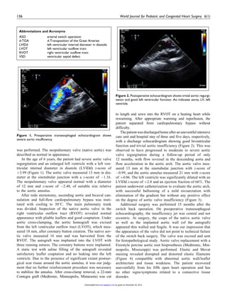
Figure 3. Image from cardiac catheterization 12 months after the
switch back operation demonstrating recurrent severe aortic valve
insufficiency.
Figure 4. Distorted and disrupted elastica is seen on the aortic side of
the aortic valve leaflet (right; elastica [Â10 original magnification]).
Brister et al 137
by guest on December 30, 2014pch.sagepub.comDownloaded from
4. echocardiogram. However, aortic insufficiency recurred and
progressed to a severe degree, which eventually required an
aortic valve replacement. Specific to our patient, no evidence
of technical failure of the switch back operation was noted at
the subsequent surgery, and the excised leaflets were found
to have distorted elastic filaments at histopathology, pointing
to a structural problem potentially leading to aortic wall and
leaflet distortion with resultant valvar dysfunction.
In conclusion, the switch back operation could be consid-
ered as an option for correction of neoaortic valve regurgitation
in patients with prior repair of d-TGA. However, reported
experience is limited. We are only aware of one previous report
of the switch back operation in a patient with previous d-TGA
and VSD. Our case provides histologic findings that suggest
abnormal elastic properties of the ‘‘native’’ aortic valve in a
patient born with d-Transposition, in whom the native aortic
valve became the neopulmonary valve after an ASO and was
later switched back into the left outflow at the time of a Switch
Back operation, followed eventually by failure with annular
dilatation and valvar insufficiency. Given the recurrence of
aortic insufficiency in our patient, despite what was initially
an apparently successful outcome, we suggest thorough consid-
eration of all risk factors before performing the Switch Back
operation in patients with d-TGA and VSD.
Author’s Note
This report has not been presented at any meeting.
Declaration of Conflicting Interests
The author(s) declared no potential conflicts of interest with respect to
the research, authorship, and/or publication of this article.
Funding
The author(s) received no financial support for the research, authorship,
and/or publication of this article.
References
1. Losay J, Touchot A, Capderou A, et al. Aortic valve regurgitation
after arterial switch operation for transposition of the great arteries.
J Am Coll Cardiol 2006;47(10): 2057-2062.
2. Bove´ T, De Meulder F, Vandenplas G, et al. Midterm Assessment
of the reconstructed arteries after the arterial switch operation. Ann
Thorac Surg. 2008;85(3): 825-830.
3. Raju V, Burkhart HM, Durham LA III, et al. Reoperation after
arterial switch: a 27-year experience. Ann Thorac Surg. 2013;
95(6): 2105-2112.
4. Hazekamp MG, Schoof PH, Suys BE, et al. Switch back: using the
pulmonary autograft to replace the aortic valve after arterial switch
operation. J Thoracic Cardiovasc Surg. 1997;114(5): 844-846.
5. Vicente W, Ferreria CA, Klamt JG, et al. The switch back ross
operation: report of two cases with good medium-to-long-term fol-
low-up. World J Ped Congenital Heart Surg. 2012;3(2): 244-248.
6. Lalezari S, Hazekamp MG, Bartelings MM, Schoof PH, Gittenber-
ger-de-Groot AC. Pulmonary artery remodeling in transposition of
the great arteries: relevance for neoaortic root dilatation. J Thorac
Cardiovasc Surg. 2003;126(4): 1053-1060.
138 World Journal for Pediatric and Congenital Heart Surgery 6(1)
by guest on December 30, 2014pch.sagepub.comDownloaded from