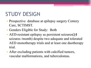
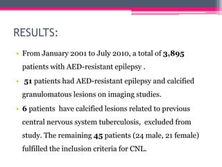
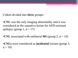
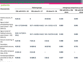
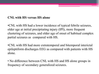
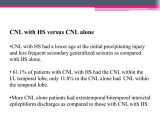
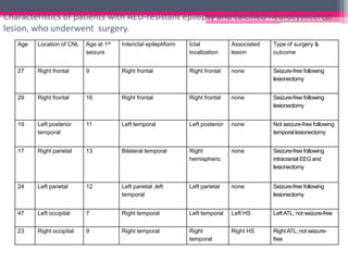
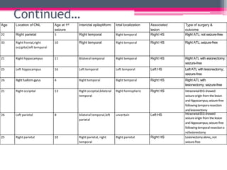
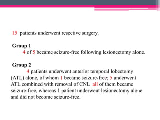
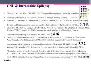
Epilepsia
Calcified neurocysticercosis lesions and
antiepileptic drug- resistant epilepsy: A
surgically remediable syndrome?
Chaturbhuj Rathore, Bejoy Thomas,
Chandrasekharan Kesavadas, Mathew
Abraham, and Kurupath Radhakrishnan
Epilepsia, 54(10):1815-1822, 2013
doi: 10.1111/epi.12349
Publised october, 2013.
BACKGROUND
• In contrast to association between acute symptomatic
seizures and neurocysticercosis, the association between
antiepileptic drug (AED)-resistant epilepsy and calcified
neurocysticercosis lesions (CNLs) is poorly understood

and controversial.
• The association between AED-resistant epilepsy and
CNLs, including the feasibility and outcome of resective
surgery.
Objectives
• To assess the causative role of CNL in AEDresistant epilepsy

• To study the factors that contribute to AED-resistant
epilepsy in patients with CNL
• To define the role of resective surgery in patients
with CNL and AED-resistant epilepsy.
STUDY DESIGN
• Prospective database at epilepsy surgery Centery
Care, SCTIMST.
• Genders Eligible for Study: Both
• AED-resistant epilepsy as persistent seizures(≥1
seizures /month) despite two adequate and tolerated
AED monotherapy trials and at least one duotherapy
trial.
• After excluding patients with calcified tumors,
vascular malformations, and tuberculomas.
Patients and Methods
• All the patients evaluated for AED-resistant epilepsy
from January 2001 to July 2010 at SCIMT and found to
have calcified lesion/s on imaging studies were selected
for this study.
• Clinical, neuroimaging, and interictal, ictal, and
intracranial electroencephalography (EEG) findings to
determine the association between CNL and epilepsy.
PARTICIPANTS:
• 45 patients who had CNL in a resident of an
endemic region.
• 18 patients had CNL plus unilateral HS and were
classified as the CNL with HS group.
• 27 patients who did not have HS, 10 patients had
causes other than CNL for their seizures (five had
malformations of cortical development, two had
nonepileptic seizures, two had severe mental
retardation, and one had benign rolandic epilepsy).
Continued…….
• 17 patients, in whom CNL was established to be
the causative lesion for AED-resistant seizures,
formed the CNL without HS (CNL alone) group.
• For the HS without CNL (HS alone) group,
randomly selected 36 patients with mesial
temporal lobe epilepsy with HS (MTLE-HS) who
had no other lesions on magnetic resonance
imaging (MRI), pathologically verified HS after
anterior temporal lobectomy, and were seizurefree for at least 5 years following surgery.
 CNL was diagnosed as the presence of solid, dense, supratentorial
calcification of 10 mm in diameter in a resident of an endemic
region
 Clinical evaluation
Age at onset of seizures.
Age at initial precipitating injury (IPI) its nature, duration of
epilepsy.
Clinical semiology of all seizure types, and any change in seizure
semiology during the course of illness.
• EEG
• At least 2 habitual seizures recorded.
• Minimum of 10 min of every hour during a 24-h
period.
• Only definite spikes or sharp waves were considered as
IEDs.
• F7, F8, T1, T2, T3, and T4 defined as temporal IEDs.
CT and MRI evaluation
•NCCT&CECT HEAD and a 1.5 T MRI

•Number and site of calcified lesion(s); size of lesions;
presence of surrounding gliosis as determined on T2weighted and FLAIR sequences; and presence of any

other potentially epileptogenic lesion including
hippocampal sclerosis (HS).
• HS defined as the presence of unequivocal atrophy
of the hippocampus with a corresponding increase in
the signal on T2-weighted and FLAIR sequences.
Additional tests
•SPECT, and intracranial monitoring.
•For calcified lesions, removal of the lesion along with one
centimeter of surrounding margin.

•All specimens histopathologically examined.
• Defined HS as the loss of neuronal cell population of ≥30%
CA1 sector of the hippocampal formation with or without

neuronal loss and gliosis involving other mesial temporal
structures.
Follow-up and seizure outcome
• At 3 months,1 year following surgery then yearly
afterwards.
•Seizure outcome at each year classified as seizurefree (free of all seizures and auras) or not seizure-free.
•All the operated patients had a minimum follow-up of
2 years.
RESULTS:
• From January 2001 to July 2010, a total of 3,895
patients with AED-resistant epilepsy .

• 51 patients had AED-resistant epilepsy and calcified
granulomatous lesions on imaging studies.
• 6 patients have calcified lesions related to previous
central nervous system tuberculosis, excluded from
study. The remaining 45 patients (24 male, 21 female)
fulfilled the inclusion criteria for CNL.
Cohort divided into three groups:
•CNL was the only imaging abnormality and it was
considered as the causative lesion for AED-resistant
epilepsy (group 1, n = 17)
•CNL associated with unilateral HS (group 2, n = 18)

•CNLs were considered as incidental lesions (group 3,
n = 10)
Comparison of group characteristics of
patients
Patient groups

Characteristic
CNL with HS N = 18

Intergroup comparisons p-valu

CNL alone N = 17

Febrile seizures, N
(%)

4 (22.2)

0

Age at initial
precipitating injury,
years (mean ± SD)
(95% CI)

5.8 ± 5.7 (3.0–8.6)

Age at onset of
habitual seizures,
years (mean ± SD)
(95% CI)

15.8 ± 6.7 (12.5–
19.1)

HS alone N = 36

19 (52.8)

CNL with HS vs. CNL
alone

CNL with HS
alone

0.103

0.043

12.7 ± 6.8 (9.2–16.2) 1.4 ± 1.0 (1.1–1.7)

0.003

0.041

12.7 ± 6.8 (9.2–16.2) 9.8 ± 5.8 (7.8–11.8)

0.183

0.002

Seizure clustering, N
5 (27.8)
(%)

7 (41.2)

2 (5.6)

0.488

0.034

Secondary
generalized seizures, 4 (22.2)
N (%)b

14 (82.4)

10 (27.8)

0.001

0.751

CNL within temporal
11 (61.1)
lobe, N (%)

2 (11.8)

–

0.004

–

Extratemporal/bitem
poral IED on EEG, N 9 (50.0)
(%)

15 (88.2)

3 (8.3)

0.027

0.001
CNL with HS versus HS alone
•CNL with HS had a lower incidence of typical febrile seizures,
older age at initial precipitating injury (IPI), more frequent
clustering of seizures, and older age of onset of habitual complex
partial seizures as compared with HS.
•CNL with HS had more extratemporal and bitemporal interictal
epileptiform discharges EEG as compared with patients with HS
alone.
• No difference between CNL with HS and HS alone groups in
frequency of secondary generalized seizures.
CNL with HS versus CNL alone
•CNL with HS had a lower age at the initial precipitating injury
and less frequent secondary generalized seizures as compared
with HS alone.
• 61.1% of patients with CNL with HS had the CNL within the
I/L temporal lobe, only 11.8% in the CNL alone had CNL within
the temporal lobe.
•More CNL alone patients had extratemporal/bitemporal interictal
epileptiform discharges as compared to those with CNL with HS.
Characteristics of patients with AED-resistant epilepsy and calcified neurocysticercal
lesion, who underwent surgery.
Age

Location of CNL

Age at 1st
seizure

Interictal epileptiform

Ictal
localization

Associated
lesion

Type of surgery &
outcome

27

Right frontal

9

Right frontal

Right frontal

none

Seizure-free following
lesionectomy

29

Right frontal

16

Right frontal

Right frontal

none

Seizure-free following
lesionectomy

19

Left posterior
temporal

11

Left temporal

Left posterior

none

Not seizure-free following
temporal lesionectomy

17

Right parietal

13

Bilateral temporal

Right
hemispheric

none

Seizure-free following
intracranial EEG and
lesionectomy

24

Left parietal

12

Left parietal ,left
temporal

Left parietal

none

Seizure-free following
lesionectomy

47

Left occipital

7

Right temporal

Left temporal

Left HS

Left ATL; not seizure-free

23

Right occipital

9

Right temporal

Right
temporal

Right HS

Right ATL; not seizurefree
Continued…
Age at 1st
seizure
5

Interictal epileptiform

Ictal localization

Right temporal

Right frontal,right
occipital,left temporal

10

21

Right hippocampus

25

Age

Location of CNL

Right temporal

Associated
lesion
Right HS

Type of surgery &
outcome
Right ATL; not seizure-free

22

Right parietal

33

Right temporal

Right temporal

Right HS

Right ATL; seizure-free

11

Bilateral temporal

Right temporal

Right HS

Right ATL with esionectomy;
seizure-free

Left hippocampus

16

Left temporal

Left temporal

Left HS

Left ATL with lesionectomy;
seizure-free

26

Right fusiform gyrus

4

Right temporal

Right temporal

Right HS

Right ATL with
lesionectomy; seizure-free

21

Right occipital

13

Right occipital,bilateral
temporal

Right hemispheric

Right HS

26

Left parietal

8

bilateral temporal,left
parietal

uncertain

Left HS

25

Right parietal

10

Right parietal, right
temporal

Right parietal

Right HS

Intracranial EEG showed
seizure origin from the lesion
and hippocampus; seizure-free
following tempora resection
and lesionectomy
Intracranial EEG showed
seizure origin from the lesion
and hippocampus; seizure-free
following temporal resection a
nd lesionectomy
Lesionectomy alone;, not
seizure-free
15 patients underwent resective surgery.
Group 1
4 of 5 became seizure-free following lesionectomy alone.
Group 2
4 patients underwent anterior temporal lobectomy
(ATL) alone, of whom 1 became seizure-free; 5 underwent
ATL combined with removal of CNL all of them became
seizure-free, whereas 1 patient underwent lesionectomy alone
and did not become seizure-free.
Pathology
• H/E of lesions showed dense calcifications ,chronic
inflammatory infiltrates and reactive gliosis.
• All the hippocampal specimens showed classical
hippocampal sclerosis without inflammation.
DISCUSSION
• Genetic, parasitic, and environmental factors contribute to the
epileptogenesis in CNL,
• Intense inflammation and perilesional gliosis associated with
a higher risk of seizures and epilepsy.
• These results shows DRE caused by CNLs is a potentially
surgically remediable syndrome with a very good chance of

seizure freefollowing lesionectomy.
• Epileptogenicity in these patients is usually restricted to the
perilesional tissue.
LACUNAE
•
•
•
•
•

Highly selected group of patients.
These results cannot be generalized.
Only presence of CNL not responsible for epilepsy.
Not establish the definite cause-effect relationship.
Not provide the true estimate of AED-resistant epilepsy caused
by CNL.
• Longitudinal long-term follow-up studies of patients with
acute neurocysticercosis in endemic areas are required to
estimate of the AED-resistant epilepsy caused by CNL.
TAKE HOME MESSAGE
• This study highlights and supports surgical therapy in
patients of AED-drug resistant epilepsy with CNL
with HS and calcified NCC.
Applicability and application
• Is the study population relevant to my practice
and patients?
Yes
• Are the patients in my practice similar to those
in the study?
Yes
Relevance
• Is this problem common in my practice?
Yes

• Will this information require to change my
current practice?
• Yes
CRITICAL APPRAISAL
• Did study address the focused question ?
yes
• Was there a control group?
yes
• Were the groups comparable at baseline ?
No
• Was outcome measured by blind assessors ?
no
CRITICAL APPRAISAL
• Did the surgery make any significant difference
in the study?
yes
• How precise were the results ?
Precise enough.
• Are all clinically relevant outcome considered?
yes
• Can results be applied on our population ?
yes
CNL & HS: Surgical Outcome
Author

Country

Follow up

HS-CNL
(n)

HS
alone(n)

Leite et al
2000

Brazil

30 months

26 / 32
(81%)

23 / 30

Velasco et
.al 2006

Brazil

2 yrs

72%

78%

Chandra
et al 2010

India

?

4/4
(100%)

Ooi et al
2011

USA

5 yrs

Chung et
1998

Korea

2yrs

CNL
alone(n)

2/2

0/1

1 /1
CNL & Intractable Epilepsy
•

Chung, C.K., Lee, S.K., Chi, J.G., 1998. Temporal lobe epilepsy caused by intrahippocampal

•
•

calcified cysticercus: a case report. Journal of Korean medical science 13, 445-448.
Rathore, C., Thomas, B., Kesavadas, C., Radhakrishnan, K., 2012. Calcified neurocysticercosis

•

lesions and hippocampal sclerosis: potential dual pathology? Epilepsia 53, e60-62.
Chandra, P.S., Bal, C., Garg, A., Gaikwad, S., Prasad, K., Sharma, B.S., Sarkar, C., Singh, M.B.,
Padma, V.M., Tripathi, M., 2010. Surgery for medically intractable epilepsy due to

•

postinfectious etiologies. Epilepsia 51, 1097-1100.
Leite, J.P., Terra-Bustamante, V.C., Fernandes, R.M., Santos, A.C., Chimelli, L., Sakamoto,
A.C., Assirati, J.A., Takayanagui, O.M., 2000. Calcified neurocysticercotic lesions and

;1

;6 /18.

; 4 /6

.; 3/32

•

postsurgery seizure control in temporal lobe epilepsy. Neurology 55, 1485-1491
Velasco, T.R., Zanello, P.A., Dalmagro, C.L., Araujo, D., Jr., Santos, A.C., Bianchin, M.M.,

Alexandre, V., Jr., Walz, R., Assirati, J.A., Carlotti, C.G., Jr., Takayanagui, O.M., Sakamoto,
A.C., Leite, J.P., 2006. Calcified cysticercotic lesions and intractable epilepsy: a cross sectional
study of 512 patients. Journal of neurology, neurosurgery, and psychiatry 77, 485-488.;

cases.

some
Calcified neurocysticercotic lesions and postsurgery
seizure control in temporal lobe epilepsy
1.J.P. Leite, MD, PhD, V.C. Terra–Bustamante, MD, R.M. F. Fernandes, MD, PhD,
A.C. Santos, MD, PhD, L. Chimelli, MD, PhD, A.C. Sakamoto, MD, PhD, J.A. Assirati, MD and O.M. Takayanagui,
MD, PhD

doi: 10.1212/WNL.55.10.1485Neurology November 28, 2000 vol. 55 no.
10 1485-1491
The presence of CCL does not influence the clinical and pathologic profile of
patients with hippocampal atrophy. Clinical histories and postsurgical outcomes
were similar to those of patients with classic HS, suggesting that the CCL is
probably, in this set of patients, a coincidental pathology and does not have a role
in epileptogenesis.
Epilepsia ppt

Epilepsia ppt

  • 1.
    Epilepsia Calcified neurocysticercosis lesionsand antiepileptic drug- resistant epilepsy: A surgically remediable syndrome? Chaturbhuj Rathore, Bejoy Thomas, Chandrasekharan Kesavadas, Mathew Abraham, and Kurupath Radhakrishnan Epilepsia, 54(10):1815-1822, 2013 doi: 10.1111/epi.12349 Publised october, 2013.
  • 2.
    BACKGROUND • In contrastto association between acute symptomatic seizures and neurocysticercosis, the association between antiepileptic drug (AED)-resistant epilepsy and calcified neurocysticercosis lesions (CNLs) is poorly understood and controversial. • The association between AED-resistant epilepsy and CNLs, including the feasibility and outcome of resective surgery.
  • 3.
    Objectives • To assessthe causative role of CNL in AEDresistant epilepsy • To study the factors that contribute to AED-resistant epilepsy in patients with CNL • To define the role of resective surgery in patients with CNL and AED-resistant epilepsy.
  • 4.
    STUDY DESIGN • Prospectivedatabase at epilepsy surgery Centery Care, SCTIMST. • Genders Eligible for Study: Both • AED-resistant epilepsy as persistent seizures(≥1 seizures /month) despite two adequate and tolerated AED monotherapy trials and at least one duotherapy trial. • After excluding patients with calcified tumors, vascular malformations, and tuberculomas.
  • 5.
    Patients and Methods •All the patients evaluated for AED-resistant epilepsy from January 2001 to July 2010 at SCIMT and found to have calcified lesion/s on imaging studies were selected for this study. • Clinical, neuroimaging, and interictal, ictal, and intracranial electroencephalography (EEG) findings to determine the association between CNL and epilepsy.
  • 6.
    PARTICIPANTS: • 45 patientswho had CNL in a resident of an endemic region. • 18 patients had CNL plus unilateral HS and were classified as the CNL with HS group. • 27 patients who did not have HS, 10 patients had causes other than CNL for their seizures (five had malformations of cortical development, two had nonepileptic seizures, two had severe mental retardation, and one had benign rolandic epilepsy).
  • 7.
    Continued……. • 17 patients,in whom CNL was established to be the causative lesion for AED-resistant seizures, formed the CNL without HS (CNL alone) group. • For the HS without CNL (HS alone) group, randomly selected 36 patients with mesial temporal lobe epilepsy with HS (MTLE-HS) who had no other lesions on magnetic resonance imaging (MRI), pathologically verified HS after anterior temporal lobectomy, and were seizurefree for at least 5 years following surgery.
  • 8.
     CNL wasdiagnosed as the presence of solid, dense, supratentorial calcification of 10 mm in diameter in a resident of an endemic region  Clinical evaluation Age at onset of seizures. Age at initial precipitating injury (IPI) its nature, duration of epilepsy. Clinical semiology of all seizure types, and any change in seizure semiology during the course of illness.
  • 9.
    • EEG • Atleast 2 habitual seizures recorded. • Minimum of 10 min of every hour during a 24-h period. • Only definite spikes or sharp waves were considered as IEDs. • F7, F8, T1, T2, T3, and T4 defined as temporal IEDs.
  • 10.
    CT and MRIevaluation •NCCT&CECT HEAD and a 1.5 T MRI •Number and site of calcified lesion(s); size of lesions; presence of surrounding gliosis as determined on T2weighted and FLAIR sequences; and presence of any other potentially epileptogenic lesion including hippocampal sclerosis (HS).
  • 11.
    • HS definedas the presence of unequivocal atrophy of the hippocampus with a corresponding increase in the signal on T2-weighted and FLAIR sequences.
  • 12.
    Additional tests •SPECT, andintracranial monitoring. •For calcified lesions, removal of the lesion along with one centimeter of surrounding margin. •All specimens histopathologically examined. • Defined HS as the loss of neuronal cell population of ≥30% CA1 sector of the hippocampal formation with or without neuronal loss and gliosis involving other mesial temporal structures.
  • 13.
    Follow-up and seizureoutcome • At 3 months,1 year following surgery then yearly afterwards. •Seizure outcome at each year classified as seizurefree (free of all seizures and auras) or not seizure-free. •All the operated patients had a minimum follow-up of 2 years.
  • 14.
    RESULTS: • From January2001 to July 2010, a total of 3,895 patients with AED-resistant epilepsy . • 51 patients had AED-resistant epilepsy and calcified granulomatous lesions on imaging studies. • 6 patients have calcified lesions related to previous central nervous system tuberculosis, excluded from study. The remaining 45 patients (24 male, 21 female) fulfilled the inclusion criteria for CNL.
  • 15.
    Cohort divided intothree groups: •CNL was the only imaging abnormality and it was considered as the causative lesion for AED-resistant epilepsy (group 1, n = 17) •CNL associated with unilateral HS (group 2, n = 18) •CNLs were considered as incidental lesions (group 3, n = 10)
  • 16.
    Comparison of groupcharacteristics of patients Patient groups Characteristic CNL with HS N = 18 Intergroup comparisons p-valu CNL alone N = 17 Febrile seizures, N (%) 4 (22.2) 0 Age at initial precipitating injury, years (mean ± SD) (95% CI) 5.8 ± 5.7 (3.0–8.6) Age at onset of habitual seizures, years (mean ± SD) (95% CI) 15.8 ± 6.7 (12.5– 19.1) HS alone N = 36 19 (52.8) CNL with HS vs. CNL alone CNL with HS alone 0.103 0.043 12.7 ± 6.8 (9.2–16.2) 1.4 ± 1.0 (1.1–1.7) 0.003 0.041 12.7 ± 6.8 (9.2–16.2) 9.8 ± 5.8 (7.8–11.8) 0.183 0.002 Seizure clustering, N 5 (27.8) (%) 7 (41.2) 2 (5.6) 0.488 0.034 Secondary generalized seizures, 4 (22.2) N (%)b 14 (82.4) 10 (27.8) 0.001 0.751 CNL within temporal 11 (61.1) lobe, N (%) 2 (11.8) – 0.004 – Extratemporal/bitem poral IED on EEG, N 9 (50.0) (%) 15 (88.2) 3 (8.3) 0.027 0.001
  • 17.
    CNL with HSversus HS alone •CNL with HS had a lower incidence of typical febrile seizures, older age at initial precipitating injury (IPI), more frequent clustering of seizures, and older age of onset of habitual complex partial seizures as compared with HS. •CNL with HS had more extratemporal and bitemporal interictal epileptiform discharges EEG as compared with patients with HS alone. • No difference between CNL with HS and HS alone groups in frequency of secondary generalized seizures.
  • 18.
    CNL with HSversus CNL alone •CNL with HS had a lower age at the initial precipitating injury and less frequent secondary generalized seizures as compared with HS alone. • 61.1% of patients with CNL with HS had the CNL within the I/L temporal lobe, only 11.8% in the CNL alone had CNL within the temporal lobe. •More CNL alone patients had extratemporal/bitemporal interictal epileptiform discharges as compared to those with CNL with HS.
  • 19.
    Characteristics of patientswith AED-resistant epilepsy and calcified neurocysticercal lesion, who underwent surgery. Age Location of CNL Age at 1st seizure Interictal epileptiform Ictal localization Associated lesion Type of surgery & outcome 27 Right frontal 9 Right frontal Right frontal none Seizure-free following lesionectomy 29 Right frontal 16 Right frontal Right frontal none Seizure-free following lesionectomy 19 Left posterior temporal 11 Left temporal Left posterior none Not seizure-free following temporal lesionectomy 17 Right parietal 13 Bilateral temporal Right hemispheric none Seizure-free following intracranial EEG and lesionectomy 24 Left parietal 12 Left parietal ,left temporal Left parietal none Seizure-free following lesionectomy 47 Left occipital 7 Right temporal Left temporal Left HS Left ATL; not seizure-free 23 Right occipital 9 Right temporal Right temporal Right HS Right ATL; not seizurefree
  • 20.
    Continued… Age at 1st seizure 5 Interictalepileptiform Ictal localization Right temporal Right frontal,right occipital,left temporal 10 21 Right hippocampus 25 Age Location of CNL Right temporal Associated lesion Right HS Type of surgery & outcome Right ATL; not seizure-free 22 Right parietal 33 Right temporal Right temporal Right HS Right ATL; seizure-free 11 Bilateral temporal Right temporal Right HS Right ATL with esionectomy; seizure-free Left hippocampus 16 Left temporal Left temporal Left HS Left ATL with lesionectomy; seizure-free 26 Right fusiform gyrus 4 Right temporal Right temporal Right HS Right ATL with lesionectomy; seizure-free 21 Right occipital 13 Right occipital,bilateral temporal Right hemispheric Right HS 26 Left parietal 8 bilateral temporal,left parietal uncertain Left HS 25 Right parietal 10 Right parietal, right temporal Right parietal Right HS Intracranial EEG showed seizure origin from the lesion and hippocampus; seizure-free following tempora resection and lesionectomy Intracranial EEG showed seizure origin from the lesion and hippocampus; seizure-free following temporal resection a nd lesionectomy Lesionectomy alone;, not seizure-free
  • 21.
    15 patients underwentresective surgery. Group 1 4 of 5 became seizure-free following lesionectomy alone. Group 2 4 patients underwent anterior temporal lobectomy (ATL) alone, of whom 1 became seizure-free; 5 underwent ATL combined with removal of CNL all of them became seizure-free, whereas 1 patient underwent lesionectomy alone and did not become seizure-free.
  • 22.
    Pathology • H/E oflesions showed dense calcifications ,chronic inflammatory infiltrates and reactive gliosis. • All the hippocampal specimens showed classical hippocampal sclerosis without inflammation.
  • 23.
    DISCUSSION • Genetic, parasitic,and environmental factors contribute to the epileptogenesis in CNL, • Intense inflammation and perilesional gliosis associated with a higher risk of seizures and epilepsy. • These results shows DRE caused by CNLs is a potentially surgically remediable syndrome with a very good chance of seizure freefollowing lesionectomy. • Epileptogenicity in these patients is usually restricted to the perilesional tissue.
  • 24.
    LACUNAE • • • • • Highly selected groupof patients. These results cannot be generalized. Only presence of CNL not responsible for epilepsy. Not establish the definite cause-effect relationship. Not provide the true estimate of AED-resistant epilepsy caused by CNL. • Longitudinal long-term follow-up studies of patients with acute neurocysticercosis in endemic areas are required to estimate of the AED-resistant epilepsy caused by CNL.
  • 25.
    TAKE HOME MESSAGE •This study highlights and supports surgical therapy in patients of AED-drug resistant epilepsy with CNL with HS and calcified NCC.
  • 26.
    Applicability and application •Is the study population relevant to my practice and patients? Yes • Are the patients in my practice similar to those in the study? Yes
  • 27.
    Relevance • Is thisproblem common in my practice? Yes • Will this information require to change my current practice? • Yes
  • 28.
    CRITICAL APPRAISAL • Didstudy address the focused question ? yes • Was there a control group? yes • Were the groups comparable at baseline ? No • Was outcome measured by blind assessors ? no
  • 29.
    CRITICAL APPRAISAL • Didthe surgery make any significant difference in the study? yes • How precise were the results ? Precise enough. • Are all clinically relevant outcome considered? yes • Can results be applied on our population ? yes
  • 31.
    CNL & HS:Surgical Outcome Author Country Follow up HS-CNL (n) HS alone(n) Leite et al 2000 Brazil 30 months 26 / 32 (81%) 23 / 30 Velasco et .al 2006 Brazil 2 yrs 72% 78% Chandra et al 2010 India ? 4/4 (100%) Ooi et al 2011 USA 5 yrs Chung et 1998 Korea 2yrs CNL alone(n) 2/2 0/1 1 /1
  • 32.
    CNL & IntractableEpilepsy • Chung, C.K., Lee, S.K., Chi, J.G., 1998. Temporal lobe epilepsy caused by intrahippocampal • • calcified cysticercus: a case report. Journal of Korean medical science 13, 445-448. Rathore, C., Thomas, B., Kesavadas, C., Radhakrishnan, K., 2012. Calcified neurocysticercosis • lesions and hippocampal sclerosis: potential dual pathology? Epilepsia 53, e60-62. Chandra, P.S., Bal, C., Garg, A., Gaikwad, S., Prasad, K., Sharma, B.S., Sarkar, C., Singh, M.B., Padma, V.M., Tripathi, M., 2010. Surgery for medically intractable epilepsy due to • postinfectious etiologies. Epilepsia 51, 1097-1100. Leite, J.P., Terra-Bustamante, V.C., Fernandes, R.M., Santos, A.C., Chimelli, L., Sakamoto, A.C., Assirati, J.A., Takayanagui, O.M., 2000. Calcified neurocysticercotic lesions and ;1 ;6 /18. ; 4 /6 .; 3/32 • postsurgery seizure control in temporal lobe epilepsy. Neurology 55, 1485-1491 Velasco, T.R., Zanello, P.A., Dalmagro, C.L., Araujo, D., Jr., Santos, A.C., Bianchin, M.M., Alexandre, V., Jr., Walz, R., Assirati, J.A., Carlotti, C.G., Jr., Takayanagui, O.M., Sakamoto, A.C., Leite, J.P., 2006. Calcified cysticercotic lesions and intractable epilepsy: a cross sectional study of 512 patients. Journal of neurology, neurosurgery, and psychiatry 77, 485-488.; cases. some
  • 33.
    Calcified neurocysticercotic lesionsand postsurgery seizure control in temporal lobe epilepsy 1.J.P. Leite, MD, PhD, V.C. Terra–Bustamante, MD, R.M. F. Fernandes, MD, PhD, A.C. Santos, MD, PhD, L. Chimelli, MD, PhD, A.C. Sakamoto, MD, PhD, J.A. Assirati, MD and O.M. Takayanagui, MD, PhD doi: 10.1212/WNL.55.10.1485Neurology November 28, 2000 vol. 55 no. 10 1485-1491 The presence of CCL does not influence the clinical and pathologic profile of patients with hippocampal atrophy. Clinical histories and postsurgical outcomes were similar to those of patients with classic HS, suggesting that the CCL is probably, in this set of patients, a coincidental pathology and does not have a role in epileptogenesis.

Editor's Notes

  • #27 Relevance- available, affordable, acceptable, feasible