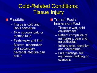
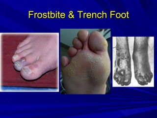
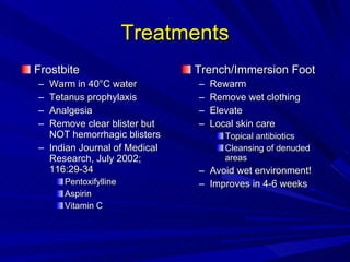
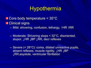
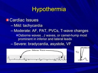
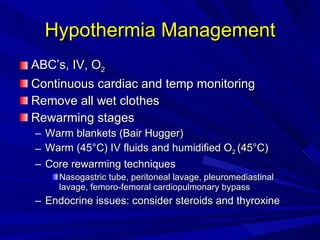
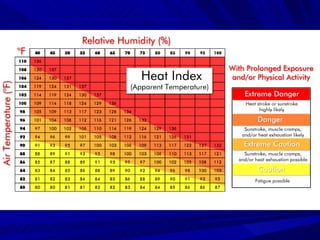
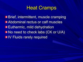
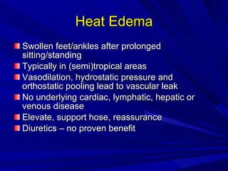
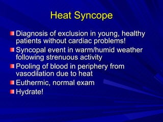
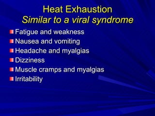
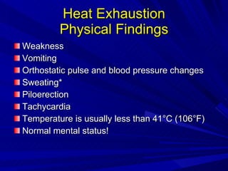
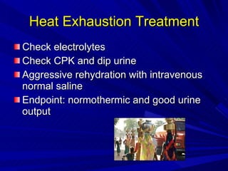
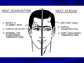
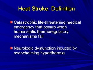
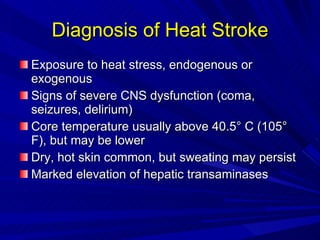
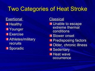
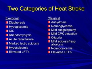
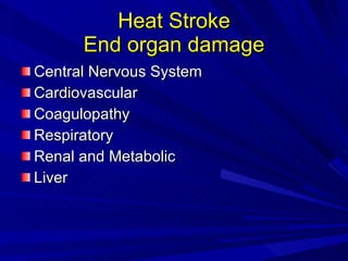
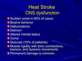
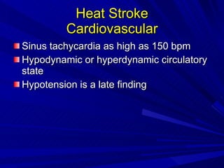
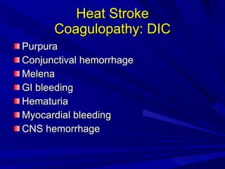
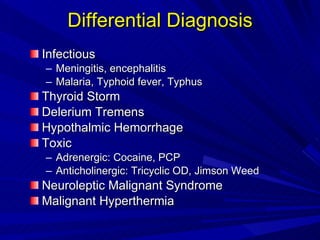
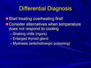
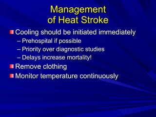
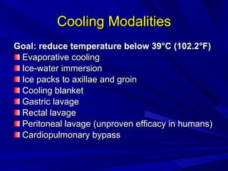
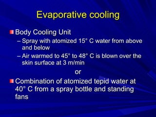
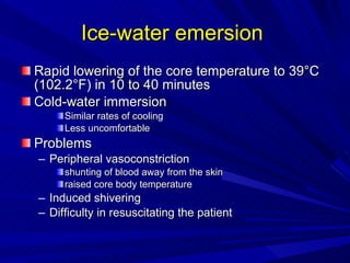
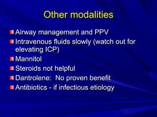
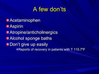
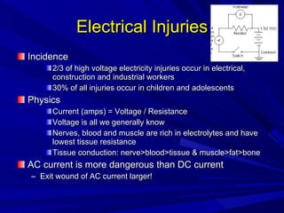
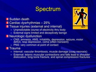
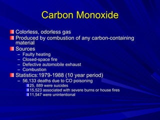
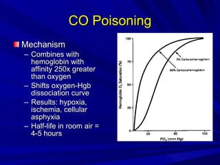
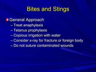
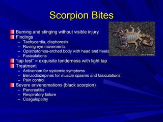
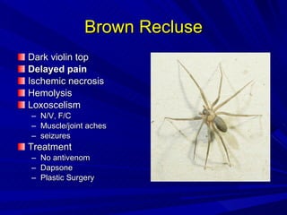
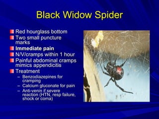
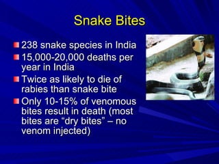
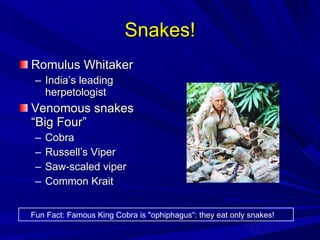
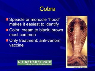
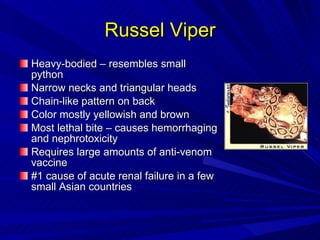
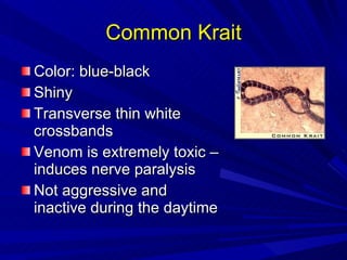
Environmental Injuries Cold-related Injuries Heat-related Injuries Electrical Injuries Submersion/Drowning CO Poisoning Bites and Stings
Cold-related Injuries
Cold-Related Conditions: Tissue Injury Frostbite Tissue is cold and lacks sensation Skin appears pale or mottled blue Feels waxy and firm Blisters, maceration and secondary bacterial infection can occur Trench Foot / Immersion Foot Tissue in wet, cold environment Patient complains of numbness, pain and paresthesias Initially pale, sensitive and edematous Later findings are erythema, mottling or cyanosis
Frostbite & Trench Foot
Treatments Frostbite Warm in 40 °C water Tetanus prophylaxis Analgesia Remove clear blister but NOT hemorrhagic blisters Indian Journal of Medical Research, July 2002; 116:29-34 Pentoxifylline Aspirin Vitamin C Trench/Immersion Foot Rewarm Remove wet clothing Elevate Local skin care Topical antibiotics Cleansing of denuded areas Avoid wet environment! Improves in 4-6 weeks
Hypothermia Core body temperature < 35 °C Clinical signs Mild: shivering, confusion, lethargy, ↑HR ↑RR Moderate: Shivering stops < 32°C, disoriented, stupor, ↓HR ↓BP ↓RR, decr reflexes Severe ( < 28 °C): coma, dilated unreactive pupils, absent reflexes, muscle rigidity, ↓HR ↓BP ↓RR,asystole, ventricular fibrillation
Hypothermia Cardiac Issues Mild: tachycardia Moderate: AF, PAT, PVCs, T-wave changes Osborne waves , J waves, or camel-hump most prominent in inferior and lateral leads Severe: bradycardia, asystole, VF
Osborne J Wave
Hypothermia Management ABC’s, IV, O 2 Continuous cardiac and temp monitoring Remove all wet clothes Rewarming stages Warm blankets (Bair Hugger) Warm (45 °C)  IV fluids and humidified O 2  (45 °C) Core rewarming techniques Nasogastric tube, peritoneal lavage, pleuromediastinal lavage, femoro-femoral cardiopulmonary bypass Endocrine issues: consider steroids and thyroxine
Hypothermia Key Concept: Afterdrop or Rewarming Shock During rewarming, the core temperature may drop! Theory Core is warmer than periphery As you warm periphery, blood vessels dilate More cold blood moves to core Prevention Surface and core rewarming should occur simultaneously
Heat-Related Conditions Internal Heat Emergencies Malignant hyperthermia Neuroleptic malignant syndrome Serotonin syndrome External Heat Emergencies Heat cramps Heat edema Heat syncope Heat exhaustion Heat stroke
Heat: A Few Facts #2 environmental killer after hypothermia USA 1936 - 1975, nearly 20,000 deaths in U.S. 1979 – 1997, nearly 7,000 deaths Great heat wave of 1980: 1,250 deaths Average year: 175 deaths in the U.S. India 1998 – heat wave killed 2600 in 10 weeks (official #) 2003 – heat wave (50 °C or 122°F)  killed > 1300 Future expectations Global warming Aging of the population Increase frequency and intensity of heat waves predicted
Risk Factors Children up to four years of age  >65 years of age  Mental or Physical Disability Overweight  Overexertion during work or exercise
 
Heat Cramps Brief, intermittent, muscle cramping Abdominal rectus or calf muscles Euthermic, mild dehydration No need to check labs (CK or U/A) IV Fluids rarely required
Heat Edema Swollen feet/ankles after prolonged sitting/standing Typically in (semi)tropical areas Vasodilation, hydrostatic pressure and orthostatic pooling lead to vascular leak No underlying cardiac, lymphatic, hepatic or venous disease Elevate, support hose, reassurance Diuretics – no proven benefit
Heat Syncope Diagnosis of exclusion in young, healthy patients without cardiac problems! Syncopal event in warm/humid weather following strenuous activity Pooling of blood in periphery from vasodilation due to heat Euthermic, normal exam Hydrate!
Heat Exhaustion Similar to a viral syndrome Fatigue and weakness Nausea and vomiting Headache and myalgias Dizziness Muscle cramps and myalgias Irritability
Heat Exhaustion Physical Findings Weakness Vomiting Orthostatic pulse and blood pressure changes Sweating* Piloerection Tachycardia Temperature is usually less than 41°C (106°F) Normal mental status!
Heat Exhaustion Treatment Check electrolytes Check CPK and dip urine Aggressive rehydration with intravenous normal saline Endpoint: normothermic and good urine output
 
Heat Stroke: Definition Catastrophic life-threatening medical emergency that occurs when homeostatic thermoregulatory mechanisms fail Neurologic dysfunction induced by overwhelming hyperthermia
Diagnosis of Heat Stroke Exposure to heat stress, endogenous or exogenous Signs of severe CNS dysfunction (coma, seizures, delirium) Core temperature usually above 40.5° C (105° F), but may be lower Dry, hot skin common, but sweating may persist Marked elevation of hepatic transaminases
Exertional  Healthy  Younger  Exercise Athletes/military recruits Sporadic  Classical Unable to escape extreme thermal conditions Slower onset Predisposing factors Older, chronic illness Sedentary Heat wave occurrence Two Categories of Heat Stroke
Two Categories of Heat Stroke Exertional Diaphoresis  Hypoglycemia  DIC  Rhabdomyolysis  Acute renal failure  Marked lactic acidosis Hypocalcemia Elevated LFT’s Classical Anhydrosis Normoglycemia Mild coagulopathy Mild CPK elevation Oliguria Mild acidosis/resp alkalosis Normocalcemia Elevated LFT’s
Heat Stroke End organ damage Central Nervous System Cardiovascular Coagulopathy Respiratory Renal and Metabolic Liver
Heat Stroke   CNS dysfunction Sudden onset in 80% of cases Bizarre behavior Hallucinations Delirium Altered mental status Coma Seizures (75% of patients) Muscle rigidity with tonic contractions, tremors, and dystonic movements  Permanent damage is common
Heat Stroke Cardiovascular Sinus tachycardia as high as 150 bpm Hypodynamic or hyperdynamic circulatory state Hypotension is a late finding
Heat Stroke Coagulopathy: DIC Purpura Conjunctival hemorrhage Melena GI bleeding Hematuria Myocardial bleeding CNS hemorrhage
Heat Stroke Respiratory Tachypnea Respiratory alkalosis Respiratory decompensation  Acute respiratory distress syndrome
Heat Stroke Renal Failure 25% of exertional and 5% of classic Splanchnic constriction Diminished renal blood flow Rhabdomyolisis Myoglobinuria Hyperuricemia and urinary acidification DIC Glomerular damage
Heat Stroke Electrolyte Disorders Respiratory alkalosis followed by severe metabolic acidosis Early hypokalemia (response to respiratory alkalosis) Later hyperkalemia (induced by rhabdomyolisis and renal failure) Hyponatremia Hypocalcemia
Heat Stroke Hepatic Damage Consistent finding Transaminase rise > 10,000 Jaundice within 24-36 hours Recovery usual but case reports of liver failure requiring transplantation
Differential Diagnosis Infectious Meningitis, encephalitis Malaria, Typhoid fever, Typhus Thyroid Storm Delerium Tremens Hypothalmic Hemorrhage Toxic Adrenergic: Cocaine, PCP Anticholinergic: Tricyclic OD, Jimson Weed Neuroleptic Malignant Syndrome Malignant Hyperthermia
Differential Diagnosis Start treating overheating first! Consider alternatives when temperature does not respond to cooling Shaking chills (rigors) Enlarged thyroid gland Mydriasis (anticholinergic poisoning)
Work-Up CT Scan Head Tox Screen LFTs Coagulation profile Electrolytes and renal function ABG Thyroid function tests
Management of Heat Stroke Cooling should be initiated immediately Prehospital if possible Priority over diagnostic studies Delays increase mortality! Remove clothing Monitor temperature continuously
Cooling Modalities Goal: reduce temperature below 39°C (102.2°F)   Evaporative cooling  Ice-water immersion Ice packs to axillae and groin Cooling blanket Gastric lavage Rectal lavage Peritoneal lavage (unproven efficacy in humans) Cardiopulmonary bypass
Evaporative cooling Body Cooling Unit Spray with atomized 15° C water from above and below Air warmed to 45° to 48° C is blown over the skin surface at 3 m/min or Combination of atomized tepid water at  40° C from a spray bottle and standing fans
Ice-water emersion  Rapid lowering of the core temperature to 39°C (102.2°F) in 10 to 40 minutes Cold-water immersion  Similar rates of cooling  Less uncomfortable Problems Peripheral vasoconstriction  shunting of blood away from the skin raised core body temperature Induced shivering Difficulty in resuscitating the patient
Other modalities Airway management and PPV Intravenous fluids slowly (watch out for elevating ICP) Mannitol Steroids not helpful Dantrolene:  No proven benefit Antibiotics - if infectious etiology
A few don’ts Acetaminophen Aspirin Atropine/anticholinergics Alcohol sponge baths Don’t give up easily Reports of recovery in patients with T 115.7 ºF
Electrical Injuries Incidence 2/3 of high voltage electricity injuries occur in electrical, construction and industrial workers 30% of all injuries occur in children and adolescents Physics Current (amps) = Voltage / Resistance Voltage is all we generally know Nerves, blood and muscle are rich in electrolytes and have lowest tissue resistance Tissue conduction: nerve>blood>tissue & muscle>fat>bone AC current is more dangerous than DC current Exit wound of AC current larger!
Spectrum Sudden death Cardiac dysrhythmias – 25% Tissue injuries (external and internal) Unpredictable course of electricity thru body External signs limited and deceptively benign Neurologic dysfunction CNS: amnesia, AMS, irritability, depression, seizure, motor deficit, resp depression, coma (often transient) PNS: very common at point of contact Trauma Internal: vascular thrombosis, muscle damage (coag necrosis) External: tetanic muscular contractions and falls lead to shoulder dislocation, long bone fracture, and spinal compression fractures
Management of High-Tension Electrical Injury Evaluate as if multiple trauma patient ABCs Immobilize cervical spine IV fluids, O 2 , monitor 12-lead ECG Extensive resuscitative measures for cardiopulmonary arrest Limb dysfunction Consider compartment syndrome Ischemic limb Tetanus
Lightning Massive DC electrical shock: 2,000 to 2 billion volts 75-300 deaths and 1500 injuries per year (USA) 20% mortality – cardiac asystole followed by resp arrest “ Flash-over” phenomenon Current passes over outside of body because of very short duration Entrance and exit burns rare 50% with structural eye lesions Cataracts, retinal detachment Hyphema, iritis Unreactive, dilated pupil from autonomic instability (not brain damage!) 50% with ruptured TM (blast/thunder component) Paraplegia and amnesia are relatively common
Lichtenberg Figures Due to Lightning Strike
Few Rules Delayed labial artery bleeding! Exposure to > 600 Volts should be monitored overnight
Submersion/Drowning Injuries Drowning: death from suffocation due to submersion Wet drowning (85-90%): fluid is aspirated Dry drowning: fluid is not aspirated; laryngospam occurs Secondary drowning: ARDS/non-cardiogenic pulmonary edema 8,000 deaths/year in the U.S. Highest incidence in 10-19 age group 40% of victims are less than 4 years old Male:Female ratio = 5:1 600x more submersion injuries than deaths Key factors Alcohol and drug use Inability to swim Seizures
Near Drowning Injuries Hypoxia Diffusion abnormalities and decreased lung compliance Surfactant inactivated/diluted by aspirated water Intrapulmonary shunting Atelectasis Bronchospasm Obstruction of alveoli by water Acidosis Respiratory cause: hypoventilation Metabolic cause: hypoperfusion leads to lactate production Cardiovascular complications Cardiac arrest and hypotension >> VF Hypothermia Rare complications Renal failure due to hypoxia or rhabdomyolysis DIC
Neurologic Injury 15-25% of near-drowning victims Cerebral edema Hypoxia Acidosis Favorable outcome Immediate CPR Hypothermia Key Fact: Of patients that present in coma with fixed and dilated pupils, 10-20% have complete recovery!!!
Submersion Injuries Evaluation History: as detailed as possible Focused physical Exam Vitals: rectal temperature! Cardiopulmonary and neuro status Evidence of trauma ECG Labs, including ABG Head CT
Submersion Injuries Management IV, O2, monitor, remove wet clothing CPR Airway Clear debris Intubate and place on PEEP Persistent hypoxia may require suctioning and bronchoscopy Albuterol for bronchospasm Consider sodium bicarbonate empirically Gastric decompression Not proven to be helpful Steroids Antibiotics Induced hypothermia
Carbon Monoxide Colorless, odorless gas Produced by combustion of any carbon-containing material Sources Faulty heating Closed-space fire Defective automobile exhaust Combustion Statistics:1979-1988 (10 year period) 56,133 deaths due to CO poisoning 25, 889 were suicides 15,523 associated with severe burns or house fires 11,547 were unintentional
CO Poisoning Mechanism Combines with hemoglobin with affinity 250x greater than oxygen Shifts oxygen-Hgb dissociation curve Results: hypoxia, ischemia, cellular asphyxia Half-life in room air = 4-5 hours
CO Poisoning CO Level Signs & Symptoms 0-10 none 10-20 headache, dyspnea on exertion 20-30 headache, dyspnea, nausea, dizziness 30-40 severe HA, vomiting, fatigue, poor judgement 40-50 confusion, syncope, tachycardia, tachypnea 50-60 syncope, seizure, coma 60-70 coma, hypotension, arrhythmias, death >70 rapidly fatal
CO Poisoning Evaluation and Management Multiple patients have similar complaints of headache and dizziness Oxygen saturation by pulse oximetry will be falsely normal! Give 100% oxygen! CO poisoning causes a metabolic acidosis No need to correct mild acidosis because shifts the curve to the right Check an ECG Consider hyperbaric oxygen therapy
Indications for Hyperbaric Oxygen Syncope Coma CO level > 40% Persistent neurologic disturbances Cardiovascular dysfunction Severe acidosis Pregnancy with CO level > 15% or signs of fetal distress
Bites and Stings General Approach Treat anaphylaxis Tetanus prophylaxis Copious irrigation with water Consider x-ray for fracture or foreign body Do not suture contaminated wounds
Bites: Cats, Dogs, Humans Infection rates Cats: 40-80% Humans: 20-40% Dogs: 5-10% Delayed primary closure Consider prophylactic antibiotics, rabies shots Special cases Cat-scratch disease “ Fight-bite”
Scorpion Bites Burning and stinging without visible injury Findings Tachycardia, diaphoresis Roving eye movements Opisthotomos-arched body with head and heels bent back Fasiculations “ tap test” = exquisite tenderness with light tap Treatment Antivenom for systemic symptoms Benzodiazepines for muscle spasms and fasiculations Pain control Severe envenomations (black scorpion) Pancreatitis Respiratory failure Coagulopathy
Spiders Brown recluse and Black widow spiders are very uncommon in India Sacred Heart College, Kerala, India Department of Zoology Southindianspiders.com
Brown Recluse Dark violin top Delayed pain Ischemic necrosis Hemolysis Loxoscelism N/V, F/C Muscle/joint aches seizures Treatment No antivenom Dapsone Plastic Surgery
Black Widow Spider Red hourglass bottom Two small puncture marks Immediate pain N/V/cramps within 1 hour Painful abdominal cramps mimics appendicitis Treatment Benzodiazepines for cramping Calcium gluconate for pain Anti-venin if severe reaction (HTN, resp failure, shock or coma)
Snake Bites 238 snake species in India 15,000-20,000 deaths per year in India Twice as likely to die of rabies than snake bite Only 10-15% of venomous bites result in death (most bites are “dry bites” – no venom injected)
Snakes! Romulus Whitaker India’s leading herpetologist Venomous snakes “Big Four” Cobra Russell’s Viper Saw-scaled viper Common Krait Fun Fact: Famous King Cobra is &quot;ophiphagus“: they eat only snakes!
Cobra Speacle or monocle “hood” makes it easiest to identify Color: cream to black; brown most common Only treatment: anti-venom vaccine
Russel Viper Heavy-bodied – resembles small python Narrow necks and triangular heads Chain-like pattern on back Color mostly yellowish and brown Most lethal bite – causes hemorrhaging and nephrotoxicity Requires large amounts of anti-venom vaccine #1 cause of acute renal failure in a few small Asian countries
Common Krait Color: blue-black Shiny Transverse thin white crossbands Venom is extremely toxic – induces nerve paralysis Not aggressive and inactive during the daytime
Saw Scaled Viper Triangular head typical of the viper Brownish with white markings Chain-like pattern common Smallest of the “big four” - about 1 foot in length Bite rarely fatal because small venom quantity

Environmental injuries

  • 1.
    Environmental Injuries Cold-relatedInjuries Heat-related Injuries Electrical Injuries Submersion/Drowning CO Poisoning Bites and Stings
  • 2.
  • 3.
    Cold-Related Conditions: TissueInjury Frostbite Tissue is cold and lacks sensation Skin appears pale or mottled blue Feels waxy and firm Blisters, maceration and secondary bacterial infection can occur Trench Foot / Immersion Foot Tissue in wet, cold environment Patient complains of numbness, pain and paresthesias Initially pale, sensitive and edematous Later findings are erythema, mottling or cyanosis
  • 4.
  • 5.
    Treatments Frostbite Warmin 40 °C water Tetanus prophylaxis Analgesia Remove clear blister but NOT hemorrhagic blisters Indian Journal of Medical Research, July 2002; 116:29-34 Pentoxifylline Aspirin Vitamin C Trench/Immersion Foot Rewarm Remove wet clothing Elevate Local skin care Topical antibiotics Cleansing of denuded areas Avoid wet environment! Improves in 4-6 weeks
  • 6.
    Hypothermia Core bodytemperature < 35 °C Clinical signs Mild: shivering, confusion, lethargy, ↑HR ↑RR Moderate: Shivering stops < 32°C, disoriented, stupor, ↓HR ↓BP ↓RR, decr reflexes Severe ( < 28 °C): coma, dilated unreactive pupils, absent reflexes, muscle rigidity, ↓HR ↓BP ↓RR,asystole, ventricular fibrillation
  • 7.
    Hypothermia Cardiac IssuesMild: tachycardia Moderate: AF, PAT, PVCs, T-wave changes Osborne waves , J waves, or camel-hump most prominent in inferior and lateral leads Severe: bradycardia, asystole, VF
  • 8.
  • 9.
    Hypothermia Management ABC’s,IV, O 2 Continuous cardiac and temp monitoring Remove all wet clothes Rewarming stages Warm blankets (Bair Hugger) Warm (45 °C) IV fluids and humidified O 2 (45 °C) Core rewarming techniques Nasogastric tube, peritoneal lavage, pleuromediastinal lavage, femoro-femoral cardiopulmonary bypass Endocrine issues: consider steroids and thyroxine
  • 10.
    Hypothermia Key Concept:Afterdrop or Rewarming Shock During rewarming, the core temperature may drop! Theory Core is warmer than periphery As you warm periphery, blood vessels dilate More cold blood moves to core Prevention Surface and core rewarming should occur simultaneously
  • 11.
    Heat-Related Conditions InternalHeat Emergencies Malignant hyperthermia Neuroleptic malignant syndrome Serotonin syndrome External Heat Emergencies Heat cramps Heat edema Heat syncope Heat exhaustion Heat stroke
  • 12.
    Heat: A FewFacts #2 environmental killer after hypothermia USA 1936 - 1975, nearly 20,000 deaths in U.S. 1979 – 1997, nearly 7,000 deaths Great heat wave of 1980: 1,250 deaths Average year: 175 deaths in the U.S. India 1998 – heat wave killed 2600 in 10 weeks (official #) 2003 – heat wave (50 °C or 122°F) killed > 1300 Future expectations Global warming Aging of the population Increase frequency and intensity of heat waves predicted
  • 13.
    Risk Factors Childrenup to four years of age >65 years of age Mental or Physical Disability Overweight Overexertion during work or exercise
  • 14.
  • 15.
    Heat Cramps Brief,intermittent, muscle cramping Abdominal rectus or calf muscles Euthermic, mild dehydration No need to check labs (CK or U/A) IV Fluids rarely required
  • 16.
    Heat Edema Swollenfeet/ankles after prolonged sitting/standing Typically in (semi)tropical areas Vasodilation, hydrostatic pressure and orthostatic pooling lead to vascular leak No underlying cardiac, lymphatic, hepatic or venous disease Elevate, support hose, reassurance Diuretics – no proven benefit
  • 17.
    Heat Syncope Diagnosisof exclusion in young, healthy patients without cardiac problems! Syncopal event in warm/humid weather following strenuous activity Pooling of blood in periphery from vasodilation due to heat Euthermic, normal exam Hydrate!
  • 18.
    Heat Exhaustion Similarto a viral syndrome Fatigue and weakness Nausea and vomiting Headache and myalgias Dizziness Muscle cramps and myalgias Irritability
  • 19.
    Heat Exhaustion PhysicalFindings Weakness Vomiting Orthostatic pulse and blood pressure changes Sweating* Piloerection Tachycardia Temperature is usually less than 41°C (106°F) Normal mental status!
  • 20.
    Heat Exhaustion TreatmentCheck electrolytes Check CPK and dip urine Aggressive rehydration with intravenous normal saline Endpoint: normothermic and good urine output
  • 21.
  • 22.
    Heat Stroke: DefinitionCatastrophic life-threatening medical emergency that occurs when homeostatic thermoregulatory mechanisms fail Neurologic dysfunction induced by overwhelming hyperthermia
  • 23.
    Diagnosis of HeatStroke Exposure to heat stress, endogenous or exogenous Signs of severe CNS dysfunction (coma, seizures, delirium) Core temperature usually above 40.5° C (105° F), but may be lower Dry, hot skin common, but sweating may persist Marked elevation of hepatic transaminases
  • 24.
    Exertional Healthy Younger Exercise Athletes/military recruits Sporadic Classical Unable to escape extreme thermal conditions Slower onset Predisposing factors Older, chronic illness Sedentary Heat wave occurrence Two Categories of Heat Stroke
  • 25.
    Two Categories ofHeat Stroke Exertional Diaphoresis Hypoglycemia DIC Rhabdomyolysis Acute renal failure Marked lactic acidosis Hypocalcemia Elevated LFT’s Classical Anhydrosis Normoglycemia Mild coagulopathy Mild CPK elevation Oliguria Mild acidosis/resp alkalosis Normocalcemia Elevated LFT’s
  • 26.
    Heat Stroke Endorgan damage Central Nervous System Cardiovascular Coagulopathy Respiratory Renal and Metabolic Liver
  • 27.
    Heat Stroke CNS dysfunction Sudden onset in 80% of cases Bizarre behavior Hallucinations Delirium Altered mental status Coma Seizures (75% of patients) Muscle rigidity with tonic contractions, tremors, and dystonic movements Permanent damage is common
  • 28.
    Heat Stroke CardiovascularSinus tachycardia as high as 150 bpm Hypodynamic or hyperdynamic circulatory state Hypotension is a late finding
  • 29.
    Heat Stroke Coagulopathy:DIC Purpura Conjunctival hemorrhage Melena GI bleeding Hematuria Myocardial bleeding CNS hemorrhage
  • 30.
    Heat Stroke RespiratoryTachypnea Respiratory alkalosis Respiratory decompensation Acute respiratory distress syndrome
  • 31.
    Heat Stroke RenalFailure 25% of exertional and 5% of classic Splanchnic constriction Diminished renal blood flow Rhabdomyolisis Myoglobinuria Hyperuricemia and urinary acidification DIC Glomerular damage
  • 32.
    Heat Stroke ElectrolyteDisorders Respiratory alkalosis followed by severe metabolic acidosis Early hypokalemia (response to respiratory alkalosis) Later hyperkalemia (induced by rhabdomyolisis and renal failure) Hyponatremia Hypocalcemia
  • 33.
    Heat Stroke HepaticDamage Consistent finding Transaminase rise > 10,000 Jaundice within 24-36 hours Recovery usual but case reports of liver failure requiring transplantation
  • 34.
    Differential Diagnosis InfectiousMeningitis, encephalitis Malaria, Typhoid fever, Typhus Thyroid Storm Delerium Tremens Hypothalmic Hemorrhage Toxic Adrenergic: Cocaine, PCP Anticholinergic: Tricyclic OD, Jimson Weed Neuroleptic Malignant Syndrome Malignant Hyperthermia
  • 35.
    Differential Diagnosis Starttreating overheating first! Consider alternatives when temperature does not respond to cooling Shaking chills (rigors) Enlarged thyroid gland Mydriasis (anticholinergic poisoning)
  • 36.
    Work-Up CT ScanHead Tox Screen LFTs Coagulation profile Electrolytes and renal function ABG Thyroid function tests
  • 37.
    Management of HeatStroke Cooling should be initiated immediately Prehospital if possible Priority over diagnostic studies Delays increase mortality! Remove clothing Monitor temperature continuously
  • 38.
    Cooling Modalities Goal:reduce temperature below 39°C (102.2°F) Evaporative cooling Ice-water immersion Ice packs to axillae and groin Cooling blanket Gastric lavage Rectal lavage Peritoneal lavage (unproven efficacy in humans) Cardiopulmonary bypass
  • 39.
    Evaporative cooling BodyCooling Unit Spray with atomized 15° C water from above and below Air warmed to 45° to 48° C is blown over the skin surface at 3 m/min or Combination of atomized tepid water at 40° C from a spray bottle and standing fans
  • 40.
    Ice-water emersion Rapid lowering of the core temperature to 39°C (102.2°F) in 10 to 40 minutes Cold-water immersion Similar rates of cooling Less uncomfortable Problems Peripheral vasoconstriction shunting of blood away from the skin raised core body temperature Induced shivering Difficulty in resuscitating the patient
  • 41.
    Other modalities Airwaymanagement and PPV Intravenous fluids slowly (watch out for elevating ICP) Mannitol Steroids not helpful Dantrolene: No proven benefit Antibiotics - if infectious etiology
  • 42.
    A few don’tsAcetaminophen Aspirin Atropine/anticholinergics Alcohol sponge baths Don’t give up easily Reports of recovery in patients with T 115.7 ºF
  • 43.
    Electrical Injuries Incidence2/3 of high voltage electricity injuries occur in electrical, construction and industrial workers 30% of all injuries occur in children and adolescents Physics Current (amps) = Voltage / Resistance Voltage is all we generally know Nerves, blood and muscle are rich in electrolytes and have lowest tissue resistance Tissue conduction: nerve>blood>tissue & muscle>fat>bone AC current is more dangerous than DC current Exit wound of AC current larger!
  • 44.
    Spectrum Sudden deathCardiac dysrhythmias – 25% Tissue injuries (external and internal) Unpredictable course of electricity thru body External signs limited and deceptively benign Neurologic dysfunction CNS: amnesia, AMS, irritability, depression, seizure, motor deficit, resp depression, coma (often transient) PNS: very common at point of contact Trauma Internal: vascular thrombosis, muscle damage (coag necrosis) External: tetanic muscular contractions and falls lead to shoulder dislocation, long bone fracture, and spinal compression fractures
  • 45.
    Management of High-TensionElectrical Injury Evaluate as if multiple trauma patient ABCs Immobilize cervical spine IV fluids, O 2 , monitor 12-lead ECG Extensive resuscitative measures for cardiopulmonary arrest Limb dysfunction Consider compartment syndrome Ischemic limb Tetanus
  • 46.
    Lightning Massive DCelectrical shock: 2,000 to 2 billion volts 75-300 deaths and 1500 injuries per year (USA) 20% mortality – cardiac asystole followed by resp arrest “ Flash-over” phenomenon Current passes over outside of body because of very short duration Entrance and exit burns rare 50% with structural eye lesions Cataracts, retinal detachment Hyphema, iritis Unreactive, dilated pupil from autonomic instability (not brain damage!) 50% with ruptured TM (blast/thunder component) Paraplegia and amnesia are relatively common
  • 47.
    Lichtenberg Figures Dueto Lightning Strike
  • 48.
    Few Rules Delayedlabial artery bleeding! Exposure to > 600 Volts should be monitored overnight
  • 49.
    Submersion/Drowning Injuries Drowning:death from suffocation due to submersion Wet drowning (85-90%): fluid is aspirated Dry drowning: fluid is not aspirated; laryngospam occurs Secondary drowning: ARDS/non-cardiogenic pulmonary edema 8,000 deaths/year in the U.S. Highest incidence in 10-19 age group 40% of victims are less than 4 years old Male:Female ratio = 5:1 600x more submersion injuries than deaths Key factors Alcohol and drug use Inability to swim Seizures
  • 50.
    Near Drowning InjuriesHypoxia Diffusion abnormalities and decreased lung compliance Surfactant inactivated/diluted by aspirated water Intrapulmonary shunting Atelectasis Bronchospasm Obstruction of alveoli by water Acidosis Respiratory cause: hypoventilation Metabolic cause: hypoperfusion leads to lactate production Cardiovascular complications Cardiac arrest and hypotension >> VF Hypothermia Rare complications Renal failure due to hypoxia or rhabdomyolysis DIC
  • 51.
    Neurologic Injury 15-25%of near-drowning victims Cerebral edema Hypoxia Acidosis Favorable outcome Immediate CPR Hypothermia Key Fact: Of patients that present in coma with fixed and dilated pupils, 10-20% have complete recovery!!!
  • 52.
    Submersion Injuries EvaluationHistory: as detailed as possible Focused physical Exam Vitals: rectal temperature! Cardiopulmonary and neuro status Evidence of trauma ECG Labs, including ABG Head CT
  • 53.
    Submersion Injuries ManagementIV, O2, monitor, remove wet clothing CPR Airway Clear debris Intubate and place on PEEP Persistent hypoxia may require suctioning and bronchoscopy Albuterol for bronchospasm Consider sodium bicarbonate empirically Gastric decompression Not proven to be helpful Steroids Antibiotics Induced hypothermia
  • 54.
    Carbon Monoxide Colorless,odorless gas Produced by combustion of any carbon-containing material Sources Faulty heating Closed-space fire Defective automobile exhaust Combustion Statistics:1979-1988 (10 year period) 56,133 deaths due to CO poisoning 25, 889 were suicides 15,523 associated with severe burns or house fires 11,547 were unintentional
  • 55.
    CO Poisoning MechanismCombines with hemoglobin with affinity 250x greater than oxygen Shifts oxygen-Hgb dissociation curve Results: hypoxia, ischemia, cellular asphyxia Half-life in room air = 4-5 hours
  • 56.
    CO Poisoning COLevel Signs & Symptoms 0-10 none 10-20 headache, dyspnea on exertion 20-30 headache, dyspnea, nausea, dizziness 30-40 severe HA, vomiting, fatigue, poor judgement 40-50 confusion, syncope, tachycardia, tachypnea 50-60 syncope, seizure, coma 60-70 coma, hypotension, arrhythmias, death >70 rapidly fatal
  • 57.
    CO Poisoning Evaluationand Management Multiple patients have similar complaints of headache and dizziness Oxygen saturation by pulse oximetry will be falsely normal! Give 100% oxygen! CO poisoning causes a metabolic acidosis No need to correct mild acidosis because shifts the curve to the right Check an ECG Consider hyperbaric oxygen therapy
  • 58.
    Indications for HyperbaricOxygen Syncope Coma CO level > 40% Persistent neurologic disturbances Cardiovascular dysfunction Severe acidosis Pregnancy with CO level > 15% or signs of fetal distress
  • 59.
    Bites and StingsGeneral Approach Treat anaphylaxis Tetanus prophylaxis Copious irrigation with water Consider x-ray for fracture or foreign body Do not suture contaminated wounds
  • 60.
    Bites: Cats, Dogs,Humans Infection rates Cats: 40-80% Humans: 20-40% Dogs: 5-10% Delayed primary closure Consider prophylactic antibiotics, rabies shots Special cases Cat-scratch disease “ Fight-bite”
  • 61.
    Scorpion Bites Burningand stinging without visible injury Findings Tachycardia, diaphoresis Roving eye movements Opisthotomos-arched body with head and heels bent back Fasiculations “ tap test” = exquisite tenderness with light tap Treatment Antivenom for systemic symptoms Benzodiazepines for muscle spasms and fasiculations Pain control Severe envenomations (black scorpion) Pancreatitis Respiratory failure Coagulopathy
  • 62.
    Spiders Brown recluseand Black widow spiders are very uncommon in India Sacred Heart College, Kerala, India Department of Zoology Southindianspiders.com
  • 63.
    Brown Recluse Darkviolin top Delayed pain Ischemic necrosis Hemolysis Loxoscelism N/V, F/C Muscle/joint aches seizures Treatment No antivenom Dapsone Plastic Surgery
  • 64.
    Black Widow SpiderRed hourglass bottom Two small puncture marks Immediate pain N/V/cramps within 1 hour Painful abdominal cramps mimics appendicitis Treatment Benzodiazepines for cramping Calcium gluconate for pain Anti-venin if severe reaction (HTN, resp failure, shock or coma)
  • 65.
    Snake Bites 238snake species in India 15,000-20,000 deaths per year in India Twice as likely to die of rabies than snake bite Only 10-15% of venomous bites result in death (most bites are “dry bites” – no venom injected)
  • 66.
    Snakes! Romulus WhitakerIndia’s leading herpetologist Venomous snakes “Big Four” Cobra Russell’s Viper Saw-scaled viper Common Krait Fun Fact: Famous King Cobra is &quot;ophiphagus“: they eat only snakes!
  • 67.
    Cobra Speacle ormonocle “hood” makes it easiest to identify Color: cream to black; brown most common Only treatment: anti-venom vaccine
  • 68.
    Russel Viper Heavy-bodied– resembles small python Narrow necks and triangular heads Chain-like pattern on back Color mostly yellowish and brown Most lethal bite – causes hemorrhaging and nephrotoxicity Requires large amounts of anti-venom vaccine #1 cause of acute renal failure in a few small Asian countries
  • 69.
    Common Krait Color:blue-black Shiny Transverse thin white crossbands Venom is extremely toxic – induces nerve paralysis Not aggressive and inactive during the daytime
  • 70.
    Saw Scaled ViperTriangular head typical of the viper Brownish with white markings Chain-like pattern common Smallest of the “big four” - about 1 foot in length Bite rarely fatal because small venom quantity

Editor's Notes

  • #3 What are the soldiers trying to prevent? Trench foot
  • #4 Destruction of tissue due to prolonged freezing temperatures Trench foot doesn’t have to be in freezing weather
  • #6 Immediate treatment of frostbite using rapid rewarming in tea decoction followed by combined therapy of pentoxifylline, aspirin &amp; vitamin C. Purkayastha SS , Bhaumik G , Chauhan SK , Banerjee PK , Selvamurthy W . Pentoxy (trental) – lowers blood viscosity Vit C – first report in Indian Journal of Medical Research in rats;
  • #12 Use a rectal thermometer to obtain core temp; ext heat emergencies are a CONTINUUM All hyperthermia is not created equal; internal = toxic-metabolic; external = environment (continuum) Malignant hyperthermia: inappropriate calcium release when exposed to sux or inhaled anesth, usually occurs in OR, muscle rigidity and acidosis, tx: stop agent, supportive care and dantrolene NMS: muscle spasticity +/- dystonia from antipsychotic med causes heat overprod; possible dantrolene/bromocriptine/amantadine, supportive care SS: MAOI plus tyramine/SSRI leads to hyoerthermia, muscle rigidity, autonomic dysfunction (triad), benzo’s and serotonin inhibitors (cyproheptadine, methysergide); should d/c MAOI 6 weeks prior to start of SSRI
  • #13 Improved mortality due to air conditioning??? Internationally: Heatstroke is uncommon in subtropical climates. The condition is recognized increasingly in countries that experience heat waves rarely (eg, Japan), and it commonly affects people who undertake a pilgrimage to Mecca, especially when the pilgrims arrive from a cold environment. In 1998, one of the worst heat waves to strike India in 50 years resulted in more than 2600 deaths in 10 weeks. Unofficial reports described the number of deaths as almost double that figure. But weather officials warn it may yet be some days before rain reaches southern states where there have been more than 1,200 heat-related deaths. The monsoon usually reaches the south-west of India in the first few days of June 2003 before a second weather front affects other parts. But this year the vital rains are expected to be at least a week late, allowing temperatures in Andhra Pradesh to soar above 50C (122F). Deaths attributed to the heat wave in the southern state now stand at 1,211, with about 100 more heat-related fatalities reported across India. The city of Chennai in southern India experienced for  the first time a high incidence of  heat stroke in the summer of 1998 during an unprecedented 23-day long spell of hot and humid climate heat wave ever recorded in the city. (Mean maximum temperature=41.3 C and mean relative humidity=74.6%). During the 23-day spell of heat wave 81 cases of heat stroke were seen at two hospitals in the city from which this data was available to me. The emphasis of this brief report  is on the occurrence of heatstroke in schizophrenia patients. Many of these subjects (60 /81) had some pre-existing chronic medical illnesses like diabetes mellitus, organic brain disease and cardiovascular disorders and nearly all of them (90%) were receiving medication for the medical conditions. There were nine patients (6men and 3 women) on treatment for schizophrenia. Medical conditions like diabetes, organic brain syndromes and cardiovascular disorders and drugs like beta- blockers and psychotropic drugs are known to increase risk for developing heat stroke ( Keatinge 1987; Yarbrough 1992).   The mean age of the patients was 63.6 years, an age group with known vulnerability to heat stroke (Yarbrough 1992). The significant observation was that the patients of schizophrenia were  much young (mean 43.5 years) in striking contrast to 61.3 years of the diabetics, 67.2 years of those with organic brain disorders 73.4 years of those with cardiovascular disorders and 66.3 years of those with no pre-existing medical illness.   On this comparison of persons who were healthy before sunstroke and those with medical illness patients with patients of schizophrenia it appears that the vulnerability to heat stroke in the latter is not merely due to drugs and old age. Does the illness and its associated neuropathology itself increase the vulnerability of schizophrenia patient to heatstroke? A neurotransmitter substrate (e.g. dopamine) common for schizophrenia as well as dysfunctional heat regulation might account for the vulnerability of patients of schizophrenia to heat stroke even when they are young.
  • #15 The city of Chennai in southern India experienced for  the first time a high incidence of  heat stroke in the summer of 1998 during an unprecedented 23-day long spell of hot and humid climate heat wave ever recorded in the city. (Mean maximum temperature=41.3 C or 106.3 F and mean relative humidity=74.6%).
  • #16 We’ve all experienced this. Give gatorade!
  • #17 euthermic
  • #19 Hot environment, h/o inadequate PO intake
  • #20 Gradual onset Sweating is spiral downward: sweat to cool skin but losing water  dehydration inevitable
  • #22 Most important slide! Anhydrosis: Late finding. &gt;50% actually are diaphoretic; More common with exertional heat stroke
  • #23 CNS dysfunction is the hallmark of heat stroke
  • #25 Heat overproduction vs. patients who poorly dissipate heat Both acute onset relative to heat exhaustion
  • #26 Hypoglycemia from impaired gluconeogenesis
  • #27 CNS dysfunction is the hallmark!
  • #31 The whole spectrum possible
  • #39 Evaporation: spray with water mist; evaporatiion 7x more effective than ice packing; maintains vasodilation What’s the problem with ice-water immersion? Logistics! How do you resuscitate?!
  • #42 IVF: approx. 2L in first 4 hours Mannitol to decrease cerebral edema and increase renal blood flow
  • #43 Acetaminophen – liver issue Aspirin – aggravate bleeding Atropine/anticholinergics – decrease sweating Alcohol sponge baths – ineffective and toxic Don’t give up easily Reports of recovery in patients with T 115.7 ºF
  • #44 Amps or current more important than voltage. Voltage&gt;1000 considered high tension. Damage depends on resistance as well: finger worse than arm/leg which is worse than torso (make sense?) 60 Electrocuted at Wedding in India AP Online; May 26, 2002  ... they were in hit a high-voltage power wire in northern India, news reports said. Two passengers who fell out of the bus survived, Press Trust of India news agency reported. The accident happened in a village in the Bahraich district in Uttar Pradesh state, 280 miles east of New Delhi ...
  • #45 Cardiac issues when transthoracic path of electricity Muscle damage can cause rhabdo which can cause renal failure
  • #46 Lots of fluids – follow urine output and renal failure; treat like severe burn
  • #47 Resp arrest usually persists past cardiac arrest resulting in death
  • #48 Lichtenberg Figures are branching, tree or fern-like patterns that form as the result of high voltage discharges on, or within, insulating materials ( dielectrics ). The first Lichtenberg Figures were actually 2-dimensional patterns formed in dust on the surface of  a charged plate in the laboratory of their discoverer, the German physicist  Georg Christoph Lichtenberg (1742-1799
  • #51 This is what we mostly care about as emergency physicians Worse the acidosis, worse the prognosis BUT profound acidosis is still compatible with full recovery
  • #52 Kansas City drowning. Child under for 20 minutes. Died in the hospital after a few days.
  • #57 Level is weakly correlated with symptoms.
  • #58 ECG especially in important in pts with cardiac risk factors.
  • #64 Pt may not recall being bitten Lox occurs 1-3 days after envenomation
  • #67 Romulus Whitaker, India&apos;s leading herpetologist, has been doing a survey of the incidence of snakebites in India. He points out that there are basically four venomous snakes that feature in this — the cobra, the Russell&apos;s viper, the saw- scaled viper and the krait. He calls them the &amp;quot;Big Four&amp;quot;. Excerpts from an interview withS. THEODORE BASKARAN. In some parts of the word, especially in India , snake charming is a roadside show performed by a charmer. In this the &amp;quot;snake charmer&amp;quot; carries a basket that contain a snake which he &amp;quot;charms&amp;quot; by singing tunes from his flute like musical instrument, to which the snake responds. However snakes are deaf they cannot hear the charmer&apos;s unique flute. Researchers have pointed out that these snake charmers are good sleight-of-hand artists. The snakes moves corresponding to the flute movement and the vibrations from the tapping of the charmer&apos;s foot which is not noticed by the public. They rarely catch their snakes and the snakes are either nonvenomous or defanged cobras. Sometimes these people exploit the fear of snakes by released snakes into the neigbourhood and claim to rid the residence of snakes. Other snake charmers also have a snake and mongoose show where both the animals have a mock fight.
  • #69 Russell&apos;s viper, Vipera russelli (Shaw), is distributed erratically in 10 south Asian countries and is a leading cause of fatal snake bite in Pakistan, India, Bangladesh, Sri Lanka, Burma and Thailand. In Burma it has been the 5th most important cause of death. Its venom is of great interest to laboratory scientists and clinicians. The precoagulant activity of the venom was used by Macfarlane and others to elucidate the human clotting cascade. Up to 70% of the protein content is phospholipase A2, present in the form of at least 7 isoenzymes. Possible clinical effects of the enzyme include haemolysis, rhabdomyolysis, pre-synaptic neurotoxicity, vasodilatation and shock, release of endogenous autacoids and interaction with monoamine receptors. Russell&apos;s viper bite is an occupational hazard of rice farmers throughout its geographical range. Defibrination, spontaneous haemorrhage, shock and renal failure develop with frightening rapidity. In several countries, Russell&apos;s viper bite is the commonest cause of acute renal failure. There is a fascinating geographical variation in the clinical manifestations, doubtless reflecting differences in venom composition. Conjunctival oedema is unique to Burma, acute pituitary infarction to Burma and south India, and rhabdomyolysis and neurotoxicity to Sri Lanka and south India. Treatment with potent specific antivenom rapidly controls bleeding and clotting disorders, but may not reverse nephrotoxicity and shock. Causes of death include shock, pituitary and intracranial haemorrhage, massive gastrointestinal haemorrhage and acute tubular necrosis or bilateral renal cortical necrosis. The paddy farmer and the Russell&apos;s viper coexist in fragile symbiosis. The snake controls rodent pests but inevitably interacts with man, often with mutually disastrous results.