Pests and Diseases of Silkworms
•
1 like•2,246 views

1) The document discusses pests and diseases that affect silkworms, including parasitoids like the Indian uzi fly (Exorista bombycis), which lays eggs on silkworms. 2) It also discusses viral diseases like nuclear polyhedrosis virus and cytoplasmic polyhedrosis virus, which cause symptoms like sluggishness and loss of appetite in silkworms. 3) Bacterial diseases discussed are flacherie, caused by streptococci and staphylococci bacteria, and fungal diseases like white muscardine caused by the fungus Beauveria bassiana. The document provides details on the life cycles, symptoms and control measures of these pests

Recommended
Recommended
More Related Content
What's hot
What's hot (20)
Similar to Pests and Diseases of Silkworms
Similar to Pests and Diseases of Silkworms (20)
More from Asst Prof SSNAIK ENTO PJTSAU
More from Asst Prof SSNAIK ENTO PJTSAU (20)
Recently uploaded
Recently uploaded (20)
Pests and Diseases of Silkworms
- 1. Course In-Charge Mr.S.Srinivasnaik Assistant Professor Department of Entomology LECTURE No.13 PESTS AND DISEASES OF SILKWORMS ENTO 332 (1+1):Management of Beneficial Insects
- 2. PARASITOIDS 1.Uzi Fly Based on geographic distribution and hosts, four kinds of Uzi flies are known to infest and kill silkworms a) Japanese uzi fly , Crossocoamia sericariae b) Hime uzi fly, Ctenophorocera pavida c) Indian uzi fly, Exorista bombycis d) Tasar uzi fly, Blepharipa zobina
- 3. Indian uzi fly: Exorista bombycis ✓ Indian uzi fly was found parasitizing mulberry silk worm in Karnataka during May, 1980. It has been causing 40-75% loss of cocoon crop. The Indian uzi fly is known to infest 44 species of Lepidopteran caterpillars belonging to 36 genera including silk worms.
- 4. The uzi fly complete 2 generations within the host larval period. It prefers III, IV and early V instar larvae for oviposition. It does not lay eggs on silk worms settled for moult and spinning worms (ripened) on mountages. ✓ The fly lays 300-1000 creamy white eggs singly mostly on ventral surface of inter segmental regions. The eggs hatch in 2-3 days and the maggots bore into the body of the silkworm. The maggots have 3 instars and the maggot period is 5-8 days and the mature maggot pierces the integument and pupates outside on the floor in 10-20 days. The pupal period lasts 10-12 days.
- 6. Symptoms: ✓ Black spots can be observed on the inter segmental surface of larvae from where the maggots enter into the body of the silk worm. White creamy oval shaped eggs can also be observed on the skin of infested larva.
- 7. Control measures: a) Use of Nylon net (40-70 mesh) to prevent entry of uzi fly into rearing house. b) Application of uzicide as mentioned below
- 8. c) Dusting with levigated china clay with a muslin cloth @ 3g/100 larvae before mounting and 4 grams /sq. ft on bamboo mountages to prevent uzi fly attack during spinning. d) Collection and destruction of uzi fly affected silkworm larvae and cocoons. e) Destruction of uzi fly maggots and pupae collected from rearing trays, mountages, cracks and crevices in the floor of rearing house. f) Application of Diflubenzuron (Dimilin 25 WP) with levigated China clay as diluent (1:9) on third instar maggots
- 11. PREADATORS 1.Dermestid beetles Anhrenus sp Attagenus sp. D.Valpinus Dermestes cadverinus Trogoderma versicolor (Dermestidae: Coleoptera) Stored cocoons after stiffling Control Cleaning of rearing house and cocoon store room. Do not store rejected cocoons and perished eggs for long time. Fumigate the dried cocoon storage rooms with Methyl bromide @ 0.5 kg/283 m2 for a day or with chloropicrin @ 0.5 kg/283m2 for 3 days.
- 12. 2. Mites, Pediculoides ventricosus Larvae, pupae and adults-death. Black specks Loose appetite, Inactive and have difficulty in excreting Excreta is attached bead like to the anus. Vomit yellowish green fluid. The mite takes nutrition from silk worm and gives out a toxic substance which kills the silkworm Control Symptoms Avoid the storage of wheat/rice straw near rearing house, Treat the building and thatched materials with acaricide or fumigant before use
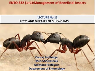
- 13. 3.) Ants They attack silkworms in rearing trays. They can be prevented by placing ant wells with water below the rearing shelves. 4.) Nematode, Hexamermis microamphidis Silkworms of late autumn rearing. The nematode attacks the young stage larvae and penetrates into their bodies. The head of affected silkworm becomes transparent and the body milky white
- 14. DISEASES OF MULBERRY SILKWORM Pebrine, grasserie, flacherie and muscardine 1. Pebrine : Protozoan parasite, Nosema bombycis Pebrine disease is also known as Pepper disease or corpuscle disease De Quatrifuges (1860) gave the popular name pebrine Pasteur (1885) a) By contact with the diseased silkworms. b) By ingestion of contaminated food. c) By transovarial transmission
- 15. Symptoms of infection Egg stage a) Few number of eggs on egg cards b) Overlapping of eggs one over the other, instead of closely side by side c) Easy to detach eggs from egg card due to lack of adhesiveness. Eggs to egg card. d) Poor/less egg hatching. Larval Stage a) No external symptoms in early stages of disease b) With advance in disease, larvae become sluggish and dull. c) Poor apatite, retarded growth resulting in irregular moulting. d) Presence of larvae in unequal size in the rearing bed. e) Appearance of irregular dark brown spots or black spots on the body of larvae. f) Hanging down of the head, instead of holding up. g) Infected larvae may die before spinning or it may spin only a poor and flimsy cocoon. h) Affected larvae lack luster and in later instars turn rusty brown.
- 16. Pupal stage a) The abdominal area is soft, swollen and dark in colour. b) Black spots present on sides of the abdomen. c) Pupa looses its luster and become dull in its movements. Adult Stage a) Discolouration of scales on abdominal area b) Black spots may be seen on abdomen c) Deformed wings d) Distorted antennae e) Low fecundity
- 17. Prevention and control : a) Production and supply of disease free layings b) Surface sterilization of disease free layings by dipping the egg cards in 2% formalin and washing in running water. c) Maintenance of strict sanitation, hygienic rearing. d) Destruction of diseased material. e) Disinfection of rearing rooms and appliances 3 methods of moth examination a) Individual moth examination b) Sample testing c) Mass examination of moths
- 18. VIRAL DISEASES Silkworm viruses are two types a) Occluded (Inclusion type) Nuclear polyhedrosis virus (NPV) – Baculoviridae Cytoplasmic polyhedrosis virus (CPV) - Reoviridae b) Non occluded (Non inclusion type) Inflections flacherie (IFV) – Unclassified small RNA viruses Densonucleosis (DNV) – Parvoviridae Kenchu virus
- 19. Nuclear polyhedrosis virus Caused by borrelina virus Polyhedrons are hexagonal or octagonal Measure 0.5 to 8.0 μ in size. Virion measures 400 x 80 nm The polyhedra engulfed along with food will get dissolved in midgut and virions infect the midgut cells. Later, they enter haemocoel and invade fat bodies, silk glands, trachea, haemocytes and the polyhedral formation occurs inside the nucleus of the target cells. Fourth and fifth instar larvae are more susceptible
- 20. Symptoms No external symptoms in early stage of disease Larvae become sluggish and lose appetite Swelling of inter segmental region Shining yellowish, fragile skin without elasticity Larvae become restless Crawling out of larvae from rearing tray and falling down, thus resulting in rupturing of fragile skin, there by releasing turbid dirty whitish haemolymph containing numerous Polyhedra The diseased larvae lose the holding power of legs except the last pair with which they hang head downwards.
- 21. Predisposing factors High temperature, high humidity and their fluctuation in rearing room. b) Excess moisture in leaf and rearing bed c) Insufficient ventilation d) Overcrowding during rearing e) Inferior quality mulberry leaves f) Feeding mature leaf followed by tender leaf g) Feeding leaf with accumulated dew drops and rain drops Prevention and control 1. Strictly avoiding the predisposing factors 2. Surface sterilization of eggs with 2% formalin 3. Use of bed disinfectants like dusting reshamkeetoushad @ 2 2.5 g/sq. ft during chawki rearing and @ 3.5-4.5 g/sq. ft during late age rearing once after each moult.
- 22. Cytoplasmic polyhedrosis virus (CPV) : (Bilisappe, Sappe) Caused by smithia virus Polyhedra of this virus are tetragonal or hexagonal and measure 3-10 μ in size. Virions are spherical and measure 60 nm in diameter Virus invades the posterior part of midgut epithelium. The cylindrical/columnar cells are infected. Polyhedra are formed in the cytoplasm showing characteristic chalky white appearance of the whole midgut. Due to increased pressure, the cell walls break and numerous polyhedra are released into the lumen of midgut which pass through excreta and further contaminate the mulberry leaves in bed Symptoms Early instar larvae more susceptible. Loss of appetite Retarded growth and development followed by vomiting gastric juice and diarrhea.
- 23. Prevention and control Thorough disinfection of rearing room and equipment Rearing under hygienic conditions and feeding good quality mulberry leaves Densonucleosis virus (DV) Caused by a parvo virus measuring 20 nm in diameter. It invades the nucleus of the cylindrical cells of midgut epithelium. Late age worms more susceptible Symptoms Infected nucleus gets swollen considerably.
- 24. Gattine Caused by a non inclusion type virus (Sub microscopic and a bacterium, Streptococcus bombycis). Also called as Luzette or Clarette. Popular names are Salpa (W.Bengal) and Hasirumoto (Karnataka). Virus affects the epithelial cells of midgut followed by bacterial infection Predisposing factors : Fecal matter containing virus excreted by infected larvae cause contamination. Symptoms : 1. Infected larvae show translucent cephalothorax. (Since the gut is devoid of mulberry leaf) 2. Diarrhoea 3. Vomit alkaline liquid
- 25. Prevention and control : 1) Rearing under hygienic conditions 2) Proper disinfection Infectious Flacherie : Caused by mortar virus It is spherical and measures 27± 2 nm in diameter. It invades the goblet cells of anterior midgut epithelium and multiplies in cytoplasm Symptoms : Stunted larval growth Translucent cephalothorax and shrinkage of body Flaccidity of the body Frequent evacuation of semisolid and whitish fecal matter Soiling of anal region Excreta in the form of chain with beads Rectal protrusion Whitish midgut
- 26. Predisposing factors : Mulberry leaves with poor nutrition Feeding with yellowish/soiled leaves or leaves grown in shade/contaminated with pesticides High temperature and humidity of rearing room Improper ventilation and accumulation of poisonous gases Prevention and control : Avoid predisposing factors Disinfection of beds with RKO Disinfection of rearing room and appliances with 2-4 per cent formalin Destruction of infected larvae, fecal matter and bed refuse by burning
- 27. These are referred to as Flacherie It is of three types a) Bacterial diseases of digestive organs – Caused by Streptococci Coli aerogenous bacilli Proteus group bacilli b) Bacterial toxicosis - Also called as sotto disease - Caused by Bacillus thuringiensis whose spores produce toxic substances and affect nervous system leading to spasm and paralysis. C) Septicemia – Caused by Sreptococci and Staphylocci BACTERIAL DISEASES
- 28. Predisposing factors : a) Diseased silkworms b) Faecal matter c) Contaminated mulberry leaves d) Rearing appliances e) Wide fluctuation in temperature and humidity Symptoms : a) Larvae become sluggish b) Poor appetite c) Retarded growth d) Body shrinkage e) Vomiting of gut juices f) Excretion in the form of beads/chains g) Body of dead larva turns black and emit a foul smell
- 29. Prevention and control : a) Maintenance of hygienic condition in rearing room b) Avoid the predisposing factors c) Avoid fluctuation of temperature and humidity FUNGAL DISEASES White muscardine – Beauviria bassiana Also known as Calcino. Infection occurs through skin Life cycle : 4-10 days Symptoms a) Presence of oily specks on body b) Infected larvae lose appetite and become sluggish c) After death, larvae become mummified and gets hardened d) Body covered with white powdery conidia
- 30. Predisposing factors Diseased worms, faecal matter Prevention and control : 1) Disinfection of room/equipment with 2% formalin / 5% bleaching powder 2) Reduce humidity in bed with lime powder 3) Application of formalin chaff @ 0.4, against I and II instars and 0.5, 0.6 and 0.8%, against III, IV and V instars, respectively Other muscardine diseases a)Green muscadine : Metarrhizium anisopliae b) Yellow muscardine : Paeciliomyces farinosus c) Brown muscardine : Aspergillus falvus..
- 36. REFERENCES: •Abrol, D.P.2010. Bees and Bee keeping in India. Kalyani Publishers, Ludhiana. Pp450 •David, B.V and Kumara Swami, T. 2016. Elements of Economic Entomology, Popular Book Depot, Madras. Pp536 •Ganga, G and Sulochana Chetty, J. 2008. An introduction to sericulture. Oxford and IBH Publishing Co.Pvt.Ltd., New Delhi. Pp160 •Gautam, R.D.2008. Biological Pest Suppression •Ghorai, N. 1995. Lac culture in India. International Books & Periodicals Supply Service. •Jolly, M.S. 1987. Appropriate sericulture techniques . International center for training and research in tropical sericulture, Mysore. Pp209 •Krishnaswami, S., Narasimma, M.N., Suryanarayan, S.K and Kumararaj,S. 1995. Silkworm Rearing. Sericulture Manual 2. Oxford and IBH Publishing Co.Pvt.Ltd., New Delhi. Pp150 •Mishra, R.C.1995. Honeybees and their management in India. ICAR, New Delhi. •Patnaik, R.K.2008. Mulberry Cultivation •Rangaswami, G., Narasimhanna, M.N., Kasiviswanatham, K., Sastry, C.R and Jolly, M.S. 1995. Mulberry Cultivation. Sericulture Manual 2. Oxford and IBH Publishing Co.Pvt.Ltd.,New Delhi. Pp150 •Sailesh Chattopadhyay. 2011. Introduction to lac and lac culture. Tech. Bulletin.FBTI:01/2011 •Abrol, D.P.2010. Bees and Bee keeping in India. Kalyani Publishers, Ludhiana. Pp450 •David, B.V and Kumara Swami, T. 2016. Elements of Economic Entomology, Popular Book Depot, Madras. Pp536 •Ganga, G and Sulochana Chetty, J. 2008. An introduction to sericulture. Oxford and IBH Publishing Co.Pvt.Ltd., New Delhi. Pp160 •Gautam, R.D.2008. Biological Pest Suppression
- 37. MARKS ALLOTMENT 1. Mid semester Examination : 50M 2. Final Practical Examination : 25M 3. Class Work : 25M Record: 10M Observation Notes: 5M Attendance: 3M Discipline and attentive and answering: 2M Internal Tests: 5M 4. Assignment : 25M Power Point Presentation:10M Model preparation:5M Photo frame/Laminations :5M Internal Write ups:5M
- 38. “Strive hard to make farming profitable and sustainable for the prosperous Nation”