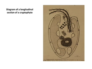
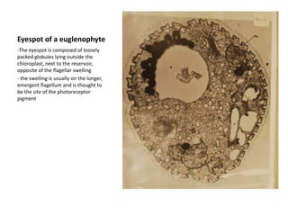
Diagram of a longitudinal section of a cryptophyte
Sieve elements of Nereocystis
Diagram of a brown algal cell
Pyrenoids of a brown algal cell
Chloroplast of Fucus
Electron micrograph of a cryptophyteThe large ejectosomes seen here line the wall of the gullet
Part of a smaller ejectosome is visible just beneath the plasmalemma on the lower left side of the cellA Cryptophyte eyespotThese eyespots may be one or several layers thick
they are located with in the chloroplast but not close to the flagella
the eyespot operates by either intercepting light (shading) or reflecting light (increasing the illumination) onto the photoreceptor pigment
which is probably localized in either the plasma membrane or chloroplast membranes over the eyespotInterphase nucleus and chromosomes of a dinoflagellateThe chromosomes lack histones, are permanently condensed and have a characteristic banded appearance
There is a large nucleolus within the nucleusInterphase nuclei of a green alga and a euglenophyteNote the condense chromosomes of the euglenophyteEyespot of a euglenophyteThe eyespot is composed of loosely packed globules lying outside the chloroplast, next to the reservoir, opposite of the flagellar swelling
the swelling is usually on the longer, emergent flagellum and is thought to be the site of the photoreceptor pigmentEuglena- Diagrammatic longitudinal section
Euglena longitudinal section- Note the distinctive outer covering, the pellicle
Outer view of a haptophyte- The cell surface is completely encased in coccoliths , special organic scales encrusted with calcium carbonate
Electron micrograph of Chrysochromulina- This photograph shows the two flagella and a haptonema, the latter lightly coiled at the tip
Several cross-sections through a haptonema- Note the outer sheath of three concentric membranes and the inner circle of 6 or 7 microtubules
Sections through parts of chloroplasts from 3 algal groups-upper photo: chrysochromulina (haptophyte)PER surrounds chloroplast
chloroplast envelope has 2 membranes
Middle photo: dinobryon (chrysophyte)
has a girdle lamella
PER surrounds chloroplast
chrloroplast envelope has 2 membranes

EMG photos

  • 1.
    Diagram of alongitudinal section of a cryptophyte
  • 2.
  • 3.
    Diagram of abrown algal cell
  • 4.
    Pyrenoids of abrown algal cell
  • 5.
  • 6.
    Electron micrograph ofa cryptophyteThe large ejectosomes seen here line the wall of the gullet
  • 7.
    Part of asmaller ejectosome is visible just beneath the plasmalemma on the lower left side of the cellA Cryptophyte eyespotThese eyespots may be one or several layers thick
  • 8.
    they are locatedwith in the chloroplast but not close to the flagella
  • 9.
    the eyespot operatesby either intercepting light (shading) or reflecting light (increasing the illumination) onto the photoreceptor pigment
  • 10.
    which is probablylocalized in either the plasma membrane or chloroplast membranes over the eyespotInterphase nucleus and chromosomes of a dinoflagellateThe chromosomes lack histones, are permanently condensed and have a characteristic banded appearance
  • 11.
    There is alarge nucleolus within the nucleusInterphase nuclei of a green alga and a euglenophyteNote the condense chromosomes of the euglenophyteEyespot of a euglenophyteThe eyespot is composed of loosely packed globules lying outside the chloroplast, next to the reservoir, opposite of the flagellar swelling
  • 12.
    the swelling isusually on the longer, emergent flagellum and is thought to be the site of the photoreceptor pigmentEuglena- Diagrammatic longitudinal section
  • 13.
    Euglena longitudinal section-Note the distinctive outer covering, the pellicle
  • 14.
    Outer view ofa haptophyte- The cell surface is completely encased in coccoliths , special organic scales encrusted with calcium carbonate
  • 15.
    Electron micrograph ofChrysochromulina- This photograph shows the two flagella and a haptonema, the latter lightly coiled at the tip
  • 16.
    Several cross-sections througha haptonema- Note the outer sheath of three concentric membranes and the inner circle of 6 or 7 microtubules
  • 17.
    Sections through partsof chloroplasts from 3 algal groups-upper photo: chrysochromulina (haptophyte)PER surrounds chloroplast
  • 18.
  • 19.
  • 20.
    has a girdlelamella
  • 21.
  • 22.