Anatomy of the Spleen: Structure, Blood Supply, and Development
•Download as PPTX, PDF•
10 likes•19,600 views

Anatomy

Recommended
More Related Content
What's hot
What's hot (20)
Similar to Anatomy of the Spleen: Structure, Blood Supply, and Development
Similar to Anatomy of the Spleen: Structure, Blood Supply, and Development (20)
More from Dr. Mohammad Mahmoud
More from Dr. Mohammad Mahmoud (20)
Recently uploaded
Recently uploaded (20)
Anatomy of the Spleen: Structure, Blood Supply, and Development
- 1. Anatomy of spleen Dr. Mohammed Mahmoud Mosaed
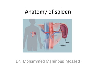
- 2. Spleen • The spleen is reddish in color • It is the largest lymphatic organ in the body. • Site Spleen is located in left upper part of abdomen (left hypochondrium) just beneath the left half of the diaphragm
- 3. Surface anatomy of the spleen • The long axis of spleen lies along the shaft of the 10th rib, vertically it is situated between the upper border of the 9th and the lower border of the 11th ribs. • The highest point (upper extremity) is at the level of the tip of the ninth thoracic spine and the lowest point (lower extremity) is in the midaxillary line at the level of the first lumbar spine. 9th 10th 11th
- 5. Coverings and ligaments of Spleen The spleen is covered by fibroelastic capsule and is surrounded by peritoneum except at the splenic hilum Ligaments of spleen: 1. gastrosplenic omentum (ligament) The spleen is surrounded by peritoneum, which passes from it at the hilum to the greater curvature of the stomach (carrying the short gastric and left gastroepiploic vessels). 2. Splenicorenal (leinorenal) ligament It is the fold of peritoneum that connects spleen to the left kidney. (carrying the splenic vessels and the tail of the pancreas).
- 7. Anatomical description of spleen • The spleen have: • 2 borders: Superior and inferior • 2 surfaces: diaphragmatic and visceral • 2 poles: posterior (medial) and anterior (lateral)
- 8. Borders of spleen • Superior border is notched • Inferior border is smooth • Posterior extremity or pole is the uppermost extremity • Anterior extremity is the lower pole of spleen
- 9. Diaphragmatic surface of the spleen • Diaphragmatic surface is the posterolateral surface of spleen and is related to the diaphragm, left pleura, left lung and 9th , 10th , and 11th ribs spleen
- 10. Visceral surface of the spleen • Visceral surface is related to viscera of the abdomen so the name visceral • The visceral surface related to: The stomach, tail of the pancreas, and left colic flexure. The left kidney • The gastric impression below the superior border • The renal impression above the inferior border • The colic impression related to the lower extremity • The pancreatic impression related to the hilum
- 13. Hilum of the spleen • The hilum is the part on the visceral surface of the spleen, giving passage to the splenic vessels and nerves.
- 14. Blood Supply of the spleen • Arteries • The large splenic artery is the largest branch of the celiac artery. • It has a tortuous course as it runs along the upper border of the pancreas. The splenic artery then divides into about six branches, which enter the spleen at the hilum. • Veins • The splenic vein leaves the hilum and runs behind the tail and the body of the pancreas. Behind the neck of the pancreas, the splenic vein joins the superior mesenteric vein to form the portal vein.
- 16. Lymph Drainage and nerve Supply of spleen • Lymph Drainage • The lymph vessels emerge from the hilum and pass through a few lymph nodes along the course of the splenic artery and then drain into the celiac nodes. • Nerve Supply • The nerves accompany the splenic artery and are derived from the celiac plexus.
- 17. Development of the Spleen • The spleen develops as a thickening of the mesenchyme in the dorsal mesentery. • In the earliest stages, the spleen consists of a number of mesenchymal masses that later fuse. • The notches along its anterior border are permanent and indicate that the mesenchymal masses never completely fuse.
- 18. CLINICAL NOTES • SPLENIC ENLARGEMENT • As the enlarged spleen projects below the left costal margin, its notched anterior border can be recognized by palpation through the anterior abdominal wall. • An enlarged spleen, usually caused by viral mononucleosis (“mono”), liver disease, blood cancers (lymphoma and leukemia), • 2. RUPTURE OF SPLEEN • Although anatomically the spleen gives the appearance of being well protected, automobile accidents of the crushing or run-over type commonly produce laceration of the spleen. • Penetrating wounds of the lower left thorax can also damage the spleen.