Functional anatomy and physiology of liver
•Download as PPTX, PDF•
3 likes•1,176 views

biliary system

Recommended
More Related Content
What's hot
What's hot (20)
Similar to Functional anatomy and physiology of liver
Similar to Functional anatomy and physiology of liver (20)
More from DAV
More from DAV (16)
Recently uploaded
Recently uploaded (20)
Functional anatomy and physiology of liver
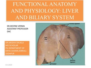
- 1. FUNCTIONALANATOMY AND PHYSIOLOGY: LIVER AND BILIARY SYSTEM 4/11/2020 1 DR.MEGHA SHUKLA MD SCHOLAR PG DEPARTMENT OF KRIYA SHARIR,DSRRAU. JODHPUR DR.DEEPAK VERMA ASSISTANT PROFESSOR DAC
- 2. INTRODUCTION ABOUT LIVER The liver is the largest gland in the body and has a wide variety of functions . It is exocrine(bile) & endocrine organ(Albumen , prothrombin & fibrinogen) . Location: Right hypochondrium + Epigastrium & extends to left hypochondrium Weight: 1/50 of body weight in adult & 1/20 of body weight in infant FUNCTIONS OF THE LIVER •Secretion of bile & bile salt •Metabolism of carbohydrate, fat and protein •Formation of heparin & anticoagulant substances •Detoxication •Storage of glycogen and vitamins •Activation of vita .D 4/11/2020 2
- 3. WHAT IS FUNCTIONAL ANATOMY • Definition (noun) the study of anatomy in its relation to function • Synonyms : morphophysiology , physiological anatomy 4/11/2020 3
- 4. Functional Anatomy of Liver 4/11/2020 4
- 5. INTRODUCTION The liver is an accessory digestive organ that produces bile, a fluid containing cholesterol and bile acids, and an alkaline compound which helps the breakdown of fat. Bile aids in digestion via the emulsification of lipids. The gallbladder, a small pouch that sits just under the liver, stores bile produced by the liver which is afterwards moved to the small intestine to complete digestion. The liver's highly specialized tissue consisting of mostly hepatocytes regulates a wide variety of high-volume biochemical reactions, including the synthesis and breakdown of small and complex molecules, many of which are necessary for normal vital functions. Estimates regarding the organ's total number of functions vary, but textbooks generally cite it being around 500. 4/11/2020 5
- 6. STRUCTURE • The liver is a reddish-brown, wedge-shaped organ with four lobes of unequal size and shape. A human liver normally weighs approximately 1.5 kg (3.3 lb), and has a width of about 15 cm (6 in).There is also considerable size variation between individuals, with the standard reference range for men being 970–1,860 g (2.14–4.10 lb) and for women 600– 1,770 g (1.32–3.90 lb). It is both the heaviest internal organ and the largest gland in the human body. • Lobules are the functional units of the liver. Each lobule is made up of millions of hepatic cells (hepatocytes), which are the basic metabolic cells. The lobules are held together by a fine, dense, irregular, fibroelastic connective tissue layer extending from the fibrous capsule covering the entire liver known as Glisson's capsule. This extends into the structure of the liver, by accompanying the blood vessels (veins and arteries), ducts, and nerves at the hepatic hilum. The whole surface of the liver except for the bare area, is covered in a serous coat derived from the peritoneum, and this firmly adheres to the inner Glisson's capsule. 4/11/2020 6
- 7. Cells Of The Liver Cells of the liver include Hepatocytes, Hepatic stellate cells - also known as perisinusoidal lipocytes, or ito cells - sinusoidal endothelial cells, Macrophages (kupffer cells), The cells of the biliary tree - Cuboidal to columnar epithelium - and Connective tissue cells of the capsule and portal tracts. 4/11/2020 7
- 8. Hepatocytes • Hepatocytes represent 60 % of the liver‘s cells and about 80 % of the liver‘s total cell mass. Most of the liver‘s synthetic and metabolic capabilities stem from the work of hepatocytes . Hepatocytes are arranged in plates only a single cell thick . • The main function of hepatocytes is to participate in lipid, carbohydrate and protein metabolism. They also produce serum proteins such as albumin and coagulation factors. • Furthermore, hepatocytes produce and secrete bile as well as detoxify and excrete cholesterol, steroid hormones and xenobiotic drugs. Numerous xenobiotics are metabolized by the mixed- functions of monooxidases found in hepatocytes. 4/11/2020 8
- 9. Endothelial Cells • The sinusoidal endothelial cells line the walls of the hepatic sinusoid and perform a function of filtration due to the presence of fenestrae. • They may also function as antigen presenting cells and secrete certain cytokines and eicosanoids. 4/11/2020 9
- 10. Stellate Cells • They are located in the space of Disse (between hepatocytes and sinusoid) and generally protrude to come into contact with several sinusoids . • The liver plays a central role in uptake and storage of vitamins A (Retinol) and stores about 95 % of retinoids found in the body. The fat storing perisinusoidal cells of the liver, stellate cells are the main vitamin A storing cells. 4/11/2020 10
- 11. Kupffer Cells • They are located within the sinusoid and are in constant contact with gut-derived particles that lead to low but constant amount of activation of these monocyte derived cells. Upon activation they are able to secrete a vast range of inflammatory mediators such as cytokines, reactive oxygen species, eicosanoids and nitric oxide • The liver harbors large amounts of kupffer cells, which represent the largest tissue resident macrophage population of the body • Kupffer cells have receptors that enable them to bind cells covered with immunoglobulins or bind to complement receptors and subsequently phagocytose cell 4/11/2020 11
- 12. HISTOLOGY Two major types of liver cell: Parenchymal cells and non parenchymal cells. About 70–85% of the liver volume is occupied by parenchymal hepatocytes. Non parenchymal cells constitute 40% of the total number of liver cells but only 6.5% of its volume. The liver sinusoids are lined with two types of cell, sinusoidal endothelial cells, and phagocytic Kupffer cells. Hepatic stellate cells are non parenchymal cells found in the perisinusoidal space, between a sinusoid and a hepatocyte. Additionally, intrahepatic lymphocytes are often present in the sinusoidal lumen. 4/11/2020 12
- 13. 4/11/2020 13
- 14. Microscopically, each liver lobe is seen to be made up of hepatic lobules. The lobules are roughly hexagonal and consist of plates of hepatocytes radiating from a central vein. In the mature liver, hepatocytes are arranged mainly in plates - or cords, Between the plates are venous sinusoids, which anastomose with each other via gaps in the hepatocyte plates. Bile secreted by the hepatocytes is collected in a network of minute tubes (canaliculi). The central vein joins to the hepatic vein to carry blood out from the liver. A distinctive component of a lobule is the portal triad, which can be found running along each of the lobule's corners. The portal triad, misleadingly named, consists of five structures: a branch of the hepatic artery, a branch of the hepatic portal vein, and a bile duct, as well as lymphatic vessels and a branch of the vagus nerve.Between the hepatocyte plates are liver sinusoids, which are enlarged capillaries through which blood from the hepatic portal vein and hepatic artery enters via the portal triads, then drains to the central vein. 4/11/2020 14
- 15. FUNCTIONALANATOMY • The central area or hepatic hilum, includes the opening known as the porta hepatis which carries the common bile duct and common hepatic artery, and the opening for the portal vein. The duct, vein, and artery divide into left and right branches, and the areas of the liver supplied by these branches constitute the functional left and right lobes. The functional lobes are separated by the imaginary plane, Cantlie's line, joining the gallbladder fossa to the inferior vena cava. The plane separates the liver into the true right and left lobes. The middle hepatic vein also demarcates the true right and left lobes. The right lobe is further divided into an anterior and posterior segment by the right hepatic vein. The left lobe is divided into the medial and lateral segments by the left hepatic vein. • The hilum of the liver is described in terms of three plates that contain the bile ducts and blood vessels. The contents of the whole plate system are surrounded by a sheath.The three plates are the hilar plate, the cystic plate and the umbilical plate and the plate system is the site of the many anatomical variations to be found in the liver.4/11/2020 15
- 16. 4/11/2020 16
- 17. SURFACES OF THE LIVER, THEIR RELATIONS & IMPRESSIONS 4/11/2020 17
- 18. The liver is the largest organ in the body. Its domed upper surface relates entirely to the diaphragm while its postero-inferior, or visceral, surface rests against the abdominal oesophagus, stomach, upper duodenum, hepatic flexure of the colon, right kidney and suprarenal gland, as well as carrying the gall bladder. Its surface relations can be marked out by joining points on: the right costal margin in the mid-axillary line, (the 10th rib) the right 5th intercostal space ditto the left 5th intercostal space in the mid-clavicular line. POSITION 4/11/2020 18
- 19. SURFACES OF LIVER •Postero - Inferior Surface= Visceral Surface •Superior Surface = Diaphragmatic Surface •Anterior Surface •Posterior Surface •Right Surface 4/11/2020 19
- 20. 4/11/2020 20
- 21. Lobes of the liver •Rt. Lobe •Lt .lobe •Quadrate lobe •Caudate lobe Rt. & Lt lobe separated by • Falciform ligament •Ligamentum Venoosum •Ligamentum teres 4/11/2020 21
- 22. Relations of the liver Anteriorly •Diaphragm •Rt & Lt pleura and lung •Costal cartilage •Xiphoid process •Ant. abdominal wall 4/11/2020 22
- 23. 4/11/2020 23
- 24. Portal Triad Branches of portal vein, hepatic artery and the biliary ducts bound together in the perivascular fibrous capsule. 4/11/2020 24
- 25. Blood Flow Distribution Total human liver blood flow represents approximately 25% of the cardiac output, up to 1500 ml/min. Hepatic °ow is subdivided in 25-30% for the hepatic artery (500 ml/min) and the major part for the portal vein (1000 ml/min). Assuming a human liver weighs 1500 g, total liver flow is 100 ml/min per 100 g liver. Comparing this normalized °ow rate to other species, it can be concluded that total liver blood flow is 100-130 ml/min per 100 g liver, independent of the species. The ratio of arterial:portal blood °ow, however, is species-dependent. The hepatic artery orig- inates directly from the descending aorta, and is therefore saturated with oxygen.It accounts for 65% of total oxygen supply to the liver. The hepatic artery also plays an important role in liver blood vessel wall and connective tissue perfusion.It also secures bile duct integrity. The blood from the portal vein is full of nutrients derived from the intestine and allows the hepatocytes to perform their tasks. Blood from the hepatic artery and the portal vein joins in the sinusoids. 4/11/2020 25
- 26. Blood supply There are 2 distinct sources that supply blood to the liver: • Oxygenated blood flows in from the hepatic artery. • Nutrient-rich blood flows in from the hepatic portal vein. Almost all blood that enters the liver via the portal tract originates from the gas-trointestinal tract as well as from the spleen, pancreas and gallbladder. A second blood supply to the liver comes from the hepatic artery, branching directly from the celiac trunc and descending aorta. The portal vein supplies venous blood under low pressure conditions to the liver, while the hepatic artery supplies high-pressured arterial blood. Since the capillary bed of the gastrointestinal tract already extracts most O2, portal venous blood has a low O2 content. Blood from the hepatic artery on the other hand, originates directly from the aorta and is, therefore, saturated with O2. Blood from both vessels joins in the capillary bed of the liver and leaves via central veins to the inferior caval vein. 4/11/2020 26
- 27. 4/11/2020 27
- 28. Biliary system • The biliary system consists of the organs and ducts (bile ducts, gallbladder, and associated structures) that are involved in the production and transportation of bile. The transportation of bile follows this sequence: When the liver cells secrete bile, it is collected by a system of ducts that flow from the liver through the right and left hepatic ducts. These ducts ultimately drain into the common hepatic duct. The common hepatic duct then joins with the cystic duct from the gallbladder to form the common bile duct. This runs from the liver to the duodenum (the first section of the small intestine). However, not all bile runs directly into the duodenum. About 50% of the bile produced by the liver is first stored in the gallbladder. This is a pear-shaped organ located directly below the liver. Then, when food is eaten, the gallbladder contracts and releases stored bile into the duodenum to help break down the fats. 4/11/2020 28
- 29. Functions of the biliary system The biliary system's main function includes the following: • To drain waste products from the liver into the duodenum • To help in digestion with the controlled release of bile 4/11/2020 29
- 30. Bile About 500 mL is secreted per day. Some of the components of the bile are reabsorbed in the intestine and then excreted again by the liver (enterohepatic circulation). The glucuronides of the bile pigments, bilirubin and biliverdin, are responsible for the golden yellow color of bile. The bile salts are sodium and potassium salts of bile acids, and all those secreted into the bile are conjugated to glycine or taurine, a derivative of cysteine. The bile acids are synthesized from cholesterol. 4/11/2020 30
- 31. CBD Extra hepatic biliary system Rt. hepatic duct + Lt hepatic duct ↓ Common hepatic duct + Cystic duct ↓ Common bile duct - 4cm - Descend in free edge of lesser omentum - Supra duodenal part Retro duodenal part Retro pancreatic part 4/11/2020 31
- 32. Composition of Human Hepatic Duct Bile 4/11/2020 32
- 33. Bile acids Primary bile acids Formed in the liver are cholic acid and chenodeoxycholic acid. Secondary bile acids In the colon, bacteria convert cholic acid to deoxycholic acid and chenodeoxycholic acid to lithocholic acid. 4/11/2020 33
- 34. • The acid-dependent fraction and the acid- independent fraction. Hepatocytes secrete • The bile acid-dependent fraction of the bile. This fraction consists of bile acids, cholesterol, lecithin (a phospholipid), and bilirubin (a bile pigment). The bile acid-independent fraction of the bile, which is secreted by the hepatocytes and epithelial cells of the bile canaliculi, is a bicarbonate-rich aqueous fluid that gives bile its alkaline pH. 4/11/2020 34
- 35. BILE SALTS • Bile salts are conjugated in the liver from primary and secondary bile acids. The primary bile acids are cholic acid and chenodeoxycholic (chenic) acid. These acids are synthesized from cholesterol by the hepatocytes. The secondary bile acids are deoxycholic acid and lithocholic acid. These acids are formed in the small intestine by the action of intestinal bacteria, after which they are absorbed and flow to the liver. • Both forms of bile acids are conjugated with amino acids in the liver to form bile salts. • Conjugation makes the bile acids more water soluble, thus restricting their diffusion from the duodenum and ileum. 4/11/2020 35
- 36. • The liver also has hemostatic functions. It synthesizes prothrombin, fibrinogen, and clotting factors. Vitamin K, a fat-soluble vitamin, is essential for the synthesis of other clotting factors. Because bile salts are needed for reabsorption of fats, vitamin K absorption depends on adequate bile production in the liver. 4/11/2020 36
- 37. Comparison of Human Hepatic Duct Bile and Gallbladder Bile. 4/11/2020 37
- 38. Major functions of liver 4/11/2020 38