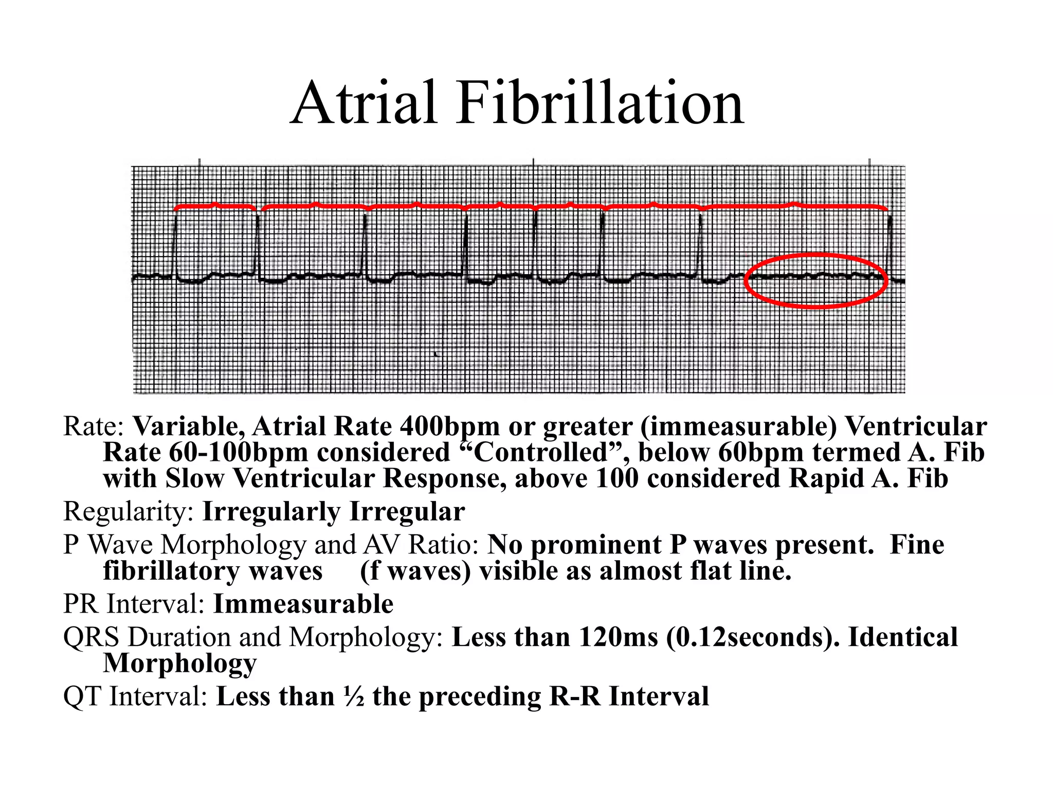
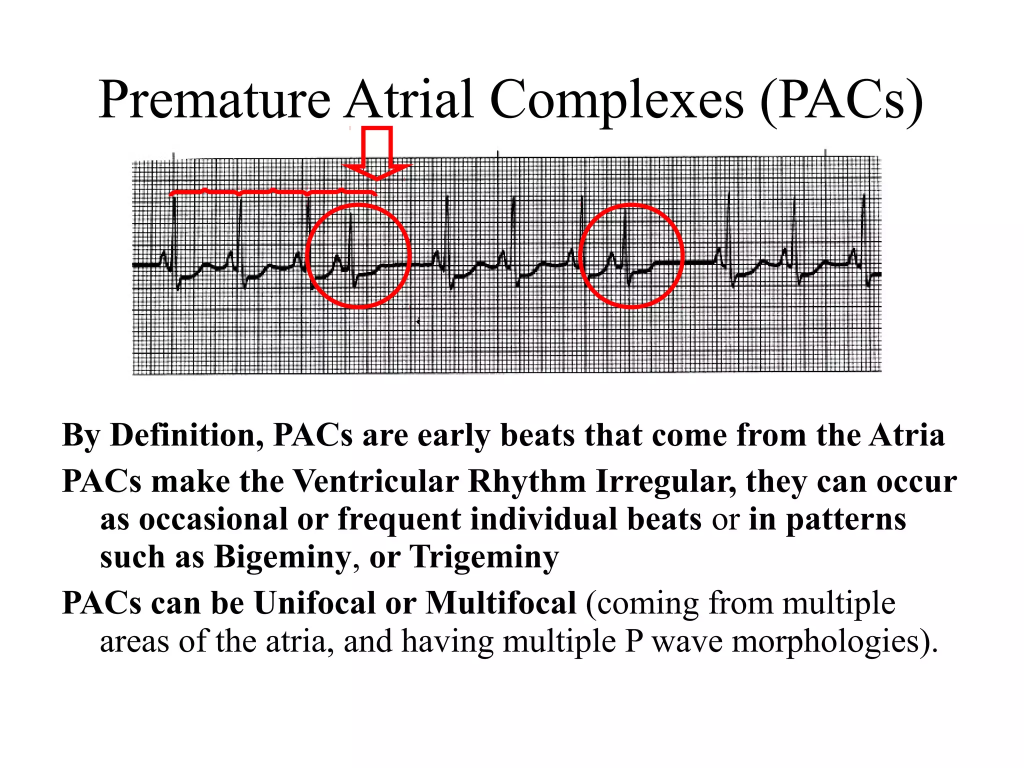
This document provides an overview of basic cardiac dysrhythmias including atrial fibrillation, atrial flutter, premature atrial complexes, wandering atrial pacemaker, junctional escape rhythms, accelerated junctional rhythms, premature junctional complexes, and supraventricular tachycardia. It describes the characteristics, causes, concerns and treatment approaches for each of these arrhythmias.
Its crucial to diagnose arrythmias quickly and treat it promptly.
Here i have made small attempt to diagnose tachyarrythmias briefly and proceeds with its immediate managenent..
Its crucial to diagnose arrythmias quickly and treat it promptly.
Here i have made small attempt to diagnose tachyarrythmias briefly and proceeds with its immediate managenent..
Tachycardias are broadly categorized based upon the width of the QRS complex on the electrocardiogram (ECG). A narrow QRS complex (<120 milliseconds) reflects rapid activation of the ventricles via the normal His-Purkinje system, which in turn suggests that the arrhythmia originates above or within the His bundle (ie, a supraventricular tachycardia). The site of origin may be in the sinus node, the atria, the atrioventricular (AV) node, the His bundle, or some combination of these sites. A widened QRS (≥120 milliseconds) occurs when ventricular activation is abnormally slow. The most common reason that a QRS is widened is because the arrhythmia originates below the His bundle in the bundle branches, Purkinje fibers, or ventricular myocardium (eg, ventricular tachycardia). Alternatively, a supraventricular arrhythmia can produce a widened QRS if there are either pre-existing or rate-related abnormalities within the His-Purkinje system (eg, supraventricular tachycardia with aberrancy), or if conduction occurs over an accessory pathway. Thus, wide QRS complex tachycardias may be either supraventricular or ventricular in origin.
TEST BANK for Operations Management, 14th Edition by William J. Stevenson, Ve...kevinkariuki227
TEST BANK for Operations Management, 14th Edition by William J. Stevenson, Verified Chapters 1 - 19, Complete Newest Version.pdf
Ethanol (CH3CH2OH), or beverage alcohol, is a two-carbon alcohol
that is rapidly distributed in the body and brain. Ethanol alters many
neurochemical systems and has rewarding and addictive properties. It
is the oldest recreational drug and likely contributes to more morbidity,
mortality, and public health costs than all illicit drugs combined. The
5th edition of the Diagnostic and Statistical Manual of Mental Disorders
(DSM-5) integrates alcohol abuse and alcohol dependence into a single
disorder called alcohol use disorder (AUD), with mild, moderate,
and severe subclassifications (American Psychiatric Association, 2013).
In the DSM-5, all types of substance abuse and dependence have been
combined into a single substance use disorder (SUD) on a continuum
from mild to severe. A diagnosis of AUD requires that at least two of
the 11 DSM-5 behaviors be present within a 12-month period (mild
AUD: 2–3 criteria; moderate AUD: 4–5 criteria; severe AUD: 6–11 criteria).
The four main behavioral effects of AUD are impaired control over
drinking, negative social consequences, risky use, and altered physiological
effects (tolerance, withdrawal). This chapter presents an overview
of the prevalence and harmful consequences of AUD in the U.S.,
the systemic nature of the disease, neurocircuitry and stages of AUD,
comorbidities, fetal alcohol spectrum disorders, genetic risk factors, and
pharmacotherapies for AUD.
Title: Sense of Smell
Presenter: Dr. Faiza, Assistant Professor of Physiology
Qualifications:
MBBS (Best Graduate, AIMC Lahore)
FCPS Physiology
ICMT, CHPE, DHPE (STMU)
MPH (GC University, Faisalabad)
MBA (Virtual University of Pakistan)
Learning Objectives:
Describe the primary categories of smells and the concept of odor blindness.
Explain the structure and location of the olfactory membrane and mucosa, including the types and roles of cells involved in olfaction.
Describe the pathway and mechanisms of olfactory signal transmission from the olfactory receptors to the brain.
Illustrate the biochemical cascade triggered by odorant binding to olfactory receptors, including the role of G-proteins and second messengers in generating an action potential.
Identify different types of olfactory disorders such as anosmia, hyposmia, hyperosmia, and dysosmia, including their potential causes.
Key Topics:
Olfactory Genes:
3% of the human genome accounts for olfactory genes.
400 genes for odorant receptors.
Olfactory Membrane:
Located in the superior part of the nasal cavity.
Medially: Folds downward along the superior septum.
Laterally: Folds over the superior turbinate and upper surface of the middle turbinate.
Total surface area: 5-10 square centimeters.
Olfactory Mucosa:
Olfactory Cells: Bipolar nerve cells derived from the CNS (100 million), with 4-25 olfactory cilia per cell.
Sustentacular Cells: Produce mucus and maintain ionic and molecular environment.
Basal Cells: Replace worn-out olfactory cells with an average lifespan of 1-2 months.
Bowman’s Gland: Secretes mucus.
Stimulation of Olfactory Cells:
Odorant dissolves in mucus and attaches to receptors on olfactory cilia.
Involves a cascade effect through G-proteins and second messengers, leading to depolarization and action potential generation in the olfactory nerve.
Quality of a Good Odorant:
Small (3-20 Carbon atoms), volatile, water-soluble, and lipid-soluble.
Facilitated by odorant-binding proteins in mucus.
Membrane Potential and Action Potential:
Resting membrane potential: -55mV.
Action potential frequency in the olfactory nerve increases with odorant strength.
Adaptation Towards the Sense of Smell:
Rapid adaptation within the first second, with further slow adaptation.
Psychological adaptation greater than receptor adaptation, involving feedback inhibition from the central nervous system.
Primary Sensations of Smell:
Camphoraceous, Musky, Floral, Pepperminty, Ethereal, Pungent, Putrid.
Odor Detection Threshold:
Examples: Hydrogen sulfide (0.0005 ppm), Methyl-mercaptan (0.002 ppm).
Some toxic substances are odorless at lethal concentrations.
Characteristics of Smell:
Odor blindness for single substances due to lack of appropriate receptor protein.
Behavioral and emotional influences of smell.
Transmission of Olfactory Signals:
From olfactory cells to glomeruli in the olfactory bulb, involving lateral inhibition.
Primitive, less old, and new olfactory systems with different path
Title: Sense of Taste
Presenter: Dr. Faiza, Assistant Professor of Physiology
Qualifications:
MBBS (Best Graduate, AIMC Lahore)
FCPS Physiology
ICMT, CHPE, DHPE (STMU)
MPH (GC University, Faisalabad)
MBA (Virtual University of Pakistan)
Learning Objectives:
Describe the structure and function of taste buds.
Describe the relationship between the taste threshold and taste index of common substances.
Explain the chemical basis and signal transduction of taste perception for each type of primary taste sensation.
Recognize different abnormalities of taste perception and their causes.
Key Topics:
Significance of Taste Sensation:
Differentiation between pleasant and harmful food
Influence on behavior
Selection of food based on metabolic needs
Receptors of Taste:
Taste buds on the tongue
Influence of sense of smell, texture of food, and pain stimulation (e.g., by pepper)
Primary and Secondary Taste Sensations:
Primary taste sensations: Sweet, Sour, Salty, Bitter, Umami
Chemical basis and signal transduction mechanisms for each taste
Taste Threshold and Index:
Taste threshold values for Sweet (sucrose), Salty (NaCl), Sour (HCl), and Bitter (Quinine)
Taste index relationship: Inversely proportional to taste threshold
Taste Blindness:
Inability to taste certain substances, particularly thiourea compounds
Example: Phenylthiocarbamide
Structure and Function of Taste Buds:
Composition: Epithelial cells, Sustentacular/Supporting cells, Taste cells, Basal cells
Features: Taste pores, Taste hairs/microvilli, and Taste nerve fibers
Location of Taste Buds:
Found in papillae of the tongue (Fungiform, Circumvallate, Foliate)
Also present on the palate, tonsillar pillars, epiglottis, and proximal esophagus
Mechanism of Taste Stimulation:
Interaction of taste substances with receptors on microvilli
Signal transduction pathways for Umami, Sweet, Bitter, Sour, and Salty tastes
Taste Sensitivity and Adaptation:
Decrease in sensitivity with age
Rapid adaptation of taste sensation
Role of Saliva in Taste:
Dissolution of tastants to reach receptors
Washing away the stimulus
Taste Preferences and Aversions:
Mechanisms behind taste preference and aversion
Influence of receptors and neural pathways
Impact of Sensory Nerve Damage:
Degeneration of taste buds if the sensory nerve fiber is cut
Abnormalities of Taste Detection:
Conditions: Ageusia, Hypogeusia, Dysgeusia (parageusia)
Causes: Nerve damage, neurological disorders, infections, poor oral hygiene, adverse drug effects, deficiencies, aging, tobacco use, altered neurotransmitter levels
Neurotransmitters and Taste Threshold:
Effects of serotonin (5-HT) and norepinephrine (NE) on taste sensitivity
Supertasters:
25% of the population with heightened sensitivity to taste, especially bitterness
Increased number of fungiform papillae
Couples presenting to the infertility clinic- Do they really have infertility...Sujoy Dasgupta
Dr Sujoy Dasgupta presented the study on "Couples presenting to the infertility clinic- Do they really have infertility? – The unexplored stories of non-consummation" in the 13th Congress of the Asia Pacific Initiative on Reproduction (ASPIRE 2024) at Manila on 24 May, 2024.
MANAGEMENT OF ATRIOVENTRICULAR CONDUCTION BLOCK.pdfJim Jacob Roy
Cardiac conduction defects can occur due to various causes.
Atrioventricular conduction blocks ( AV blocks ) are classified into 3 types.
This document describes the acute management of AV block.
micro teaching on communication m.sc nursing.pdfAnurag Sharma
Microteaching is a unique model of practice teaching. It is a viable instrument for the. desired change in the teaching behavior or the behavior potential which, in specified types of real. classroom situations, tends to facilitate the achievement of specified types of objectives.
HOT NEW PRODUCT! BIG SALES FAST SHIPPING NOW FROM CHINA!! EU KU DB BK substit...GL Anaacs
Contact us if you are interested:
Email / Skype : kefaya1771@gmail.com
Threema: PXHY5PDH
New BATCH Ku !!! MUCH IN DEMAND FAST SALE EVERY BATCH HAPPY GOOD EFFECT BIG BATCH !
Contact me on Threema or skype to start big business!!
Hot-sale products:
NEW HOT EUTYLONE WHITE CRYSTAL!!
5cl-adba precursor (semi finished )
5cl-adba raw materials
ADBB precursor (semi finished )
ADBB raw materials
APVP powder
5fadb/4f-adb
Jwh018 / Jwh210
Eutylone crystal
Protonitazene (hydrochloride) CAS: 119276-01-6
Flubrotizolam CAS: 57801-95-3
Metonitazene CAS: 14680-51-4
Payment terms: Western Union,MoneyGram,Bitcoin or USDT.
Deliver Time: Usually 7-15days
Shipping method: FedEx, TNT, DHL,UPS etc.Our deliveries are 100% safe, fast, reliable and discreet.
Samples will be sent for your evaluation!If you are interested in, please contact me, let's talk details.
We specializes in exporting high quality Research chemical, medical intermediate, Pharmaceutical chemicals and so on. Products are exported to USA, Canada, France, Korea, Japan,Russia, Southeast Asia and other countries.
Ozempic: Preoperative Management of Patients on GLP-1 Receptor Agonists Saeid Safari
Preoperative Management of Patients on GLP-1 Receptor Agonists like Ozempic and Semiglutide
ASA GUIDELINE
NYSORA Guideline
2 Case Reports of Gastric Ultrasound
Lung Cancer: Artificial Intelligence, Synergetics, Complex System Analysis, S...Oleg Kshivets
RESULTS: Overall life span (LS) was 2252.1±1742.5 days and cumulative 5-year survival (5YS) reached 73.2%, 10 years – 64.8%, 20 years – 42.5%. 513 LCP lived more than 5 years (LS=3124.6±1525.6 days), 148 LCP – more than 10 years (LS=5054.4±1504.1 days).199 LCP died because of LC (LS=562.7±374.5 days). 5YS of LCP after bi/lobectomies was significantly superior in comparison with LCP after pneumonectomies (78.1% vs.63.7%, P=0.00001 by log-rank test). AT significantly improved 5YS (66.3% vs. 34.8%) (P=0.00000 by log-rank test) only for LCP with N1-2. Cox modeling displayed that 5YS of LCP significantly depended on: phase transition (PT) early-invasive LC in terms of synergetics, PT N0—N12, cell ratio factors (ratio between cancer cells- CC and blood cells subpopulations), G1-3, histology, glucose, AT, blood cell circuit, prothrombin index, heparin tolerance, recalcification time (P=0.000-0.038). Neural networks, genetic algorithm selection and bootstrap simulation revealed relationships between 5YS and PT early-invasive LC (rank=1), PT N0—N12 (rank=2), thrombocytes/CC (3), erythrocytes/CC (4), eosinophils/CC (5), healthy cells/CC (6), lymphocytes/CC (7), segmented neutrophils/CC (8), stick neutrophils/CC (9), monocytes/CC (10); leucocytes/CC (11). Correct prediction of 5YS was 100% by neural networks computing (area under ROC curve=1.0; error=0.0).
CONCLUSIONS: 5YS of LCP after radical procedures significantly depended on: 1) PT early-invasive cancer; 2) PT N0--N12; 3) cell ratio factors; 4) blood cell circuit; 5) biochemical factors; 6) hemostasis system; 7) AT; 8) LC characteristics; 9) LC cell dynamics; 10) surgery type: lobectomy/pneumonectomy; 11) anthropometric data. Optimal diagnosis and treatment strategies for LC are: 1) screening and early detection of LC; 2) availability of experienced thoracic surgeons because of complexity of radical procedures; 3) aggressive en block surgery and adequate lymph node dissection for completeness; 4) precise prediction; 5) adjuvant chemoimmunoradiotherapy for LCP with unfavorable prognosis.
2. Atrial Fibrillation
Rate: Variable, Atrial Rate 400bpm or greater (immeasurable) Ventricular
Rate 60-100bpm considered “Controlled”, below 60bpm termed A. Fib
with Slow Ventricular Response, above 100 considered Rapid A. Fib
Regularity: Irregularly Irregular
P Wave Morphology and AV Ratio: No prominent P waves present. Fine
fibrillatory waves (f waves) visible as almost flat line.
PR Interval: Immeasurable
QRS Duration and Morphology: Less than 120ms (0.12seconds). Identical
Morphology
QT Interval: Less than ½ the preceding R-R Interval
3. Atrial Fibrillation
Causes: many different sites of both atria, and sites in
the pulmonary veins that are irritable, and firing
rapidly, and irregularly.
Causes of the irritable cells include: Hypoxia, Ischemia,
Infarction, Electrolyte disturbances Hypervolemia
(causes stretch of the atria), Cardiac Surgery (50% of
Valve surgery patients develop AF, approx. 25% of
CABG patients develop AF), Excessive adrenergic
stimulation (catecholamine surge from stress,
surgery), Cardiomyopathy, CHF, Pericarditis,
Alcohol Withdrawal, Hyperthyroidism.
4. Atrial Fibrillation
Concerns: Atrial Fibrillation is the most common
dysrhythmia.
Patients can live with permanent AF.
1) Rate control
2) Prevent Clot Formation
3) Loss of Cardiac Output form “Atrial Kick”
5. Initial Treatment of Atrial Fibrillation
Rate Control: Initial treatment is with β-adrenergic
blockers or Calcium Channel Blockers. These
medications the have negative chronotropic and/or
negative dromotropic properties (slow speed of
conduction through AV node) Amiodarone is not
recommended as a first line treatment due the drug’s
extensive side effect/adverse reaction profile.
Anticoagulation: If the underlying etiology cannot be
corrected, or the rhythm cannot be converted within
24 hours, anticoagulation is indicated to prevent
blood clot formation.
6. Conversion of Atrial fibrillation to
Sinus Rhythm
Ideally, treatment of atrial fibrillation includes
correcting the rhythm disturbance and conversion
to sinus rhythm. This is not always possible for all
patients.
Methods to achieve this goal include: Antiarrhythmic
Drugs: Propafenone, Flecainide, Sotalol,
Dofetilide, Amiodarone, Dronedarone
Cardioversion EP Procedures MAZE
8. Atrial Flutter
Rate: Atrial Rate 250-400, Ventricular Rate Variable
Regularity: Atrial Regular, Ventricular-Variable
dependent on A:V
P Wave Morphology and AV Ratio: Fast “saw-tooth”
or “picket fence” Flutter waves (F waves) Present
and Uniform. AV Ratio can be fixed as 3:1, 4:1
etc. (and the Ventricular Rhythm is regular) or
variable (Variable Conduction Ratio).
PR Interval: Immeasurable
QRS Duration and Morphology: Less than 120ms
(0.12seconds). Identical Morphology
QT Interval: Less than ½ the preceding R-R Interval
Treatment: similar to Atrial fibrillation- responds
better to cardioversion
9. Premature Atrial Complexes (PACs)
By Definition, PACs are early beats that come from the Atria
PACs make the Ventricular Rhythm Irregular, they can occur
as occasional or frequent individual beats or in patterns
such as Bigeminy, or Trigeminy
PACs can be Unifocal or Multifocal (coming from multiple
areas of the atria, and having multiple P wave morphologies).
10. PACs
QRS Duration and Morphology: Less than 120ms
(0.12seconds) if conducted through the His-
Purkinje System in a normal fashion. If the QRS
following the P wave of a PAC is wider than the
other QRS Complexes, the impulse has depolarized
the heart in an aberrant fashion. (Those beats are
called PACs with aberrant conduction)
PACs indicate that an area, or areas of the atria are
irritated. PACs are often precursor beats to Atrial
Fibrillation and Atrial Flutter.
Treatment for “high-risk” groups.
11. Wandering Atrial Pacer
Rate: Usually between 60-100bpm, termed Multiple Atrial Tachycardia if
above 100bpm
Regularity: Regular or Irregular
P Wave Morphology and AV Ratio: At least 3 different P wave
Morphologies by definition. A:V ration 1:1.
PR Interval: Will Vary Depending on Location of Ectopic Atrial Sites, and
some may be less than 120ms(0.12seconds).
QRS Duration and Morphology: Less than 120ms (0.12seconds).
QT Interval: Less than ½ the preceding R-R Interval
12. WAP
In WAP, multiple sites, including the SA node, the
Atria and the AV node compete for pacing the heart.
It is a sign of increased autorhythmicity of the various
pacing sites of the heart.
Causes: Common causes include mitral or tricuspid
valve disease, chronic lung disease, digitalis
preparations or enhanced vagal tone.
Patients usually asymptomatic. Discontinue or
change dosage of any medications thought to
be the cause of the arrhythmia.
13.
14. Junctional Escape Rhythms
Rate: 40-60bpm
Regularity: Regular
P Wave One of three morphologies as described on
previous slide
AV Ratio: Retrograde P waves before or after the
QRS complexes, or No visible P waves
PR Interval: PR Interval is less than 120ms (0.12 sec)
if P waves are prior to QRS complexes
QRS Duration and Morphology: Less than 120ms
(0.12seconds).
QT Interval: Less than ½ the preceding R-R Interval
15. Junctional Escape Rhythm
The AV junction is the second line of defense
in the hierarchy of pacemakers. evidence of
a junctional rhythm may be protective for a
patient with impaired SA node
autorhythmicity.
Common causes include damage to the SA
node from MI, or ischemia, calcification,
Valvular heart disease, and Myocarditis,
CHF and Digitalis Toxicity.
16. Accelerated Junctional and Junctional
Tachycardia Rhythms
Rate: Accelerated = 60-100 bpm Tachycardia=
greater than 100 bpm
P Wave Morphology: Same P wave morphologies as
Junctional Escape Rhythms
PR Interval: Short if Measurable
17. Accelerated Junctional Rhythm
Causes: Enhance Autorhythmicity of Junction
including Digitalis or Theophylline toxicity,
catecholamine surge from stress or stimulants,
or acid base imbalances.
Treatments: Treatment is based on removal of
cause, and control of heat rate. Meds that limit
the automaticity of the Junction include β-
Blockers, Ca Channel Blockers and
Amiodarone,
18. Premature Junctional Complexes
(PJCs)
By Definition, PJCs are early beats that originate
from the Junction
PJCs take on the same morphology as Junctional Escape
Rhythms, with either Retrograde P waves prior to or
after the QRS complexes, or No Visible P waves.
19. Supra-Ventricular Tachycardia (SVT)
Fast Rhythms:
First Discern if the Rhythm comes from the ventricle
(wide QRS complex), or from above the ventricle
(Narrow QRS complex).
Then Identify the specific origin of the Rhythm.
20. Treatment of SVTs
Serious signs and symptoms (hypotension,
acutely altered mental status, signs of shock,
ischemic chest discomfort, acute heart failure):
1. Immediate Synchronized Cardioversion
2. Consider adenosine (now also first line)
Stable without rate related CV compromise:
1.Attempt to visualize the origin of the SVT
by using vagal maneuvers or adenosine.
2. Slow the ventricular response using Ca
channel blockers or B-adrenergic blockers.
21. Synchronized Cardioversion
Goal: spontaneous depolarization of a “critical
mass” of cardiac cells to disrupt the erratic
tachycardia, with hope that the conduction
system will restore normal function.
Synchronized with the patient’s rhythm to
deliver the electrical current on the R wave to
prevent deterioration to less viable rhythm.
Painful: provide analgesia, and anxiolytics
23. Adenosine
Used as a diagnostic tool to help identify
rhythm.
Adenosine
6mg IVP, “high and fast”
May repeat with 12 mg, q 1-2 min x 2
Predictable signs and symptoms: CP, palpitation,
LOC, SOB diaphoresis – please inform patient
Editor's Notes
Atrial fibrillation is a common dysthythmia with very distinctive attributes. Most predominant is the fact that atrial fibrillation is an irregularly irregular rhythm.
In fact, if you see an irregularly irregular rhythm with narrow QRS complexes, you should think that it is a-fib until proven otherwise. You will also note that there is no evidence of organized atrial depolarization.
That is, there are no discernible p-waves. Instead, there are small fibrillatory waves called f waves. A –fib is multiple sites in the atria firing rapidly and simultaneously. Once these impulses reach the AV node, only a few are allowed to depolarize the ventricle, otherwise the patients heart rate would be 400 plus beats per minute. Since ventricular depolarization is unaltered the QRS duration and QT intervals are normal.
Common causes of atrial fibrillation include hypoxia, ischemia, electrolyte disturbances like hypokalemia and hypomagnesaemia, and hypervolemia. Any stress state can lead to a-fib, especially post-surgical catecholamine surge and drug withdrawal. It is often a presenting symptom of hyperthyroidism especially in older women.
There are three primary concerns with atrial fibrillation; rate control, clot formation and the loss of cardiac output. The ventricular rate in response to a-fib is of particular concern, and reflects the condition of the AV node. Specifically, a-fib can result in fast, slow or normal ventricular rate, and this depends on how quickly the impulses travel through the AV node. A goal is to maintain the ventricular rate between 60 and 100 beats per minute.
Clot formation is also a major concern. Because there is no organized electrical activity in the atria, there is also no organized contraction. As a result, blood pools in both the right and left atrial and begins to clot. Blood clots can be ejected from the atrial and into the pulmonary or peripheral circuits and cause serious injury. Blood begins to clot within 24-48 hours after the onset of atrial fibrillation. Therefore, patients who are in a-fib for longer than 24 hours are started on anticoagulant medications. Further, if attempts are going to be made to convert a patients rhythm from a-fib to sinus rhythm, an echocardiogram is performed to guarantee that there is no clot in the atria.
The lack of organized atrial contraction also decreases cardiac output by up to 30%. Atrial contraction contributes significantly to filling of the ventricles just before contraction, the so-called atrial kick. With such a decrease in cardiac output, patients are at risk for decreased end-organ perfusion and limited ability to augment their cardiac performance during exercise or other activity.
The management of atrial fibrillation is outlined here. Drugs that can slow the speed of conduction through the AV node have what is called negative dromotropic properties. Most medications that decrease the heart rate are also negative dromotropic drugs, and will decrease the ventricular response to atrial fibrillation in patients who have a-fibrillation with rapid ventricular response.
Amiodarone is an antiarrhythmic medication with additional beta adrenergic blocking and multiple ion channel altering qualities. Unfortunately, amiodarone also has many serious adverse reactions which make it undesirable as a first tier treatment for atrial fibrillation. It is used when other medications are ineffective and/or once the individual patient risk for adverse reactions has been quantified.
Heparin is often used as the anticoagulant until long-term anti-coagulants are at levels great enough to be therapeutic.
Ideally, treatment of atrial fibrillation includes correcting the rhythm disturbance and conversion to sinus rhythm. This is not always possible for all patients.’
Patients with newly diagnosed atrial fibrillation may respond to antiarrhythmic medications and convert back into sinus rhythm.
Cardioversion, and several types of electrophysiological procedures including the MAZE procedure are used to convert patients into more hemodynamically and hematologically stable rhythms like sinus rhythm. The MAZE procedure can also be performed surgically.
Atrial flutter is identified by rapid atrial depolarization. This can be seen as so-called picket fence, or saw tooth p-waves occurring throughout the entire rhythm strip. The top rhythm strip is atrial flutter with a fixed A-V ratio. That means that the ventricular response to this rapid atrial rhythm is regular.
Here we see the regular and very fast atrial depolarization at a rate of about 300 beats per minute. Also, we note the regular R-R interval. When both the atrial and ventricular rhythms are regular it is called a fixed AV ratio. Here there are 3 p-waves for every QRS complex, therefore this is called 3:1 A flutter.
The bottom strip is A-flutter with a variable AV ratio. Although the atrial rate is regular and also very fast, the ventricular rhythm is irregular. This is called A-flutter with variable ventricular response, or variable conduction ratio.
The atrial rate in a-flutter is usually between 250-400 beats per minute, and as discussed the ventricular rate can vary. Since ventricular depolarization and repolarization is un-affected, the QRS duration and QT interval should be normal. If they are not, then there are different pathophsiological mechanisms at play.
The treatment for atrial flutter is very similar to the treatment of atrial fibrillation. Oftentimes cardioversion is used, and there are medications that seem to work better with a-flutter than a-fib, and vise-versa
Slow ventricular response with B-adrenergic blockers, Ca channel blockers, amiodarone or digoxin.
PACs are not a rhythm but interrupt an underlying rhythm. Therefore, it is important to identify the underlying rhythm first, and then identify where the early beats come from.
To begin looking at this strip, I have circled the two early beats. After analyzing the rhythm, one can determine that it is in fact sinus rhythm.
Next, we need to discern where the early beats come from. We know that they are early because they occur before the next predicted beat, marked here by the red arrow. They can come from the atria, the junction or the ventricles. We have good evidence that these beats come from the atria, because the first waveform is an upright p-wave followed by a nice an narrow QRS complex.
That means that atrial depolarization occurred first and in a normal fashion, and the ventricular depolarization was also normal. Thus, upright p-waves and normal QRS durations help identify where these early beats come from. Pay attention to the terminology described on this slide. This strip is an example of sinus rhythm with unifocal atrial quadrigeminy.
The same conditions that cause a-fibb and a-flutter cause PACs. In fact, PAC’s are a sign that there is an area or multiple areas of the atria that are irritable, thus indicating that the patient may be at risk for converting to a-fibb or flutter. Patients who are at high risk for developing a-fibb may require treatment of PACs, which involves correcting oxygenation, electrolyte and/or fluid balance etc., to keep them from converting to a more dangerous rhythm. PACs in patients without high-risk are usually benign. In fact, if you monitored your heart rhythm continuously, you would no doubt see PACs, especially after substantial caffeine intake.
Wandering atrial pacer is not a common rhythm, but has some unique characteristics. In this rhythm, there are at least three sites between the SA node, the atria and the junction that take over pacing for the heart. This is evidenced by at least three different p-wave morphologies.
This also provides a decent rationale for determining that there uniform and upright p-waves before every QRS complex as one of the first few steps in rhythm analysis.
Clarification of the definition of wandering atrial pacer and common causes are listed here. Please take a moment to review.
Rhythms and beats that come from the junction of the AV node and Bundle of His take on one of three different morphologies. In this top strip, this junctional rhythm come from high in the junction and therefore depolarizes the atria first, and then the ventricle.
Because this impulse comes from the AV node, depolarization of the atria occurs in a retrograde fashion, meaning from the bottom up as opposed to the top down. This results in a retrograde, or upside-down p-wave in front of a QRS complex.
Junctional rhythms that originate from the middle of the junction depolarize the atria and ventricles at the same time, thus there is no visible p-wave. A key to differentiating mid-junctional rhythms from atrial-fibrillation is that junctional rhythms are always regular, where a-fibb is always irregularly irregular.
Junctional rhythms that originate from low in the junction depolarize the ventricle first, and then the atria. This results in a QRS complex followed by a retrograde p-wave. Depolarization and repolarization of the ventricles is not affected, thus the QRS duration and QT intervals should be normal. You will be asked to identify a rhythm or beat that comes from the junction based on one of these three morphologies, but will not be held to determine where in the junction the beat originates.
The three rhythms on the previous slide are all junction escape rhythms. That means that the AV node or surrounding tissue has taken over as the pacemaker of the heart. This is most often due to failure of the SA node. Junctional escape rhythms are slow, and regular, and take on one of the previously described morphologies. If the PR interval is measurable, it will be less than 120 milliseconds.
A junctional escape rhythm is evidence of healthy AV node function in response to impaired or absent SA node firing. Common causes are listed here. Please take a moment to review this slide.
Treatments: Treatment of all bradycardias is guided by symptoms. Asymptomatic junctional escape rhythms are not treated. Medications that block parasympathetic tone may not be as effective. Pacing or the use of Dopamine or Epinephrine drips may be indicated.
If a junctional escape rhythm exceeds 60 beats per minute it is called accelerated junctional rhythm, and if it exceeds 100 beats per minute is called junctional tachycardia. Regarding this strip, we have evidence that this fast rhythm comes from the junction because of the retrograde p-waves in front of each QRS complex.
If this rhythm has unwanted hemodynamic consequences, the treatment entails slowing the rate of impulse formation by administering beta blockers, calcium channel blockers or amiodarone. Accelerated junctional rhythm is often self-limiting.
This sinus bradycardia is interrupted by two early beats, which are circled. When there are premature or early beats, it is important to determine their origin. These early beats come from the junction, as we have evidence of a retrograde p-wave that occurs after each nice and narrow QRS complex. This rhythm strip would be called sinus bradycardia with unifocal junctional trigeminy.
Supraventricular tachycardia is a general term for fast rhythms that originate from above the ventricle. Because the rhythm is so fast, we cannot determine which rhythm it actually is. Instead, we can only determine that it is fast and comes from above the ventricle. Anytime you come across a fast rhythm, you should determine if the rhythms come from the ventricle, or comes from above the ventricle. We can determine this primarily by looking at the QRS duration. In general, SVTs have a narrow QRS that is less than 120 milliseconds , and rhythms that come from the ventricle have a QRS duration greater than 120 milliseconds. Once you have determined that it is a SVT, you will want to determine the area of impulse generation and provide a rhythm-specific treatment.
If a patient exhibiting SVT has serious signs and symptoms, two treatments are recommended: synchronized cardioversion or adenosine.
If the patient does not have serious signs and symptoms then attempts are made to slow the rhythm and determine the origin of the SVT. A definitive rhythm-specific treatment can then be developed.
Vagal maneuvers alone are successful in approximately 25% of all SVTs. The first line drug for most stable regular narrow complex tachycardia is Adenosine. This medication slows the heart rate and is employed as both a therapeutic and diagnostic maneuver. Adenosine is not recommended for sinus tachycardia and is contraindicated in drug induced tachycardia and second or third degree heart block.
Beta blockers or ca channel blockers are administered to slow the ventricular response.
When an SVT results in serious signs and/or symptoms, synchronized cardioversion is indicated. This is a procedure performed with a defibrillator that aims to disrupts the erratic and hemodynamlically unstable rhythm in hopes that a more normal or manageable rhythm is restored. The goal of defibrillation is to cause the spontaneous depolarization of the majority of the cells in the heart at the same time. This involves sending a large amount of energy through the patients chest, which can be extremely painful and anxiety producing.
Synchronized cardioversion is similar to defibrillation, but the two procedures differ in one important way. With synchronized cardioversion, the patient rhythm is taken into consideration. In fact, the device will start to detect and mark the patients QRS complexes and deliver the large amount of energy right on the R wave. This is the best place to try to synchronize the depolarization of a critical mass of cardiac cells, because many of the ventricular cells are already depolarized. Defibrillation is only use in patients who do not have a pulse, and does not take into account the patients underlying rhythm. These nuances will be revisited when defibrillation is discussed.
If a patient with an SVT does not have serious signs and symptoms, non-pharmacological interventions may help stimulate the parasympathetic nervous system. Doing so will slow down the rate of the SA and AV nodes, and slow down the rate of impulse conduction through the AV node. There are several so-called vagal maneuvers that may cause a decrease in ventricular response to an SVT. The most commonly practiced vagal maneuvers are listed. Carotid Sinus massage is sometimes still used by physicians or advanced practitioners but comes with a significant stroke risk especially if a practitioner is unsure of existent plaque in the carotid arteries.
Vagal maneuvers alone are successful in approximately 25% of all SVTs.
When vagal maneuvers are performed successfully, the ventricular rate slows enough to detect the underlying atrial activity to see if the rhythm is sinus tachycardia, atrial flutter, junctional tachycardia etc. The appropriate rhythm-specific treatment may then be initiated.
The first line drug for most stable regular narrow complex tachycardia is Adenosine. This medication slows the heart rate and is employed as both a therapeutic and diagnostic maneuver. Adenosine is not recommended for sinus tachycardia and is contraindicated in drug induced tachycardia and second or third degree heart block.
Adenosine has a very short half life and must be given as a rapid IV push (over 1-3 seconds). The medication must be followed immediately with a bolus of 20ml of saline and elevation of the peripheral IV extremity. Adenosine stops ventricular activity for approximately 3-6 seconds. The patient may experience transient side effects including flushing, chest pain, or tightness, brief periods of asystole, bradycardia, or ventricular ectopy. Informing the patient of potential side effects is essential.
Stopping the ventricular activity and allowing the heart to restart will enable you to determine the atrial activity, pinpointing the etiology of the SVT. The administration of Adenosine may be therapeutic, result in termination of SVT .