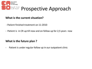
Intermediate Risk Hodgkin Lymphoma
Dr. Mahmoud Motaz Mohammed
South Egypt Cancer Institute
Assiut University
EASO Course on Paediatric Oncology
17-18 May 2013
Cairo, Egypt
History
• Female patient, 12years old, presented on 10/2009 .
• The condition started 2 weeks ago with sever diffuse abdominal
pain,abdominal distenstion, absolute constipation so patient saught
medical advice and was diagnosed as acute intestinal obstruction.Urgent
exploration done with intestinal mass found and completely excised
moderate to high grade fever mainly by night, partially responding to
medical treatment.
• The condition was associated with small right neck swelling of lemon size
surgical removal of which showed a non malignant nature.
• On 5/2010, patient developed multiple painless neck swellings with
gradual onset progressive course associated with night fever and weight
loss - no drenching night sweating.
• Surgical removal of cervical swelling was done and patient was referred to
us.
• No manifestation of other system affection was apparent.
Examination & Workup
Lymphatic system:
- Right upper deep cervical LN about 3*2 cm
- Multiple small left upper deep cervical LNs the largest one about 1 cm in diameter
- RT axillary LN about 1,5 cm in diameter in the apical group
-All of the above described lymph nodes are firm to rubbery in consistency, non tender,
freely mobile and with normal overlying skin.
- No other enlarged LN groups
Laboratory workup:
ESR: 1st hr 25
Imaging Workup:
• CT neck : Multiple bilateral upper and lower deep cervical LNs the largest one on RT side
about 3*2.5 cm, the largest one on the left side about 1 *1 cm.
• US axillae: Multiple bilateral axillary LNs the largest one on the RT side about 1.6*0.8 cm
most of them show loss of the central hilum mostly pathological LNs
• Echo: FS 36 with MR grade1
• PFT : Free
Pathology report
• Suggestive of Hodgkin lymphoma, nodular lymphocytic predominant type with tumor cells
positive for CD20
• BMB Free
histolopathological appearance of nodular lymphocytic
predominant Hodgkin lymphoma with characteristic popcorn
cells adopted from www.webpathology.com
Management
Final Diagnosis: Hodgkin lymphoma, nodular lymphocytic predominant type stage 2B,
non bulky disease
Treatment plan:
• 4 courses of ABVD /COEP then IFRTH 25 gy/17 settings on neck and axillae
Evaluation after 2 courses:
• CT neck : multiple enlarged cervical LNs the largest one about 1cm
• Axillae US: RT LN with eccenteric hilum largest 1cm
Evaluation after another 2 courses and IFRTH:
• CT neck : multiple enlarged cervical LNs the largest one about 1cm.
• Axillae US: RT LN with eccenteric hilum largest one about 1 cm.
• Other imaging investigations, Echo, PFT, BMB were free
Prospective Approach
What is the current situation?
- Patient finished treatment on 11-2010
- Patient is in CR up till now and on follow up for 2,5 years now
What is the future plan ?
- Patient is under regular follow up in our outpatient clinic
Case 1
QUESTIONS FOR DISCUSSION WITH FACULTY
1. As PET-CT is not available in my center, Can we finish treatment in the presence of
residual enlarged LNs? IF so, What's the size of acceptable LN residual after
chemotherapy and radiotherapy to finish chemotherapy on?
2. Taking into consideration the good prognosis of HL in general. In this case can we
use only ABVD instead of alternating ABVD/ COEP to avoid the late effect of
cyclophosphamide on fertility& puberty especially the female child is 8 years old
(pre pubertal)? Does this will affect overall survival?
3. What’s the rule of PET-CT in the management of HL in such a case ?

Easo workshop Hodgkin Lymphoma case presentation

  • 1.
    Intermediate Risk HodgkinLymphoma Dr. Mahmoud Motaz Mohammed South Egypt Cancer Institute Assiut University EASO Course on Paediatric Oncology 17-18 May 2013 Cairo, Egypt
  • 2.
    History • Female patient,12years old, presented on 10/2009 . • The condition started 2 weeks ago with sever diffuse abdominal pain,abdominal distenstion, absolute constipation so patient saught medical advice and was diagnosed as acute intestinal obstruction.Urgent exploration done with intestinal mass found and completely excised moderate to high grade fever mainly by night, partially responding to medical treatment. • The condition was associated with small right neck swelling of lemon size surgical removal of which showed a non malignant nature. • On 5/2010, patient developed multiple painless neck swellings with gradual onset progressive course associated with night fever and weight loss - no drenching night sweating. • Surgical removal of cervical swelling was done and patient was referred to us. • No manifestation of other system affection was apparent.
  • 3.
    Examination & Workup Lymphaticsystem: - Right upper deep cervical LN about 3*2 cm - Multiple small left upper deep cervical LNs the largest one about 1 cm in diameter - RT axillary LN about 1,5 cm in diameter in the apical group -All of the above described lymph nodes are firm to rubbery in consistency, non tender, freely mobile and with normal overlying skin. - No other enlarged LN groups Laboratory workup: ESR: 1st hr 25 Imaging Workup: • CT neck : Multiple bilateral upper and lower deep cervical LNs the largest one on RT side about 3*2.5 cm, the largest one on the left side about 1 *1 cm. • US axillae: Multiple bilateral axillary LNs the largest one on the RT side about 1.6*0.8 cm most of them show loss of the central hilum mostly pathological LNs • Echo: FS 36 with MR grade1 • PFT : Free Pathology report • Suggestive of Hodgkin lymphoma, nodular lymphocytic predominant type with tumor cells positive for CD20 • BMB Free
  • 5.
    histolopathological appearance ofnodular lymphocytic predominant Hodgkin lymphoma with characteristic popcorn cells adopted from www.webpathology.com
  • 6.
    Management Final Diagnosis: Hodgkinlymphoma, nodular lymphocytic predominant type stage 2B, non bulky disease Treatment plan: • 4 courses of ABVD /COEP then IFRTH 25 gy/17 settings on neck and axillae Evaluation after 2 courses: • CT neck : multiple enlarged cervical LNs the largest one about 1cm • Axillae US: RT LN with eccenteric hilum largest 1cm Evaluation after another 2 courses and IFRTH: • CT neck : multiple enlarged cervical LNs the largest one about 1cm. • Axillae US: RT LN with eccenteric hilum largest one about 1 cm. • Other imaging investigations, Echo, PFT, BMB were free
  • 8.
    Prospective Approach What isthe current situation? - Patient finished treatment on 11-2010 - Patient is in CR up till now and on follow up for 2,5 years now What is the future plan ? - Patient is under regular follow up in our outpatient clinic
  • 9.
    Case 1 QUESTIONS FORDISCUSSION WITH FACULTY 1. As PET-CT is not available in my center, Can we finish treatment in the presence of residual enlarged LNs? IF so, What's the size of acceptable LN residual after chemotherapy and radiotherapy to finish chemotherapy on? 2. Taking into consideration the good prognosis of HL in general. In this case can we use only ABVD instead of alternating ABVD/ COEP to avoid the late effect of cyclophosphamide on fertility& puberty especially the female child is 8 years old (pre pubertal)? Does this will affect overall survival? 3. What’s the rule of PET-CT in the management of HL in such a case ?