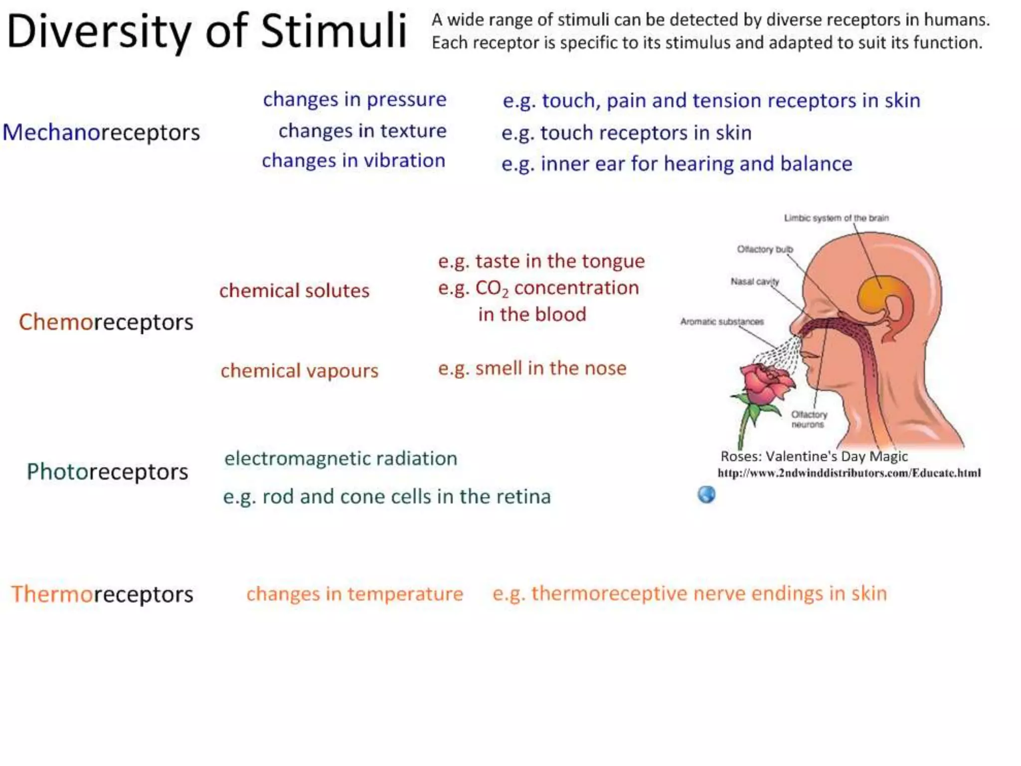
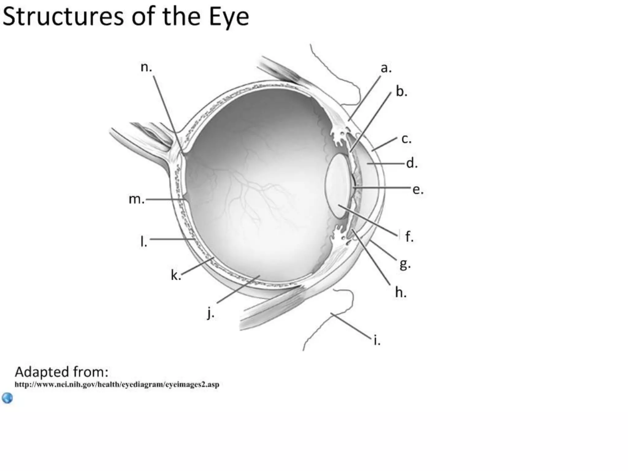
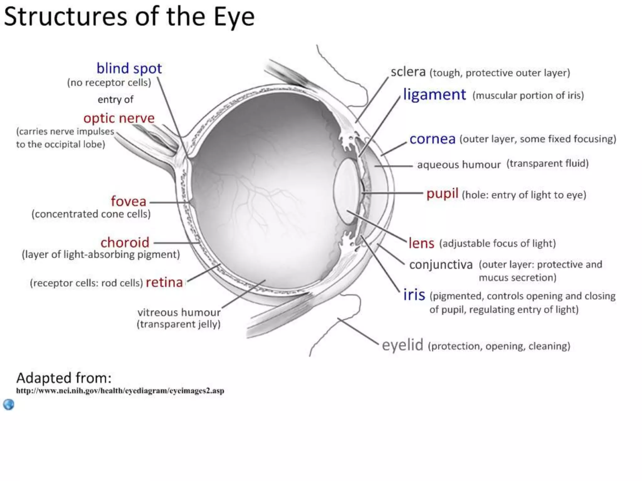
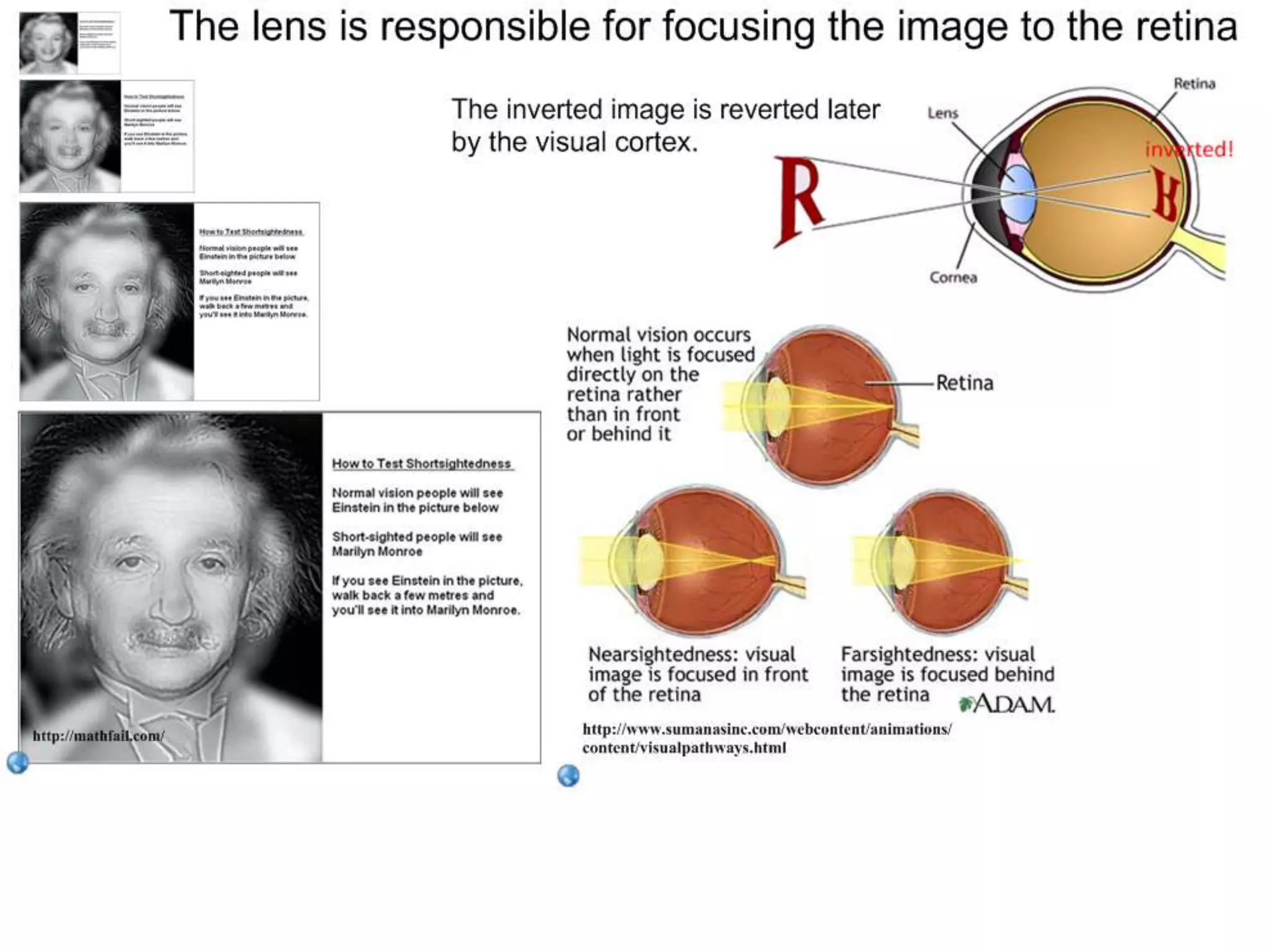
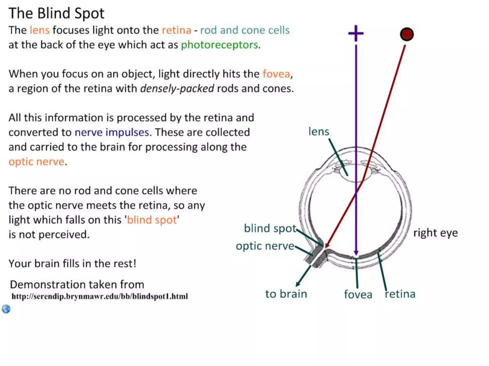
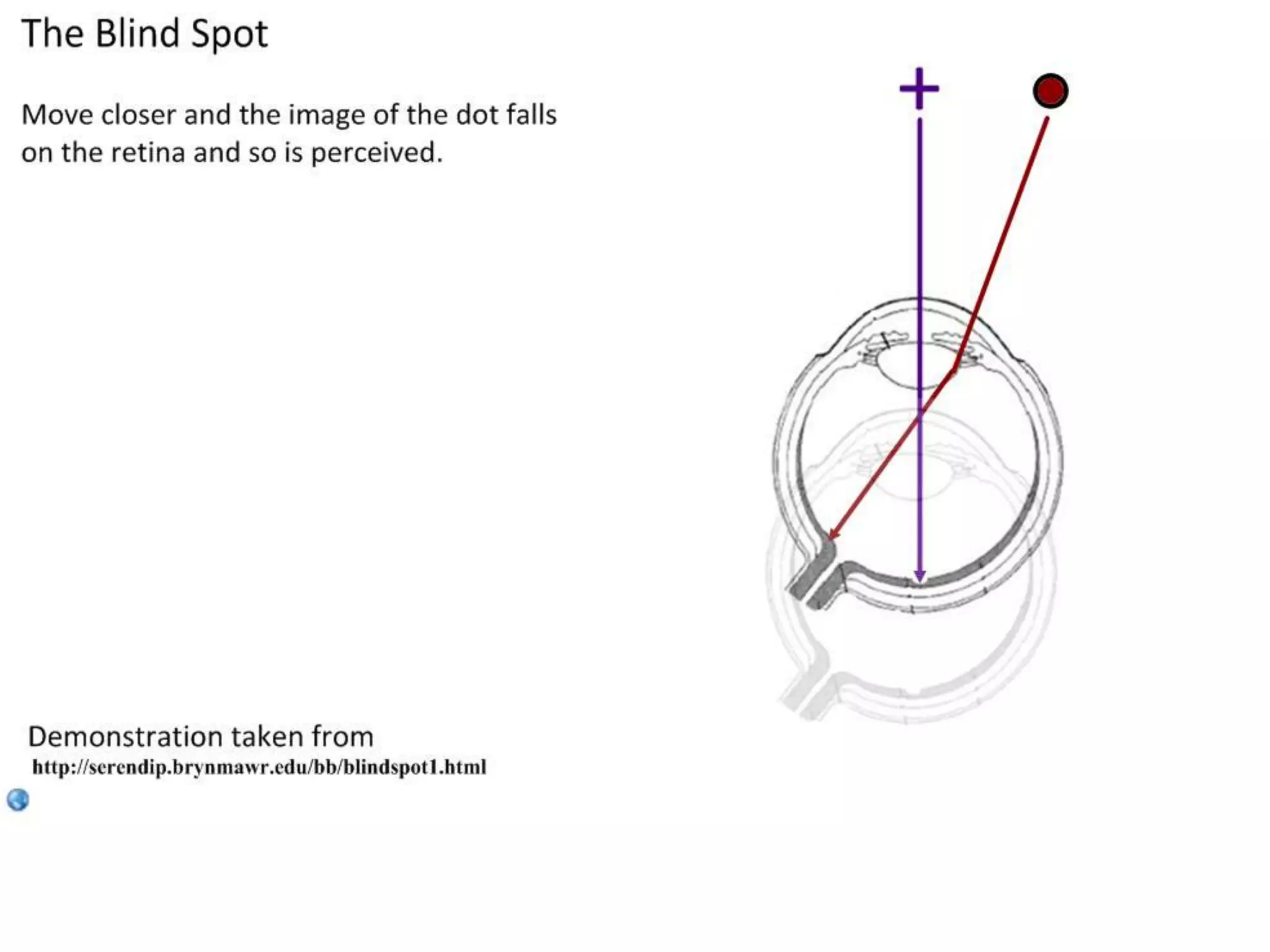
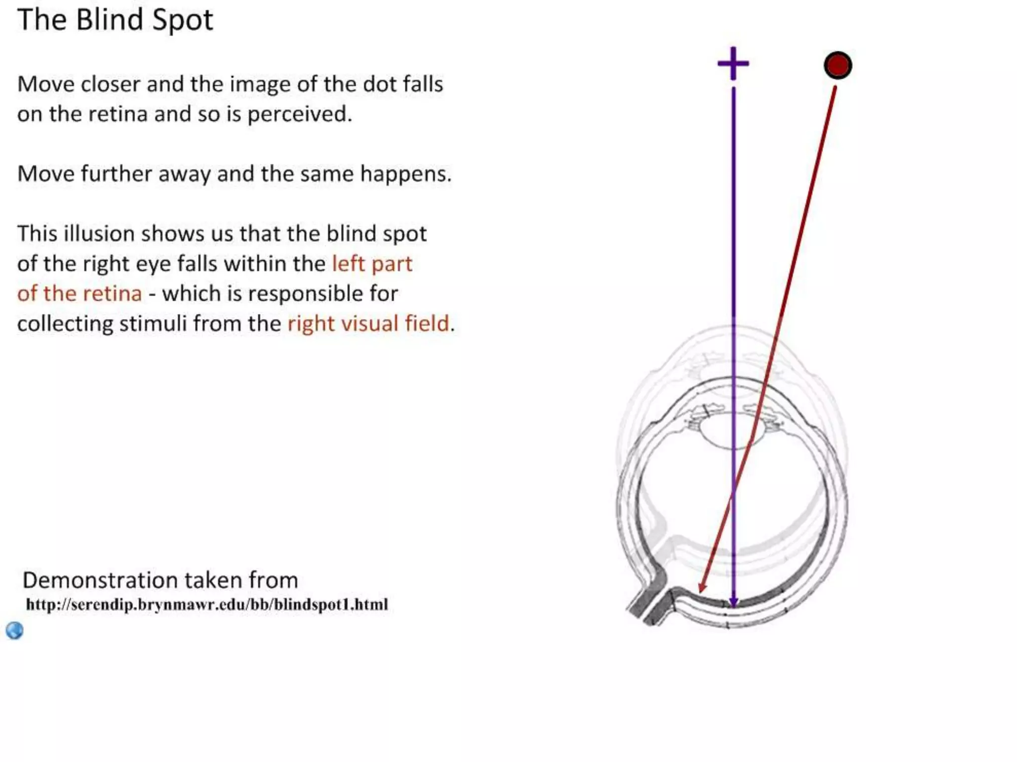
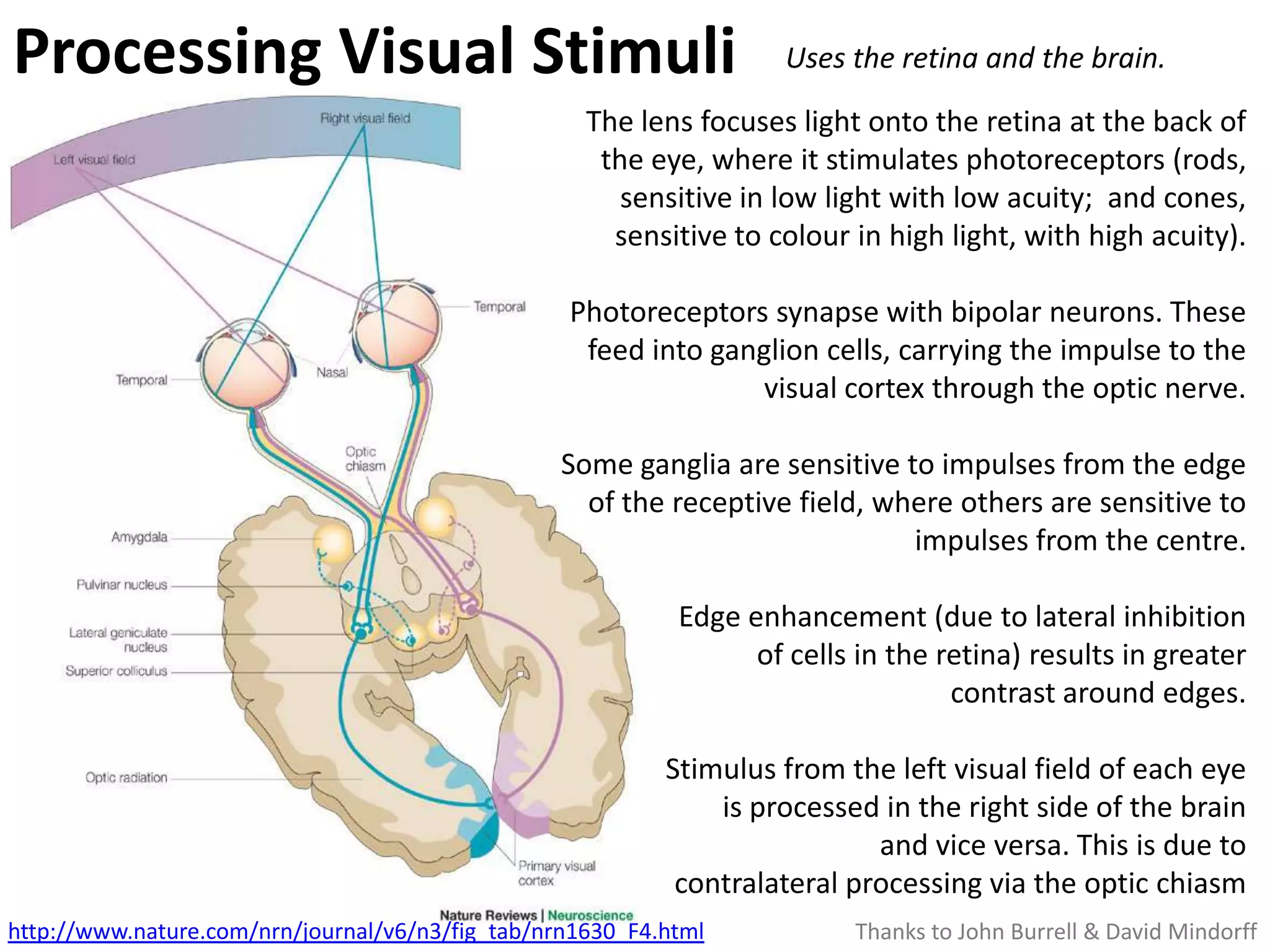
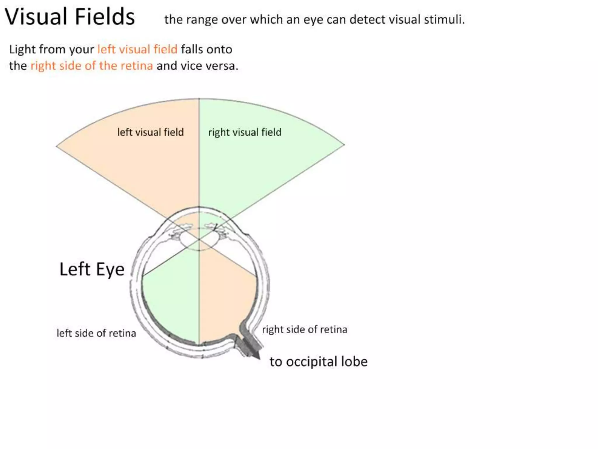
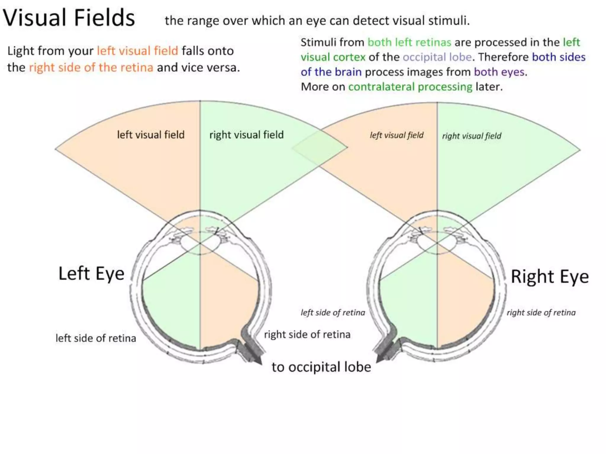
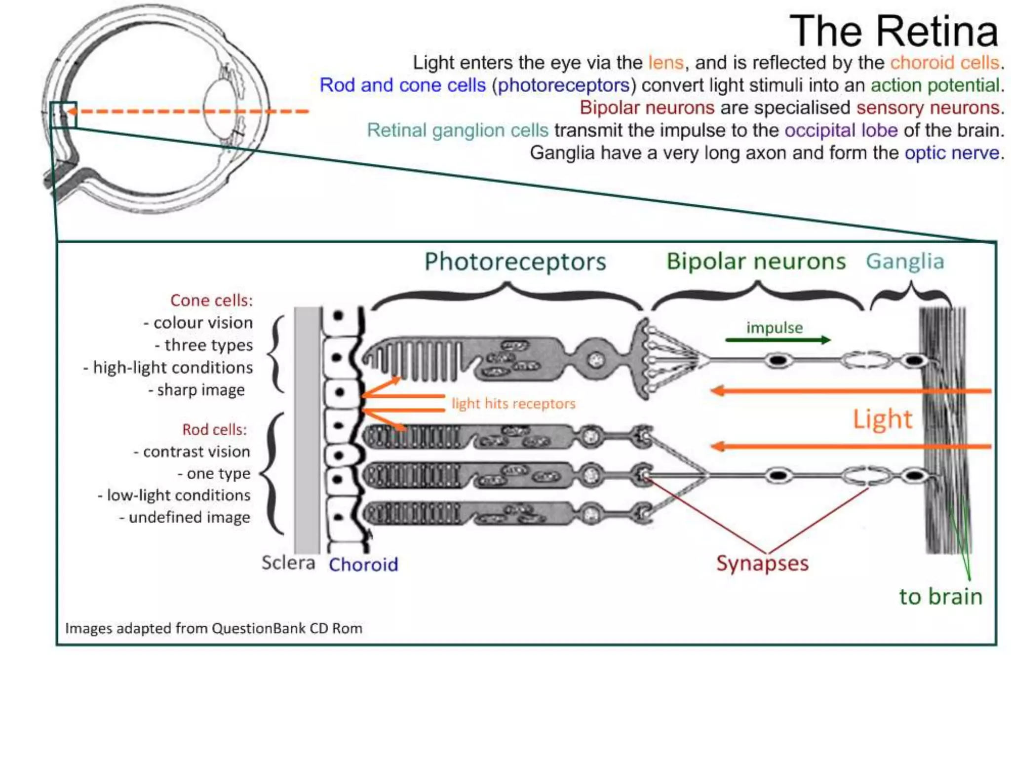
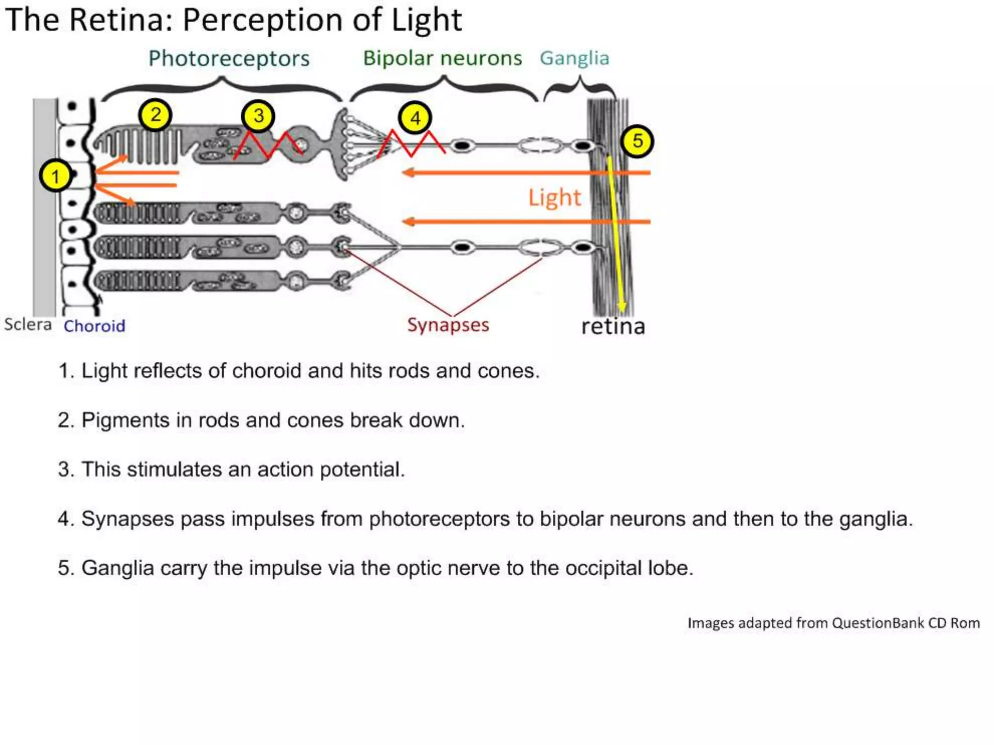
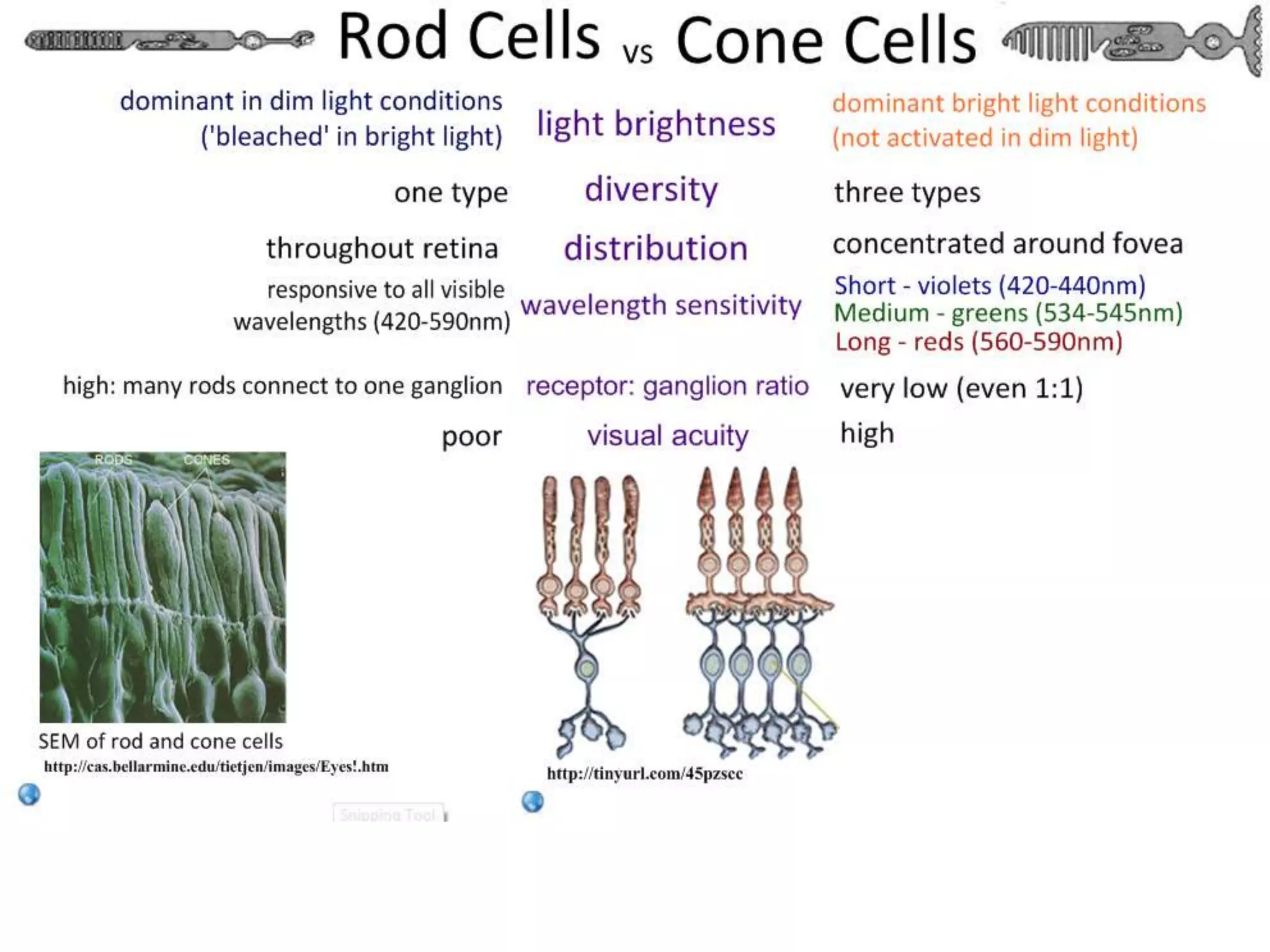
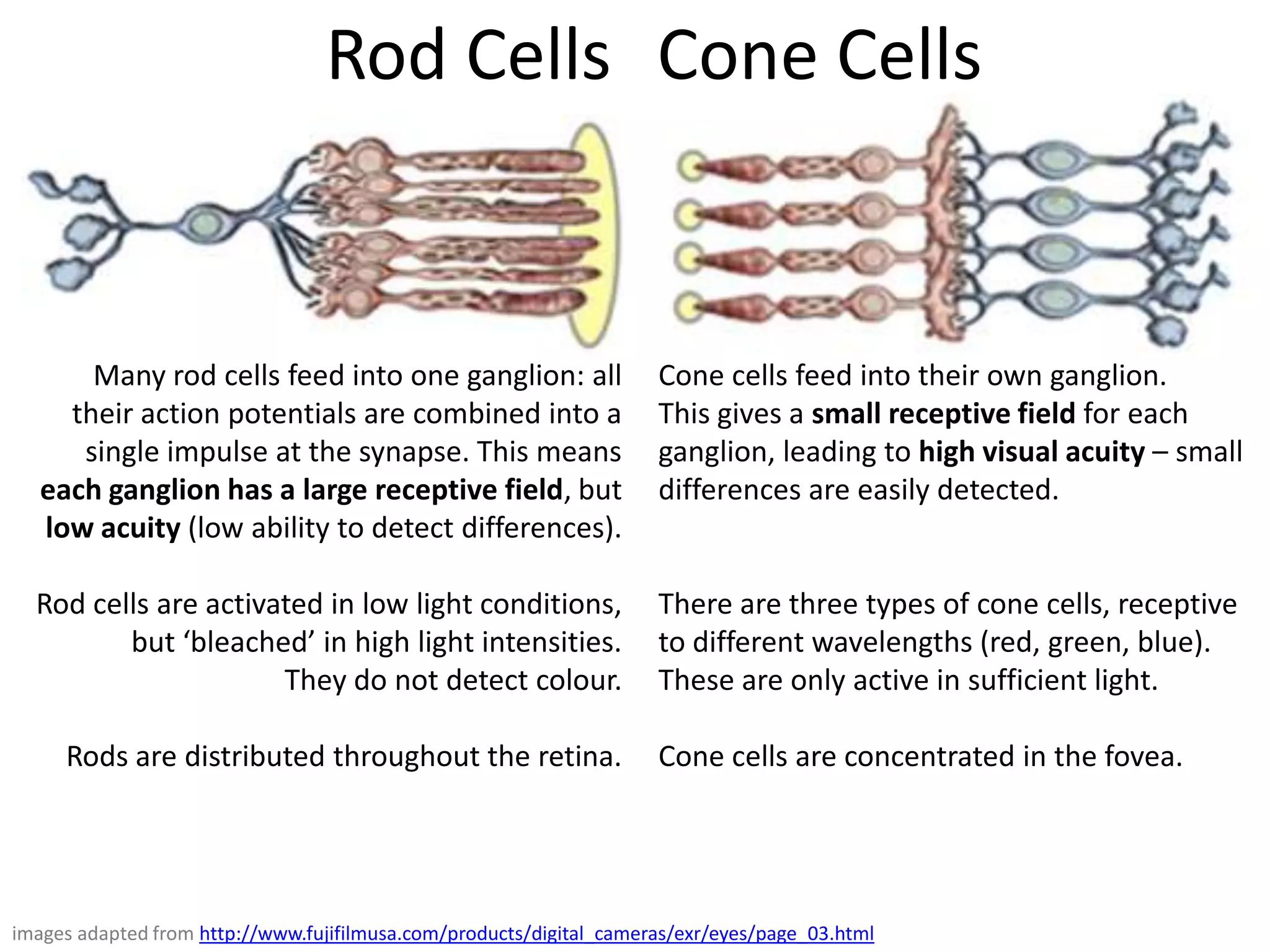
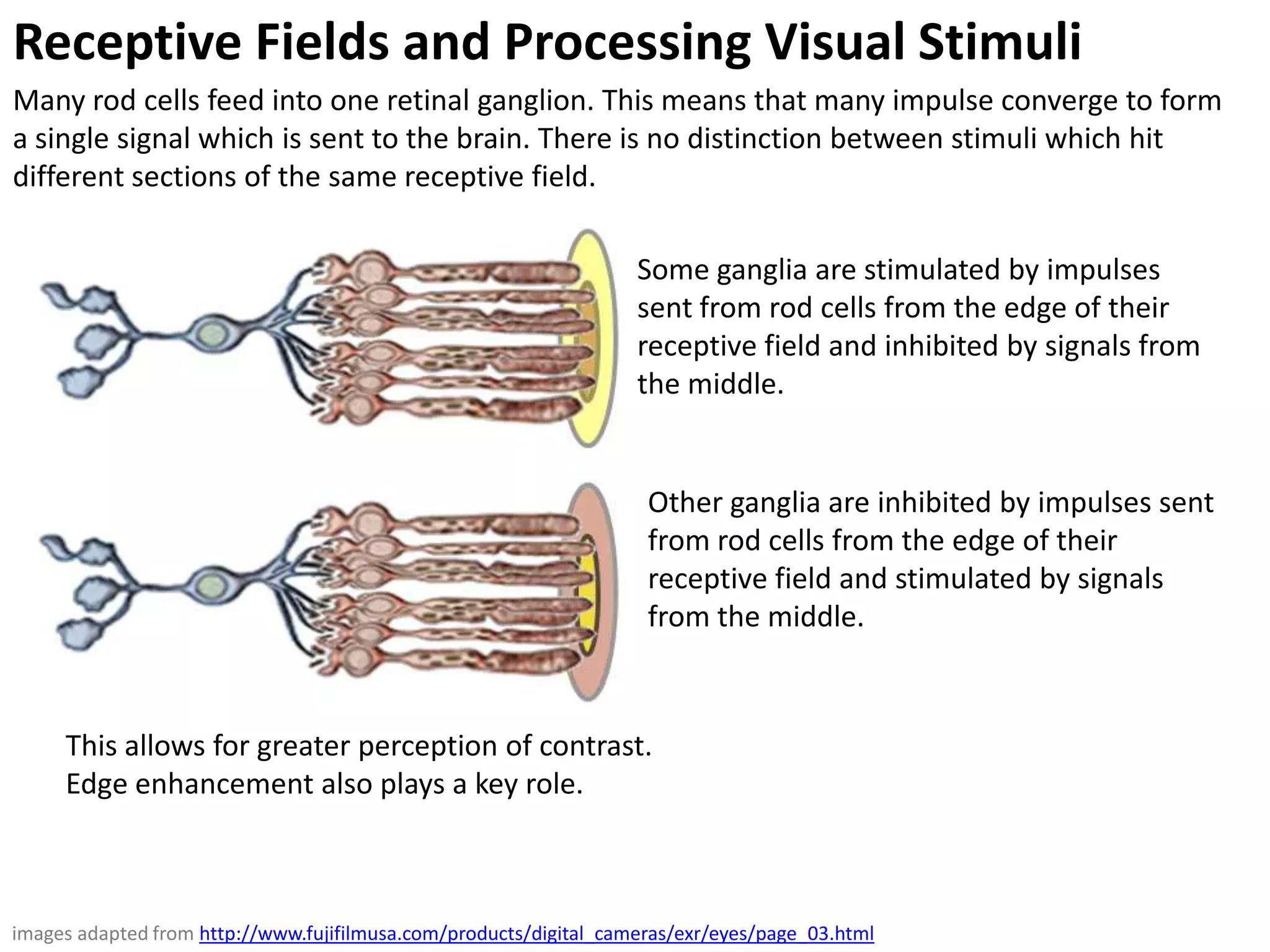
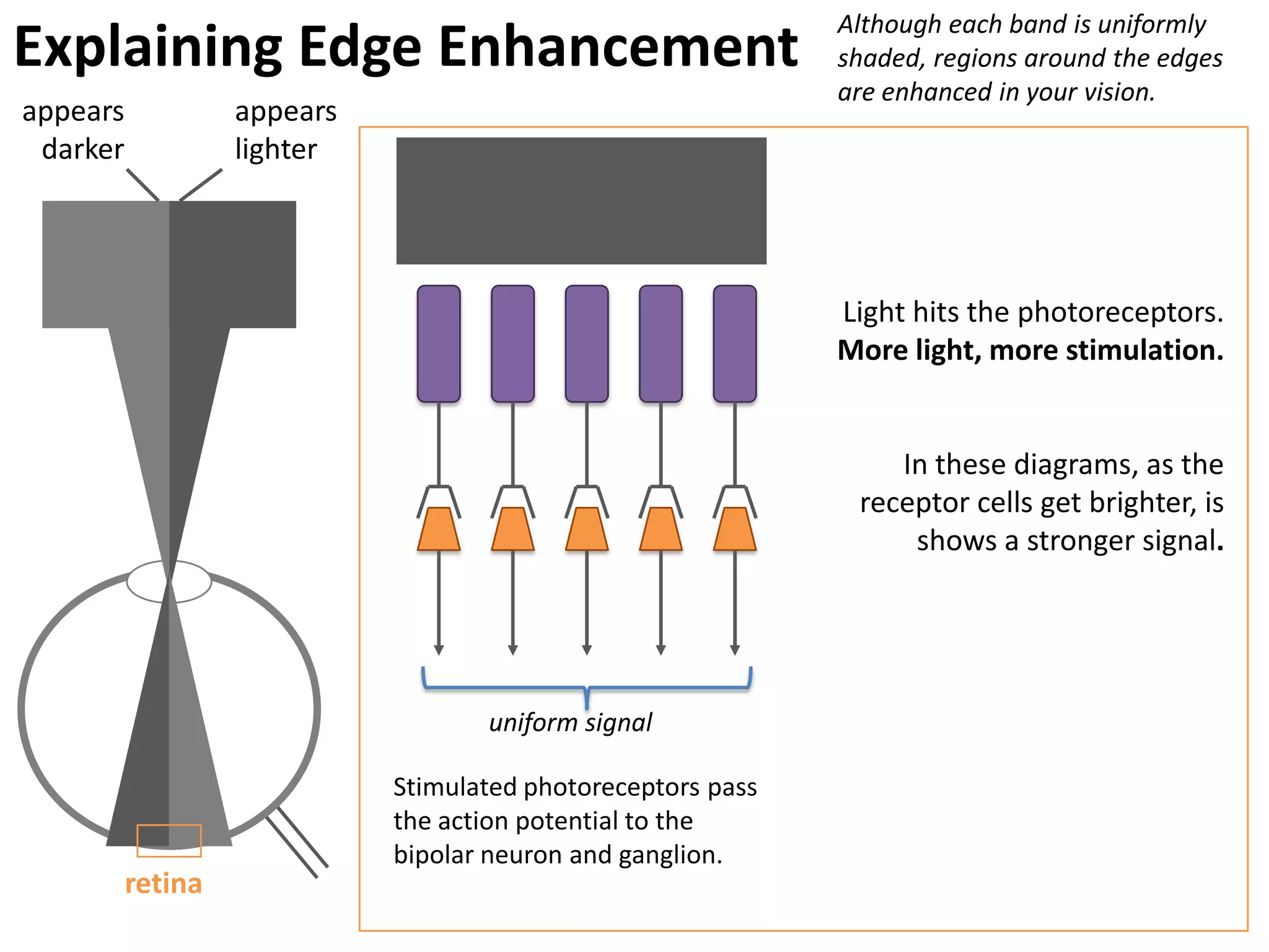
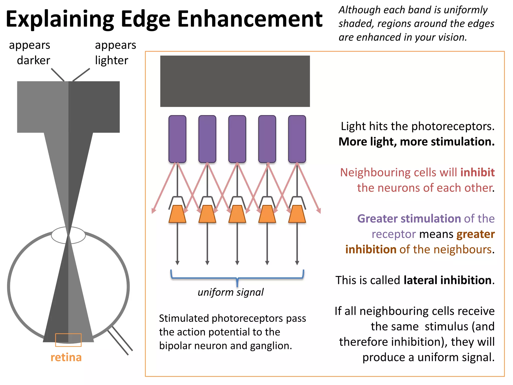
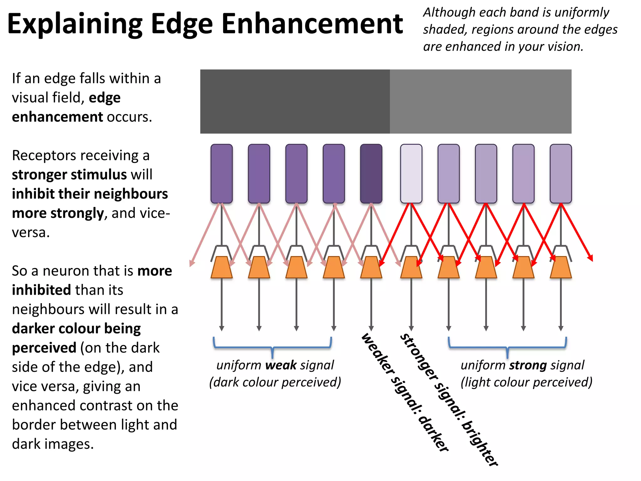
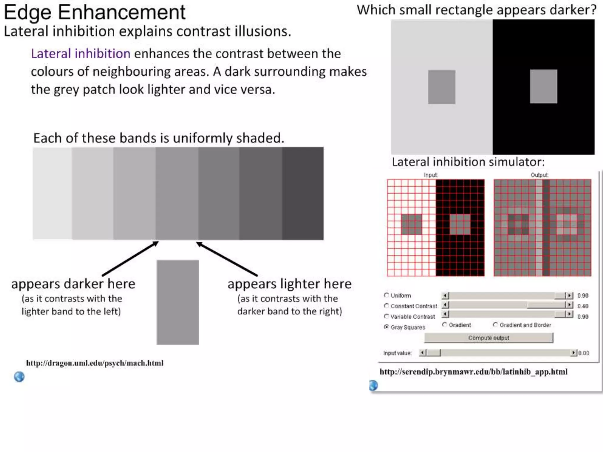
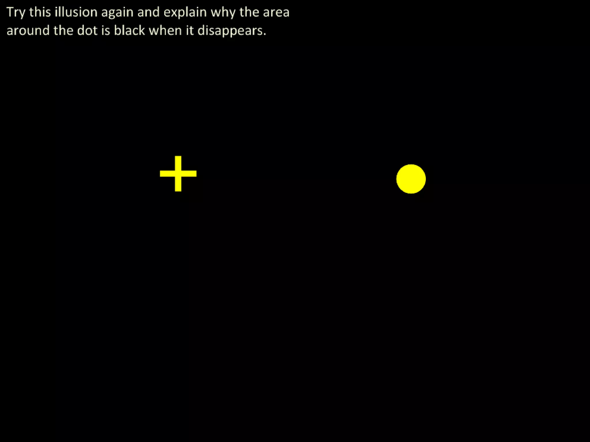
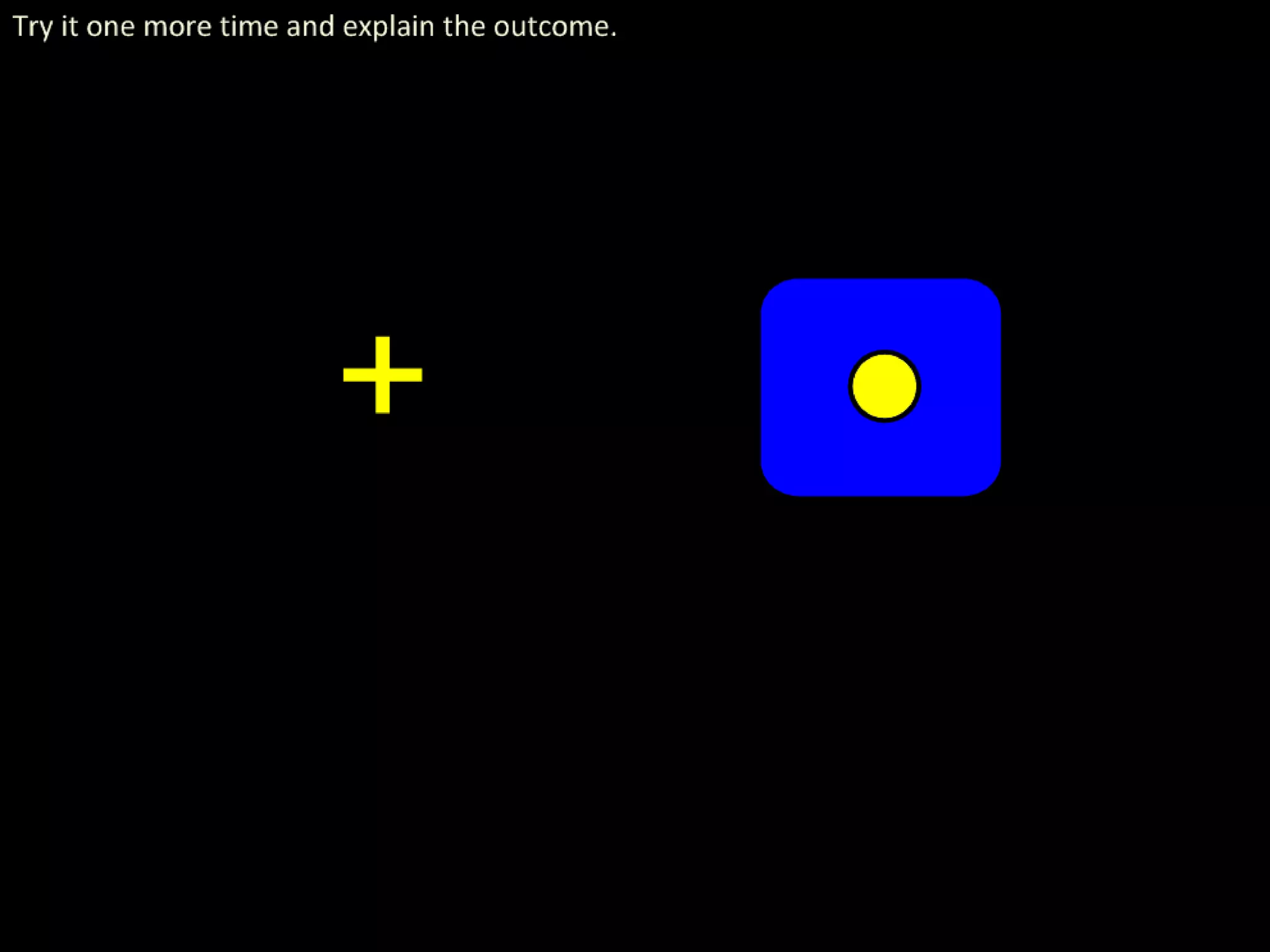
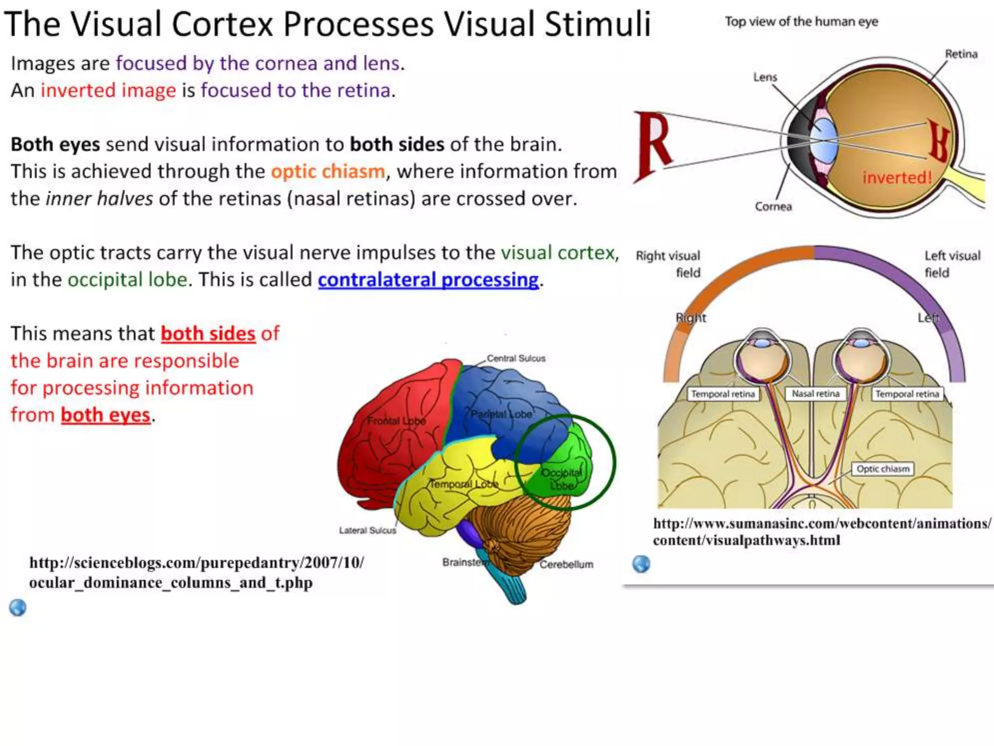
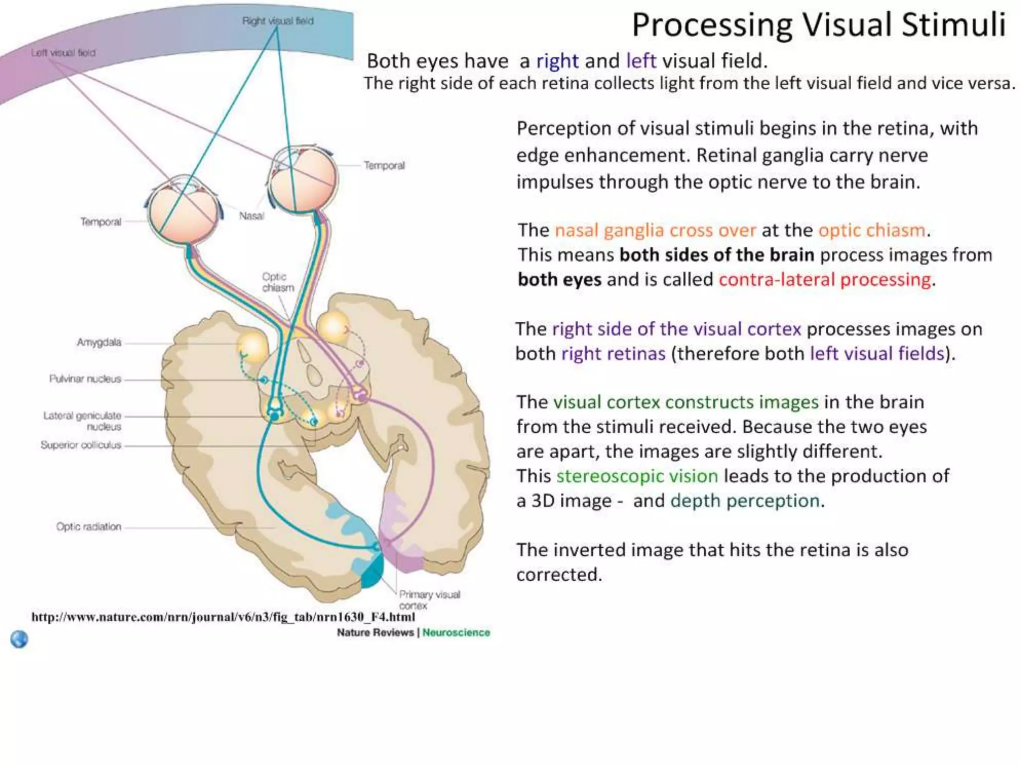
The document discusses how visual stimuli are processed. It explains that light is focused onto the retina by the lens, stimulating photoreceptors. There are rod and cone photoreceptors, with rods sensitive in low light and cones detecting color. Photoreceptors synapse with bipolar and ganglion cells, carrying signals through the optic nerve to the visual cortex. Edge enhancement occurs through lateral inhibition between photoreceptors, increasing contrast around edges. Stimuli from the left and right visual fields are processed in opposite sides of the brain.
12. Processing Visual Stimuli Uses the retina and the brain.
The lens focuses light onto the retina at the back of
the eye, where it stimulates photoreceptors
(rods, sensitive in low light with low acuity; and
cones, sensitive to colour in high light, with high
acuity).
Photoreceptors synapse with bipolar neurons. These
feed into ganglion cells, carrying the impulse to the
visual cortex through the optic nerve.
Some ganglia are sensitive to impulses from the edge
of the receptive field, where others are sensitive to
impulses from the centre.
Edge enhancement (due to lateral inhibition
of cells in the retina) results in greater
contrast around edges.
Stimulus from the left visual field of each eye
is processed in the right side of the brain
and vice versa. This is due to
contralateral processing via the & Davidchiasm
http://www.nature.com/nrn/journal/v6/n3/fig_tab/nrn1630_F4.html Thanks to John Burrell optic Mindorff
13.
14.
15.
16.
17.
18. Rod Cells Cone Cells
Many rod cells feed into one ganglion: all Cone cells feed into their own ganglion.
their action potentials are combined into a This gives a small receptive field for each
single impulse at the synapse. This means ganglion, leading to high visual acuity – small
each ganglion has a large receptive field, but differences are easily detected.
low acuity (low ability to detect differences).
Rod cells are activated in low light There are three types of cone cells, receptive
conditions, but ‘bleached’ in high light to different wavelengths (red, green, blue).
intensities. These are only active in sufficient light.
They do not detect colour.
Cone cells are concentrated in the fovea.
Rods are distributed throughout the retina.
images adapted from http://www.fujifilmusa.com/products/digital_cameras/exr/eyes/page_03.html
19. Receptive Fields and Processing Visual Stimuli
Many rod cells feed into one retinal ganglion. This means that many impulse converge to form
a single signal which is sent to the brain. There is no distinction between stimuli which hit
different sections of the same receptive field.
Some ganglia are stimulated by impulses
sent from rod cells from the edge of their
receptive field and inhibited by signals from
the middle.
Other ganglia are inhibited by impulses sent
from rod cells from the edge of their
receptive field and stimulated by signals
from the middle.
This allows for greater perception of contrast.
Edge enhancement also plays a key role.
images adapted from http://www.fujifilmusa.com/products/digital_cameras/exr/eyes/page_03.html
20. Although each band is uniformly
Explaining Edge Enhancement shaded, regions around the edges
are enhanced in your vision.
appears appears
darker lighter
Light hits the photoreceptors.
More light, more stimulation.
In these diagrams, as the
receptor cells get brighter, is
shows a stronger signal.
uniform signal
Stimulated photoreceptors pass
the action potential to the
bipolar neuron and ganglion.
retina
21. Although each band is uniformly
Explaining Edge Enhancement shaded, regions around the edges
are enhanced in your vision.
appears appears
darker lighter
Light hits the photoreceptors.
More light, more stimulation.
Neighbouring cells will inhibit
the neurons of each other.
Greater stimulation of the
receptor means greater
inhibition of the neighbours.
This is called lateral inhibition.
uniform signal
If all neighbouring cells receive
Stimulated photoreceptors pass
the action potential to the the same stimulus (and
bipolar neuron and ganglion. therefore inhibition), they will
retina produce a uniform signal.
22. Although each band is uniformly
Explaining Edge Enhancement shaded, regions around the edges
are enhanced in your vision.
If an edge falls within a
visual field, edge
enhancement occurs.
Receptors receiving a
stronger stimulus will
inhibit their neighbours
more strongly, and vice-
versa.
So a neuron that is more
inhibited than its
neighbours will result in a
darker colour being
perceived (on the dark
side of the edge), and uniform weak signal uniform strong signal
vice versa, giving an (dark colour perceived) (light colour perceived)
enhanced contrast on the
border between light and
dark images.
23. Explaining Edge Enhancement
Receptor A receives the A B C D Receptor D receives the
same light stimulus as B. same light stimulus as C.
Why is B darker than A? Why is C brighter than D?
A receives the same weak stimulus as its D receives the same strong stimulus as its
neighbours and so is inhibited equally by them. neighbours and so is inhibited equally by them.
B is next to C, which recieves a stronger stimulus C is next to B, which recieves a weaker stimulus
and therefore inhibits C more. As a result, B is and therefore inhibits C less. As a result, C is
overall more inhibited than A, so is darker. overall less inhibited than D, so is brighter.
24. It’s more like a gradient… see if you can explain why by annotating the diagram.
25.
26.
27. images adapted from http://www.fujifilmusa.com/products/digital_cameras/exr/eyes/page_03.html
28.
29.
30.
31.
32.
33.
34.
35.
36.
37. Wheels turning illusion from
http://www.newopticalillusions.com/moving-optical-illusions/two-wheels-new-optical-illusion/
@IBiologyStephen Please consider a donation to charity via Biology4Good.
Click here for more information about Biology4Good charity donations.
This is a Creative Commons presentation. It may be linked and embedded but not sold or re-hosted.