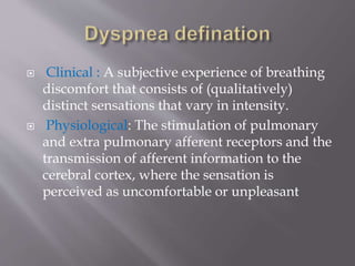
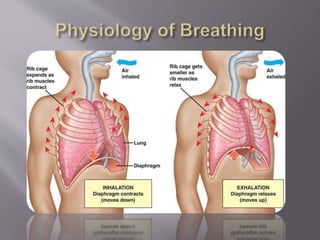
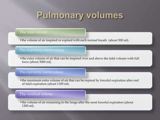
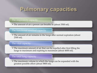
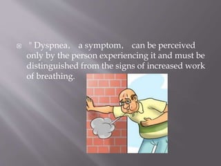
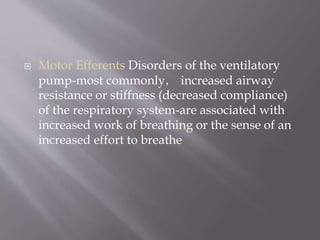
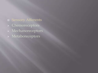
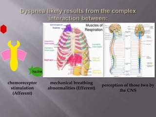
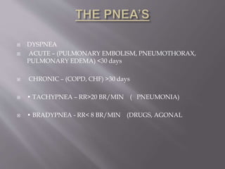
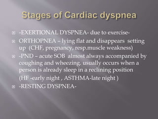
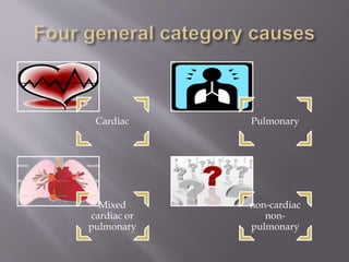
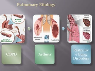
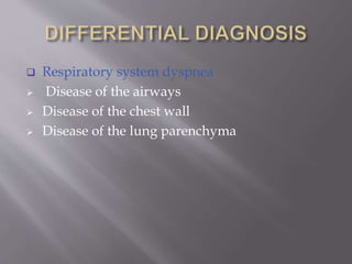
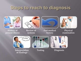
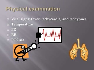
This document discusses dyspnea, or shortness of breath, from both clinical and physiological perspectives. Clinically, dyspnea is a subjective experience that varies in intensity, while physiologically it involves the stimulation of pulmonary and extra pulmonary receptors. The document outlines various lung volumes and capacities and explores the potential mechanisms of dyspnea. It also examines the evaluation, diagnosis, and treatment of different causes of dyspnea. A thorough history, physical exam, and testing are needed to diagnose the underlying condition and guide management.
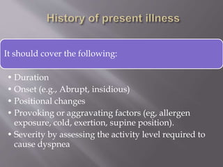
Cardiology 1.2. Dyspnea - by Dr. Farjad IkramFarjad Ikram
Introduction to one of the most common symptoms that can represent a wide range of diseases, from benign to life-threatening, covering number of systems including gastrointestinal, cardiovascular, pulmonary, musculoskeletal and psychiatric.
Template design credits - http://www.slidescarnival.com
This document discusses the approach to patients presenting with undifferentiated dyspnea. It begins by covering the pathophysiology and epidemiology of dyspnea. Important aspects of the history and physical exam are reviewed. The differential diagnosis is extensive, but the history, physical exam, chest x-ray can identify 66% of causes. The diagnostic approach involves considering if dyspnea is new, chronic, or both and if it is primarily pulmonary or cardiac in nature. Key tests include pulse oximetry, EKG, chest x-ray, and select use of labs, imaging, and biomarkers. Certain diagnoses like PE, MI and infection must be considered for every patient. A structured approach can efficiently identify the underlying cause
Dyspnea, also known as shortness of breath, is difficult or labored breathing. It is caused by conditions affecting the lungs, heart, and other organs and has pulmonary, cardiac, mixed, and non-cardiopulmonary causes. Risk factors include smoking, asthma, heart conditions, and exposure to irritants. Symptoms include cough, anxiety, chest pain, and rapid, shallow breathing. Treatment involves identifying and treating the underlying cause, using bronchodilators, steroids, oxygen therapy, and managing anxiety.
This document summarizes a seminar on dyspnea and respiratory failure. It defines dyspnea as subjective breathlessness and lists its potential causes such as cardiac, respiratory, or psychogenic issues. It describes different grading scales used to classify breathlessness. Types of dyspnea include orthopnea and paroxysmal nocturnal dyspnea. Respiratory failure is defined as impaired gas exchange shown by low oxygen and potentially high carbon dioxide levels. It discusses the mechanisms, clinical manifestations, differential diagnosis, and management of both dyspnea and respiratory failure.
Dyspnea is a subjective experience of breathing discomfort that results from interactions between physiological, psychological, social, and environmental factors. The document discusses the mechanisms, causes, evaluation, and management of dyspnea. Evaluation involves obtaining a thorough history regarding onset, timing, severity, and relieving/precipitating factors. A physical exam focuses on vital signs, respiratory exam, cardiovascular exam, and neurological exam to help identify potential causes like heart failure, COPD, pneumonia, or asthma.
This document provides an overview of the approach to evaluating and diagnosing dyspnea. It begins by defining dyspnea and describing its various causes. Mechanisms of dyspnea include stimulation of intrapulmonary nerves, mechanical loading of respiratory muscles, and hypoxia. A thorough history regarding onset, variation, and relieving/worsening factors is important. Physical exam focuses on vital signs, respiratory exam, and cardiac exam. Key investigations include chest imaging, pulmonary function tests, echocardiogram, and cardiopulmonary exercise testing to differentiate cardiovascular vs. respiratory causes. The document reviews descriptors, grading, history, physical exam findings, investigations and differential diagnosis of dyspnea in detail.
This document provides guidance on evaluating a patient presenting with shortness of breath (dyspnea). It outlines the key steps to reach a diagnosis, including taking a thorough history, physical examination, and interpreting findings from testing. The history should cover duration, onset, exacerbating factors, and severity of dyspnea. The physical exam evaluates vital signs, lung and heart exams, and looks for signs suggesting causes like heart failure. Interpretation of exam findings and testing like CXR, ECG, and blood tests guide the differential diagnosis. Likely causes in this case include pulmonary embolism given risk factors like recent immobilization, or pneumonia given imaging findings of consolidation and pleural effusion.
This document provides guidance on evaluating and diagnosing causes of acute breathlessness. It lists potential conditions based on presenting symptoms such as wheezing, stridor, crepitations, or a clear chest. Priority is given to pulse oximetry, ECG, chest x-ray, and blood tests. Common diagnoses are acute asthma, pulmonary edema, pneumonia, or COPD exacerbation. Features, urgent investigations, and expected blood gas results for each condition are outlined to aid clinical assessment.
This document provides information on evaluating and diagnosing shortness of breath. It lists various potential causes of shortness of breath including cardiac, lung, anatomical, trauma, and other issues. Specific conditions that could cause wheezing, stridor, crepitations, or a clear chest are identified. The speed of onset can help determine if the underlying cause is acute, subacute, or chronic. Guidelines for triaging patients with shortness of breath into green, yellow, or red zones based on dyspnea and oxygen saturation are also provided. The evaluation involves assessing severity, examining the chest, providing oxygen support if needed, and getting a chest x-ray.
This document discusses the diagnostic approach to patients presenting with dyspnea (shortness of breath). It outlines various potential causes of dyspnea including congestive heart failure, pulmonary edema, myocardial infarction, valvular heart disease, cardiomyopathy, chronic obstructive pulmonary disease, asthma, pulmonary embolism, pneumonia and other infections, and interstitial lung disease. It emphasizes gathering a clinical history, performing a physical exam, and utilizing ancillary tests like chest x-rays, ECGs, and blood tests to differentiate between cardiac and pulmonary causes to arrive at the correct diagnosis and guide treatment.
- A 67-year-old man presented with shortness of breath, productive cough with green sputum, feeling pale and feverish. On examination, he had reduced breath sounds and wheezing. Pulmonary function tests showed decreased lung volumes and airflow limitation.
- Differential diagnoses included COPD, asthma, pneumonia and congestive heart failure. Based on features favoring asthma like onset before age 20, variable symptoms, and response to bronchodilators, a diagnosis of asthma exacerbation was made.
- Treatment began with salbutamol via nebulizer. As symptoms were not resolving, prednisolone was added. Oxygen was given to maintain saturation above 92%. The patient's condition gradually
1. Dyspnea, or shortness of breath, can be caused by conditions affecting the heart, lungs, blood, chest wall, or nerves and muscles controlling breathing.
2. Physical examination may reveal signs related to cardiac causes like pulmonary edema, or pulmonary disorders like emphysema, chronic bronchitis, or restrictive lung diseases.
3. Investigations like chest X-ray, pulmonary function tests, CT scan of the chest and lung biopsy help to evaluate abnormalities and arrive at a diagnosis.
The document discusses various respiratory conditions that can cause dyspnea (difficult or labored breathing). It describes hyperventilation syndrome, which causes anxiety and dizziness due to overbreathing. Foreign body obstruction is a common cause of pediatric airway issues and can cause sudden dyspnea in adults. Pharyngeal edema, epiglottitis, and croup can cause upper airway swelling and obstruction. Asthma is a reversible obstructive lung disease often caused by allergens or irritants. Chronic obstructive pulmonary disease (COPD) includes chronic bronchitis and emphysema from long-term smoking. Pulmonary edema and pulmonary embolism can cause fluid or clots in the lungs resulting in
1. This case discusses a 45-year-old female patient presenting with progressive breathlessness, swelling of the lower limbs, and hard nodules on the palms and soles.
2. Differential diagnoses considered include mixed connective tissue disease, lupus, scleroderma, and rheumatoid arthritis.
3. Investigations revealed a strongly positive ANA, elevated markers of inflammation, interstitial lung disease, pulmonary hypertension, and features consistent with calcinosis cutis.
4. The final diagnosis was mixed connective tissue disease with interstitial lung disease, pulmonary hypertension, and calcinosis cutis. Management involves immunosuppressants like corticosteroids and
The patient is a 67-year-old male former smoker presenting with shortness of breath on exertion. Physical examination finds reduced breath sounds and wheezing. Tests show reduced lung function and oxygen levels. The differential diagnosis includes cardiac and pulmonary causes like COPD. Dyspnea is the medical term for shortness of breath and can result from various lung and heart conditions. Treatment focuses on the underlying cause, like using bronchodilators for COPD.
This document discusses acute dyspnea, defined as an uncomfortable need to breathe. It results from an imbalance between the perceived need and ability to breathe, often due to carbon dioxide buildup or oxygen deprivation. Common causes include pulmonary issues like pneumonia, cardiac issues like pulmonary edema, and metabolic acidosis. Diagnosis involves history, exam, and tests to identify the underlying cause. Treatment focuses on relieving symptoms like with opioids and addressing the specific condition through methods such as antibiotics for pneumonia or oxygen therapy for pulmonary edema.
Dyspnea, also known as shortness of breath, is difficult or labored breathing that can have many causes. The document outlines an approach to evaluating and treating dyspnea in patients. It describes taking a history, performing an examination, ordering relevant tests, and treating any underlying conditions found to be contributing to the dyspnea. Specific causes discussed include asthma, pulmonary edema, pneumonia, congestive heart failure, and acute coronary syndrome. For each, the document provides details on treatments aimed at opening airways, reducing fluid buildup, fighting infections, and improving blood flow.
This document defines and discusses dyspnea (shortness of breath) and cyanosis (blue discoloration of the skin). It outlines the mechanisms, causes, and characteristics of dyspnea associated with respiratory, cardiac, and other medical conditions. Key signs and symptoms that may suggest pulmonary or cardiac origins of dyspnea are provided. The document also defines and describes peripheral and central cyanosis, listing various conditions that can cause each type.
1) Dyspnea, or shortness of breath, is a subjective experience that results from interactions between physiological, psychological, and environmental factors. It can be caused by disorders of the ventilatory pump or sensory receptors in the lungs and muscles.
2) Pulmonary edema occurs when fluid accumulates in the lungs, which can be cardiogenic due to increased hydrostatic pressures from heart problems, or noncardiogenic from direct lung injury or diseases affecting the lung barrier.
3) Differentiating cardiogenic and noncardiogenic pulmonary edema involves examining physical exam findings, chest x-rays, and response to supplemental oxygen, as they have distinct mechanisms and presentations.
This document provides information on evaluating and diagnosing acute shortness of breath (dyspnea). It defines dyspnea and lists its potential causes, which include respiratory, cardiovascular, musculoskeletal, central nervous system, and gastrointestinal issues. The approach involves taking a thorough history, examining the patient, and conducting initial tests like chest x-ray, ECG, and arterial blood gas analysis to help determine the underlying cause, such as pneumonia, pulmonary edema, asthma exacerbation, or pulmonary embolism. Further tests like spirometry, echocardiogram, or cardiopulmonary exercise testing may be needed in some cases. The goal is to identify any life-threatening conditions and make a rapid diagnosis to guide treatment.
This document outlines an approach to evaluating and diagnosing dyspnea. It begins by defining dyspnea and noting its high prevalence. Types of dyspnea like orthopnea and paroxysmal nocturnal dyspnea are described. The diagnostic approach involves obtaining a detailed history regarding onset, duration, patterns and associated symptoms. A physical exam assesses respiratory effort, oxygenation, and signs of heart failure. Initial tests may include EKG, chest x-ray, and bloodwork. Further tests are guided by initial findings and may include echocardiogram, pulmonary function tests, CT, or arterial blood gas. Treatment focuses on the underlying cause identified through diagnosis.
Cough is a protective reflex that clears the lungs of secretions and foreign materials. It can be acute (<3 weeks), subacute (3-8 weeks), or chronic (>8 weeks). Coughs are classified based on duration and characterized by timing, associated symptoms, sputum production, and presence of blood. Causes include infections, lung diseases like COPD, and conditions like GERD or postnasal drip. Treatment depends on the underlying cause but may include over-the-counter medications, antibiotics, inhalers, or acid blockers.
This document provides an overview of the approach to a patient presenting with dyspnea. It begins with definitions of dyspnea and classifications including the modified Medical Research Council dyspnea scale and NYHA classification. It then discusses the receptors involved in the mechanism of dyspnea, common causes of acute, subacute and chronic dyspnea, important parts of the physical examination, red flags, and recommended initial testing.
Dyspnoea refers to undue awareness of one's own breathing and increased drive to breathe. It can be normal with strenuous exercise but is pathological if it occurs at lower thresholds. Dyspnoea has many potential causes including cardiac, respiratory, neuromuscular, metabolic issues or toxins. It is classified based on the cardiovascular and respiratory systems with grades ranging from no limitations to total confinement based on the level of physical activity one can perform without symptoms.
This patient presented with shortness of breath and was found to have pulmonary edema based on physical exam findings and chest x-ray results. Pulmonary edema can be caused by left ventricular failure from conditions like heart attack or hypertension. Key findings included elevated jugular venous pressure, crackles on lung auscultation, and hypoxemia. The patient was treated with oxygen, diuretics, and other medications to reduce pre- and afterload on the heart to relieve symptoms of pulmonary fluid buildup. Lifestyle changes and strict control of risk factors like diabetes and hypertension can help prevent further episodes.
This document discusses the approach to a patient presenting with dyspnea. It defines dyspnea and outlines its types, pathophysiology, physiological pathways, etiologies including cardiac, pulmonary, mixed and other causes. It describes the clinical features, assessment, relevant history, examinations and investigations for a patient with dyspnea. It concludes by stating that dyspnea is a common symptom that varies between individuals depending on physiological, psychological and social factors, and its management depends on identifying and treating the underlying cause.
This document discusses shortness of breath during pregnancy. It begins by noting that shortness of breath is common in pregnancy, occurring in about three quarters of women, especially in the first and third trimesters. It then explains the physiological reasons for shortness of breath in pregnancy, such as increased blood volume and the uterus pushing up on the diaphragm. The document differentiates between physiological and pathological causes. It provides tips for easing shortness of breath during pregnancy and signs that warrant a medical evaluation. The document concludes that distinguishing normal from abnormal shortness of breath is important for diagnosis and management.
histologic variants of oral squmous cell carcinoma /certified fixed orthodont...Indian dental academy
The Indian Dental Academy is the Leader in continuing dental education , training dentists in all aspects of dentistry and
offering a wide range of dental certified courses in different formats.for more details please visit
www.indiandentalacademy.com
This document provides an overview of a training course on treating acute respiratory distress. It covers identifying and treating ARD caused by conditions like congestive heart failure, COPD, asthma, and other less common causes. Treatment techniques discussed include oxygen administration, positioning, ventilation, assisting with patient medications, and using adjuncts like pulse oximetry and humidifiers. The objectives, signs and symptoms, and pre-hospital treatments are reviewed for different conditions that can cause ARD like CHF, COPD, anaphylaxis, hyperventilation, and spontaneous pneumothorax.
- Hyperventilation syndrome (HVS) was classically defined as excessive ventilation leading to chemical and physiological changes causing dysphoric symptoms, but many patients with HVS do not have low carbon dioxide levels. It is now termed behavioral breathlessness or psychogenic dyspnea.
- Symptoms of HVS and panic disorder overlap, with around half of patients with panic disorder and 60% of patients with agoraphobia experiencing HVS symptoms. Around 25% of HVS patients also meet criteria for panic disorder.
- The exact pathophysiology is unknown but theories involve abnormal respiratory responses to stress, caffeine, lactate or carbon dioxide as well as learned abnormal breathing patterns from overprotective parents in
Tobacco use is a major risk factor for oral cancer. Tobacco products contain numerous carcinogens like nitrosamines and polycyclic aromatic hydrocarbons that can cause DNA damage and lead to cancer. Oral lesions caused by tobacco include leukoplakia, erythroplakia, nicotine stomatitis and snuff keratosis which have increased risk of becoming cancerous. Squamous cell carcinoma is the most common type of oral cancer, often found on the tongue, floor of mouth and lips. Early detection of pre-cancerous lesions and treatment can help prevent oral cancer caused by tobacco.
The document discusses various verrucal-papillary lesions of the oral cavity including reactive lesions such as papillary hyperplasia, condyloma latum, squamous papilloma, condyloma acuminatum, and focal epithelial hyperplasia. It also discusses neoplasms like keratoacanthoma and verrucous carcinoma. Rare lesions of unknown etiology discussed include pyostomatitis vegetans and verruciform xanthoma. Each lesion is described in terms of etiology, clinical features, histopathology, differential diagnosis, and treatment.
Dr. Chaudhary's presentation discussed the dual wave-particle nature of X-rays and their interaction with matter. X-rays can behave as both waves, which allows them to be reflected, and particles called photons. The photoelectric effect occurs when a photon interacts with and ejects an electron from an atom, becoming absorbed. This produces characteristic radiation as the electron vacancy is filled. The photoelectric effect yields an ion, photoelectron, and photon, and is more likely with low energy photons and high atomic number elements if the photon energy exceeds the electron's binding energy. It provides excellent radiographic images with no scatter but maximum radiation exposure to the patient.
Hyperventilation syndrome (HVS) is a condition characterized by breathing that exceeds metabolic demands, resulting in chemical and physiological changes that produce dysphoric symptoms. It commonly presents with chest pain, dizziness, tingling, and anxiety. While the underlying cause is not fully understood, stress and anxiety are thought to trigger exaggerated breathing patterns. HVS exists in both acute and chronic forms, with chronic HVS being more difficult to diagnose due to subtle or absent hyperventilation. Treatment focuses on breathing retraining and stress reduction techniques.
Syncope is a transient loss of consciousness due to decreased blood flow to the brain. It has many potential causes including cardiac arrhythmias, orthostatic hypotension, and vasovagal responses. Management involves stopping any procedures, placing the patient supine with legs elevated, assessing ABCs, providing oxygen, and monitoring vitals. For presyncope, stopping and allowing recovery is usually sufficient while syncope may require interventions like atropine for bradycardia. Thorough history and evaluation of potential causes is important to prevent future episodes.
Interaction of x and gamma rays with matterVarun Babu
1. When photons interact with matter, they can be transmitted, absorbed, or scattered. Absorption and scattering are stochastic processes and it is impossible to predict the fate of individual photons.
2. The linear attenuation coefficient measures the probability that a photon interacts per unit length of material and depends on the material's density, atomic number, and photon energy. As photon energy decreases or atomic number/density increases, attenuation increases.
3. The main interaction processes are the photoelectric effect, Compton scattering, and elastic scattering. The photoelectric effect dominates for high Z materials and low energy photons, while Compton scattering is more important for low Z materials and high energy photons. Secondary electrons and ionization produced are
This document discusses the evaluation and assessment of dyspnea (shortness of breath). It defines dyspnea as a subjective experience that can have multiple physiological and psychological causes. The author examines the respiratory and cardiovascular systems and how various components (controller, ventilatory pump, gas exchanger, heart, etc.) can lead to dyspnea if deranged. Clinical assessment of dyspnea includes temporal patterns, exertion levels, descriptors used, and associated symptoms. Initial testing for chronic dyspnea involves tests like spirometry, chest X-ray, ECG, and blood tests to evaluate the most common causes of asthma, COPD, heart issues, and interstitial lung disease.
Dyspnea derives from Greek for “ “ shortness of breath hard breathing ”. It is often also described as ”. This is a subjective sensation of breathing, from mild discomfort to feelings of suffocation. It is a sign of a variety of disorders and is primarily an indication of ventilation or of inadequate insufficient amounts of oxygen in the circulating blood .
Dyspnea happens when a “mismatch” occurs between afferent and efferent signaling. As the brain receives afferent ventilation information, it is able to compare it to the current level of respiration by the efferent signals. If the level of respiration is inappropriate for the body’s status and need, then dyspnea might occur
Dyspnea, also known as shortness of breath, is a subjective sensation that results from a mismatch between the brain's perception of how much ventilation is needed and the amount of ventilation that is occurring. Three main components contribute to dyspnea: afferent signals from receptors in the lungs and airways, efferent signals from the brain to respiratory muscles, and central processing in the brain. A variety of cardiac, pulmonary, and other medical disorders can cause dyspnea through different mechanisms that impact ventilation. Management of dyspnea involves treating its underlying cause, reducing ventilatory demand, improving respiratory muscle function, and techniques to decrease the sense of respiratory effort.
Dyspnea, or shortness of breath, is a common symptom that can be caused by many cardiac and pulmonary conditions. A thorough diagnostic evaluation of dyspnea involves taking a detailed patient history, conducting a physical exam, and obtaining initial tests like an electrocardiogram, chest x-ray, and blood tests to evaluate for conditions involving the heart, lungs, blood, and other potential causes and to guide further testing if needed. Grading scales are used to characterize the severity of a patient's dyspnea. The pathophysiology of dyspnea involves an imbalance between the perceived need to breathe and the ability to breathe.
This document provides guidance on taking a history and conducting a physical exam of the cardiorespiratory system. It outlines key questions to ask about symptoms like cough, sputum production, hemoptysis, breathlessness, chest pain, edema, respiratory sounds, palpitations, syncope, and cyanosis. The physical exam section describes inspection of the general appearance, face, neck, chest, lungs, heart, veins, and peripheral circulation. Examples are given for interpreting different symptom patterns.
This document covers several topics related to respiratory pathophysiology:
1. It describes the anatomy and control of breathing, including the medullary respiratory center and pontine and apneustic areas.
2. Various types of breathing patterns are defined, such as Cheyne-Stokes respirations and Biot's respiration, along with the areas of brain injury that cause each pattern.
3. Common respiratory symptoms like cough, dyspnea, and hemoptysis are discussed alongside their typical causes.
4. Physical exam findings on chest auscultation and percussion are outlined, including vocal fremitus and lung sounds.
5. The calculation of the alveolar-arterial oxygen
The document discusses breathlessness/dyspnoea by defining it, describing its pathophysiology, types, differential diagnosis, clinical assessment, investigations, and treatment. Breathlessness has no defined receptors or localized brain representation and can be caused by health issues like exercise or diseases of the lungs, heart, or muscles. Its assessment considers consciousness, cyanosis, breathing efforts, oxygenation, speech, and cardiovascular status. Investigations may include chest X-rays, ECGs, blood gases, and tests to identify specific causes, while treatment depends on the initial diagnosis.
The document discusses the approach to evaluating and diagnosing dyspnea. It defines dyspnea and describes the mechanisms that can cause it, including afferent and efferent signals in the respiratory system. The evaluation involves taking a thorough history, performing a physical exam focusing on respiratory and cardiac systems, ordering relevant lab and imaging tests, and potentially a cardiopulmonary exercise test. A wide range of cardiac, pulmonary, neuromuscular, and other conditions can lead to acute or chronic dyspnea.
THIS PPT CONTAINS DESCRIPTION ABOUT HISTORY TAKING IN PATIENTS WITH CARDIORESPIRATORY DISEASES, EXPLAINED IN DETAILS ABOUT ALL SYMPTOMS & ITS DETAILED HISTORY.
This document discusses manifestations and disorders of the respiratory system. It covers several topics including:
- Cardinal respiratory symptoms like cough, sputum, dyspnea, and cyanosis.
- Disorders of lung mechanics including airway obstruction at different levels and abnormalities of the lung parenchyma and chest wall.
- Disorders of gas exchange that can cause hypoxemia or hypercapnia due to issues like ventilation/perfusion mismatching or overall alveolar hypoventilation.
- Other respiratory conditions addressed include cough, dyspnea, hypoxemia, cyanosis, and hypercapnia. Primary respiratory disorders can also affect other body systems like the cardiovascular system.
Atelectasis is defined as the collapse or closure of the lung resulting in reduced or absent gas exchange. It develops when alveoli become airless from absorption of air without replacement through breathing. Common causes include obstruction of the airway by secretions, diminished distention of alveoli due to chest wall issues or neuromuscular disorders, and anesthesia effects. Signs include cough, dyspnea, and hypoxemia. Treatment focuses on clearing secretions through positioning, deep breathing, and chest physical therapy. Bronchodilators and mucolytics may also be used, along with mechanical ventilation for severe cases.
This document provides information on obstructive sleep apnea (OSA), including its physiology, risk factors, symptoms, diagnosis, and treatment. OSA involves pauses in breathing during sleep due to upper airway collapse. It is diagnosed through an overnight sleep study that measures breathing, oxygen levels, and brain waves. A high number of breathing pauses or dips in oxygen (apnea-hypopnea index over 5) indicates OSA. Common symptoms include loud snoring, witnessed breathing pauses, and daytime sleepiness. Risk factors include obesity, large neck size, and family history. Treatment typically involves a CPAP machine to keep the airway open during sleep.
The document describes various respiratory conditions in children. It discusses the essential components of the respiratory system and signs of respiratory distress. Common signs include tachypnea, retractions, and altered mental status. Obstructive conditions like asthma involve airway narrowing and present with wheezing, while restrictive conditions involve stiff lungs and present with shallow, rapid breathing. Common respiratory illnesses in children such as bronchiolitis, pneumonia, and pertussis are also outlined.
Obstructive sleep apnea (OSA) is a common sleep disorder characterized by recurrent episodes of upper airway collapse during sleep, which can fragment sleep and cause daytime sleepiness. Risk factors include obesity, large neck size, and anatomical features that narrow the airway. Polysomnography is the gold standard test to diagnose OSA by measuring breathing patterns, oxygen levels, and brain waves during sleep. Left untreated, OSA is associated with increased risks of hypertension, heart disease, and stroke. Treatment involves lifestyle changes and devices like CPAP that maintain airway pressure during sleep.
The document discusses considerations for transporting patients by air, including assessing their cardiovascular, respiratory and general health status prior to flight and taking precautions to address issues like hypoxia, changes in barometric pressure, thermal stress, noise and vibration during transport. It provides guidance on stabilizing patients, securing airways, providing oxygen, monitoring vital signs, and positioning patients comfortably for air travel.
This document discusses dyspnea (shortness of breath) and asthma. It begins by defining dyspnea and describing its pathophysiology, which involves a mismatch between afferent signals from lung receptors and efferent motor signals. It then discusses the various causes and categories of dyspnea, including cardiac, pulmonary, cardiac/pulmonary, and non-cardiac/non-pulmonary. The document provides details on differentiating dyspnea through history, physical exam, and investigations. It also discusses types of dyspnea and provides a diagnostic algorithm. Later sections focus on asthma, covering etiology, risk factors, physical exam findings, investigations such as spirometry, and classifications of asthma exacerbations.
The document summarizes key aspects of the respiratory system for EMTs, including anatomy, physiology, assessment, and initial management of respiratory emergencies. It describes the respiratory system's purpose of gas exchange, relevant anatomy such as the lungs and airways, normal physiology of breathing, common pathologies affecting ventilation and gas exchange, and the ABCDE approach to assessment and initial management of patients with respiratory distress or failure.
This document provides an overview of the approach to dyspnea. It defines dyspnea and describes related terms like orthopnea. The mechanisms of orthopnea and paroxysmal nocturnal dyspnea are increased venous return when lying flat overwhelming the heart. Receptors involved in the perception of dyspnea are described. Common causes of acute and chronic dyspnea from cardiovascular, pulmonary, and other systems are listed. The approach involves assessing airway, breathing, vital signs, history, and focused physical exam. Key exam findings that can point to different diagnoses are outlined. Important investigations include spirometry, ABG, imaging, and ECG.
This document provides information on assessing and managing various respiratory emergencies. It discusses the anatomy and physiology of breathing and defines conditions such as asthma, chronic obstructive pulmonary disease (COPD), hyperventilation, epiglottitis, and pulmonary embolism. For each condition, it outlines signs and symptoms and guidelines for treatment, including administering oxygen, positioning the patient, and alerting emergency services. The overall goal is to familiarize first responders with respiratory systems and protocols for responding to breathing difficulties.
1) Management of acute myocardial infarction involves aspirin, P2Y12 inhibitors like prasugrel or ticagrelor, and revascularization with either primary PCI or fibrinolytic therapy.
2) Antiplatelet therapy with dual antiplatelet therapy (DAPT) for 1 year is indicated for all patients after drug-eluting or bare-metal stents.
3) Early use of beta blockers, ACE inhibitors, and statins provides long-term benefits in reducing mortality for patients with acute MI.
1) Thyroid storm, or a thyrotoxic crisis, is a life-threatening exacerbation of hyperthyroidism caused by an abrupt release of thyroid hormones into circulation.
2) It presents with fever, tachycardia, arrhythmias, heart failure, tremors, and gastrointestinal issues like nausea and vomiting.
3) Treatment involves controlling adrenergic symptoms with beta blockers, treating the underlying thyroid abnormality with antithyroid drugs like PTU, and providing supportive care like fluids and electrolyte replacement.
1. Hypothyroidism is a clinical syndrome where the thyroid gland fails to produce sufficient thyroid hormones.
2. It can be primary, meaning it is caused by problems with the thyroid gland itself, or central/secondary, caused by problems with the pituitary gland or hypothalamus.
3. Symptoms affect many body systems and include fatigue, weight gain, dry skin, constipation, muscle weakness, decreased heart rate, impaired cognition and more.
Heart failure is a complex clinical syndrome resulting from structural or functional impairment of the ventricles. It leads to symptoms of dyspnea and fatigue. Treatment involves correcting reversible causes, using diuretics, ACE inhibitors, beta blockers, aldosterone antagonists, and devices like ICDs and CRT. Nonpharmacological treatments include diet, exercise, and procedures like CABG or transplantation for advanced cases. The goal is to relieve symptoms, improve function, and prevent death and hospitalizations.
1) The document discusses various tests of coagulation and platelet function, including bleeding time, activated partial thromboplastin time (aPTT), prothrombin time (PT), thrombin time (TT), and INR. These tests measure different parts of the coagulation cascade.
2) Disseminated intravascular coagulation (DIC) is described as a condition where excessive blood protease activity leads to widespread fibrin formation in blood vessels. Common causes include infection, cancer, trauma, and pregnancy complications. DIC can cause both bleeding and thrombosis.
3) Laboratory findings in DIC may include low platelet count, prolonged aPTT and PT, elevated D-dimer, and presence of
1) Management of acute myocardial infarction involves aspirin, P2Y12 inhibitors like prasugrel or ticagrelor, and revascularization with either primary PCI or fibrinolytic therapy.
2) Antiplatelet therapy with dual antiplatelet therapy (DAPT) for 1 year is indicated for all patients after drug-eluting or bare-metal stents.
3) Early use of beta blockers, ACE inhibitors, and statins provides long-term benefits in reducing mortality for patients with acute MI.
This document provides an overview of acid-base disturbances, including normal values and types of metabolic and respiratory acid-base disorders. It discusses mixed acid-base disturbances and evaluating the appropriateness of the compensatory response. It describes causes and features of increased and normal anion gap metabolic acidosis, lactic acidosis, diabetic ketoacidosis, alcoholic acidosis, and uremic acidosis. It also discusses metabolic alkalosis, including saline-responsive and unresponsive types, and respiratory acidosis. Treatment approaches are outlined for different acid-base disorders.
This presentation was provided by Racquel Jemison, Ph.D., Christina MacLaughlin, Ph.D., and Paulomi Majumder. Ph.D., all of the American Chemical Society, for the second session of NISO's 2024 Training Series "DEIA in the Scholarly Landscape." Session Two: 'Expanding Pathways to Publishing Careers,' was held June 13, 2024.
Level 3 NCEA - NZ: A Nation In the Making 1872 - 1900 SML.pptHenry Hollis
The History of NZ 1870-1900.
Making of a Nation.
From the NZ Wars to Liberals,
Richard Seddon, George Grey,
Social Laboratory, New Zealand,
Confiscations, Kotahitanga, Kingitanga, Parliament, Suffrage, Repudiation, Economic Change, Agriculture, Gold Mining, Timber, Flax, Sheep, Dairying,
Leveraging Generative AI to Drive Nonprofit InnovationTechSoup
In this webinar, participants learned how to utilize Generative AI to streamline operations and elevate member engagement. Amazon Web Service experts provided a customer specific use cases and dived into low/no-code tools that are quick and easy to deploy through Amazon Web Service (AWS.)
Gender and Mental Health - Counselling and Family Therapy Applications and In...PsychoTech Services
A proprietary approach developed by bringing together the best of learning theories from Psychology, design principles from the world of visualization, and pedagogical methods from over a decade of training experience, that enables you to: Learn better, faster!
Chapter wise All Notes of First year Basic Civil Engineering.pptxDenish Jangid
Chapter wise All Notes of First year Basic Civil Engineering
Syllabus
Chapter-1
Introduction to objective, scope and outcome the subject
Chapter 2
Introduction: Scope and Specialization of Civil Engineering, Role of civil Engineer in Society, Impact of infrastructural development on economy of country.
Chapter 3
Surveying: Object Principles & Types of Surveying; Site Plans, Plans & Maps; Scales & Unit of different Measurements.
Linear Measurements: Instruments used. Linear Measurement by Tape, Ranging out Survey Lines and overcoming Obstructions; Measurements on sloping ground; Tape corrections, conventional symbols. Angular Measurements: Instruments used; Introduction to Compass Surveying, Bearings and Longitude & Latitude of a Line, Introduction to total station.
Levelling: Instrument used Object of levelling, Methods of levelling in brief, and Contour maps.
Chapter 4
Buildings: Selection of site for Buildings, Layout of Building Plan, Types of buildings, Plinth area, carpet area, floor space index, Introduction to building byelaws, concept of sun light & ventilation. Components of Buildings & their functions, Basic concept of R.C.C., Introduction to types of foundation
Chapter 5
Transportation: Introduction to Transportation Engineering; Traffic and Road Safety: Types and Characteristics of Various Modes of Transportation; Various Road Traffic Signs, Causes of Accidents and Road Safety Measures.
Chapter 6
Environmental Engineering: Environmental Pollution, Environmental Acts and Regulations, Functional Concepts of Ecology, Basics of Species, Biodiversity, Ecosystem, Hydrological Cycle; Chemical Cycles: Carbon, Nitrogen & Phosphorus; Energy Flow in Ecosystems.
Water Pollution: Water Quality standards, Introduction to Treatment & Disposal of Waste Water. Reuse and Saving of Water, Rain Water Harvesting. Solid Waste Management: Classification of Solid Waste, Collection, Transportation and Disposal of Solid. Recycling of Solid Waste: Energy Recovery, Sanitary Landfill, On-Site Sanitation. Air & Noise Pollution: Primary and Secondary air pollutants, Harmful effects of Air Pollution, Control of Air Pollution. . Noise Pollution Harmful Effects of noise pollution, control of noise pollution, Global warming & Climate Change, Ozone depletion, Greenhouse effect
Text Books:
1. Palancharmy, Basic Civil Engineering, McGraw Hill publishers.
2. Satheesh Gopi, Basic Civil Engineering, Pearson Publishers.
3. Ketki Rangwala Dalal, Essentials of Civil Engineering, Charotar Publishing House.
4. BCP, Surveying volume 1
Beyond Degrees - Empowering the Workforce in the Context of Skills-First.pptxEduSkills OECD
Iván Bornacelly, Policy Analyst at the OECD Centre for Skills, OECD, presents at the webinar 'Tackling job market gaps with a skills-first approach' on 12 June 2024
A Visual Guide to 1 Samuel | A Tale of Two HeartsSteve Thomason
These slides walk through the story of 1 Samuel. Samuel is the last judge of Israel. The people reject God and want a king. Saul is anointed as the first king, but he is not a good king. David, the shepherd boy is anointed and Saul is envious of him. David shows honor while Saul continues to self destruct.
Philippine Edukasyong Pantahanan at Pangkabuhayan (EPP) CurriculumMJDuyan
(𝐓𝐋𝐄 𝟏𝟎𝟎) (𝐋𝐞𝐬𝐬𝐨𝐧 𝟏)-𝐏𝐫𝐞𝐥𝐢𝐦𝐬
𝐃𝐢𝐬𝐜𝐮𝐬𝐬 𝐭𝐡𝐞 𝐄𝐏𝐏 𝐂𝐮𝐫𝐫𝐢𝐜𝐮𝐥𝐮𝐦 𝐢𝐧 𝐭𝐡𝐞 𝐏𝐡𝐢𝐥𝐢𝐩𝐩𝐢𝐧𝐞𝐬:
- Understand the goals and objectives of the Edukasyong Pantahanan at Pangkabuhayan (EPP) curriculum, recognizing its importance in fostering practical life skills and values among students. Students will also be able to identify the key components and subjects covered, such as agriculture, home economics, industrial arts, and information and communication technology.
𝐄𝐱𝐩𝐥𝐚𝐢𝐧 𝐭𝐡𝐞 𝐍𝐚𝐭𝐮𝐫𝐞 𝐚𝐧𝐝 𝐒𝐜𝐨𝐩𝐞 𝐨𝐟 𝐚𝐧 𝐄𝐧𝐭𝐫𝐞𝐩𝐫𝐞𝐧𝐞𝐮𝐫:
-Define entrepreneurship, distinguishing it from general business activities by emphasizing its focus on innovation, risk-taking, and value creation. Students will describe the characteristics and traits of successful entrepreneurs, including their roles and responsibilities, and discuss the broader economic and social impacts of entrepreneurial activities on both local and global scales.
2. Clinical : A subjective experience of breathing
discomfort that consists of (qualitatively)
distinct sensations that vary in intensity.
Physiological: The stimulation of pulmonary
and extra pulmonary afferent receptors and the
transmission of afferent information to the
cerebral cortex, where the sensation is
perceived as uncomfortable or unpleasant
3.
4. •the volume of air inspired or expired with each normal breath (about 500 ml).
The tidal volume
•the extra volume of air that can be inspired over and above the tidal volume with full
force (about 3000 ml).
The inspiratory reserve volume
•the maximum extra volume of air that can be expired by forceful expiration after end
of tidal expiration (about 1100 ml).
The expiratory reserve volume
•the volume of air remaining in the lungs after the most forceful expiration (about
1200 ml).
The residual volume
5. • The amount of air a person can breathe in (about 3500 ml).
The inspiratory capacity
• The amount of air remains in the lungs after normal expiration (about
2300 ml).
The functional residual capacity
• The maximum amount of air that can be expelled after first filling the
lungs to maximum and expiring to maximum (about 4600 ml).
The vital capacity
• The maximum volume to which the lungs can be expanded with the
greatest possible effort (about 5800 ml).
The total lung capacity
6. " Dyspnea, a symptom, can be perceived
only by the person experiencing it and must be
distinguished from the signs of increased work
of breathing.
7. The pathophysiology is poorly understood.
There are no specialized receptors for dyspnea.
Recent MRI studies have identified a few
specific areas in the midbrain that may mediate
perception of dyspnea
Mechanism of dyspnea
8. Dyspnea results when a
"mismatch" occurs in CNS
between afferent & efferent
signaling.
As the brain receives
afferent ventilation
information, it is able to
compare it to the current
level of respiration by the
efferent signals.
If the level of respiration is
inappropriate for the body's
status then dyspnea might
occur.
9. A given disease state may lead to dyspnea by
one or more mechanisms, some of which may
be operative under some circumstances (e.g.
exercise) but not others (e.g., a change in
position).
An increase in breathing occurs normally
during exercise and a high altitudes
10. Motor Efferents Disorders of the ventilatory
pump-most commonly, increased airway
resistance or stiffness (decreased compliance)
of the respiratory system-are associated with
increased work of breathing or the sense of an
increased effort to breathe
13. Acute anxiety or fear may increase the severity
of dyspnea either by altering the interpretation
of sensory data
14.
15. modified Borg scale or visual analogue scale
can be utilized to measure dyspnea
at rest
immediately following exercise
or on recall of a reproducible physical task
such as climbing the stairs at home.
16. An alternative approach is to gain a sense of
the patient's disability by inquiring about what
activities are possible
non respiratory factors, such as leg arthritis or
weakness
19. -EXERTIONAL DYSPNEA- due to exercise-
ORTHOPNEA – lying flat and disappears setting
up (CHF, pregnancy, resp.muscle weakness)
-PND – acute SOB almost always accompanied by
coughing and wheezing. usually occurs when a
person is already sleep in a reclining position
(HF -early night , ASTHMA-late night )
-RESTING DYSPNEA-
29. Causes of dyspnea as assessed by
Spirometry Echocardiography, & EKG in
129 SubjectsOnly 69% of
patients were
diagnosed by
these 3 tests
* Heart Disease
defined as AF, LV
systolic
dysfunction or
valve disease
Lung Disease
defined as
FEV1% < 70%
Obesity
defined as
BMI > 30 kg/m2
Pedersen et al., Int J Clin Pract, 2007, 61, 9, 1481–1491
30. Respiratory system dyspnea
Disease of the airways
Disease of the chest wall
Disease of the lung parenchyma
34. Diseases of the heart
Disease of the mycardium (CAD)
Non ischemic cardiomyopathies
LV diastolic dysfunction
Myocarditis
Dysrhythmia
Left atrial myxoma
43. It should cover the following:
• Duration
• Onset (e.g., Abrupt, insidious)
• Positional changes
• Provoking or aggravating factors (eg, allergen
exposure, cold, exertion, supine position).
• Severity by assessing the activity level required to
cause dyspnea
44. Past medical history should cover disorders known to
cause dyspnea, including asthma, COPD, and heart
disease.
You should look for risk factors for the different
etiologies (next slide).
Occupational exposures (eg, gases, smoke, asbestos)
should be investigated
45. In this step, you should look for symptoms of
possible causes.
For
example:
chest pain
or pressure
suggests
pulmonary
embolism
[PE],
myocardial
ischemia,
or
pneumonia
dependent
edema,
orthopnea,
and
paroxysmal
nocturnal
dyspnea
suggests
heart
failure
fever,
chills,
cough, and
sputum
production
suggests
pneumonia
46. •Smoking history
For cancer,
COPD, and
heart disease
•Family history, hypertension, and high cholesterol
levels
For coronary
artery disease
•Recent immobilization , trauma or surgery, recent
long-distance travel, prior or family history of clotting,
pregnancy, oral contraceptive use, calf pain, leg
swelling, and known deep venous thrombosis
For PE
47. Vital signs: fever, tachycardia, and tachypnea.
Temperature
PR
RR
PO2 sat
51. Response to stress, anxiety
Patient exhales CO2 faster than
metabolism produces it
Blood vessels in brain constrict
Anxiety, dizziness, lightheadedness
Seizures, unconsciousness
52. Chest pains, dyspnea
Numbness, tingling of fingers, toes, area
around mouth, nose
Carpopedal spasms of hands, feet
53. Suspect in any child with
Sudden onset of dyspnea
Decreased LOC
Suspect in any adult who develops
dyspnea or loses consciousness while
eating
54. Associated with:
Prolonged bed rest or immobilization
Casts or orthopedic traction
Pelvic or lower extremity surgery
Phlebitis
Use of BCPs
58. Diagnosis Features
Acute asthma Wheeze with reduced peak flow rate
Previous similar episodes responding to bronchodilator therapy
Diurnal and seasonal variation in symptoms
Symptoms provoked by allergen exposure or exercise
Sleep disturbance by breathlessness and wheeze
Pulmonary
oedema Cardiac disease
Abnormal ECG
Bilateral interstitial or alveolar shadowing on chest x-ray
59. Pneumonia Fever
Productive cough
Pleuritic chest pain
Focal shadowing on
chest X-ray
Exacerbation of chronic
obstructive pulmonary
disease
Increase in sputum volume,
tenacity or purulence
Previous chronic bronchitis: sputum production
daily for 3 months of the year,
for 2 or more consecutive years
Wheeze with reduced peak
flow rate
60. Pulmonary
embolism
Pleuritic or non-pleuritic chest
pain
Haemoptysis
Risk factors for venous thromboembolism present (signs of
DVT commonly absent)
Pneumothorax
Sudden breathlessness in young
otherwise fit adult
Breathlessness following invasive procedure e.g
subclavian vein puncture
Pleuritic chest pain
Visceral pleural line on chest x-ray, with absent lung markings
between this line and the chest wall
61. Cardiac
tamponade Raised JVP
Pulsus paradoxus >
20mmHg
Enlarged cardiac silhouette on chest
X-ray
Known carcinoma of bronchus or
breast
Laryngeal
obstruction
History of smoke inhalation or the ingestion of
corrosives
Palatal or tongue oedema
Anaphylaxis
62. Tracheobronchial
obstruction
Stridor (inspiratory noise) or mnophonic
wheeze (expiratory 'squeak')
Known carcinoma of the bronchus
History of inhaled foreign body
PaCo2>5 kPa in the absence of chronic
obstructive pulmonary disease
Wheeze unresponsive to bronchodilators
63. Large pleural
effusion
Distinguished from pulmonary consolidation
on the chest x-ray by:
Shadowing higher laterally than medially
Shadowing does not conform to that of a
lobe or segment
No air bronchogram
Trachea and mediastinum pushed to
opposite side
64. Arterial blood gases and pH in breathlessness with a normal chest X-ray
Disorder PaO2 PaCO2 PHa
Acute asthma Normal/low Low High
Acute exacerbation of
COPD Usually low
May be
high
Normal or
low
Pulmonary embolism
Normal/low (without pre-existing
cardiopulmonary disease) Low High
Pre-radiological pneumonia Low Low High
Sepsis syndrome Normal/low Low Low
Metabolic acidosis Normal Low Low
Hyperventilation without
organic disease High/normal Low High
65. Causes of dyspnea that can be managed
without chest radiography are few: ingestions
causing lactic acidosis, anemia,
methemoglobinemia, and carbon monoxide
poisoning.
68. If no clear diagnosis obtained from chest x-ray
and ECG and patient is at moderate or high
risk of having PE, he should undergo
CT angiography
ventilation/perfusion scanning.
• Patients who are at low risk may have
d-dimer testing (a normal d-dimer level effectively rules
out PE in a low-risk patient).
72. The treatment of urgent or emergent causes of
dyspnea should aim to relieve the underlying
cause.
Pending diagnosis, immediately provided
supplemental oxygen
Opioid therapy, anxiolytics, and
corticosteroids can provide substantial relief
independent of the severity of hypoxemia
Pulmonary rehabilitation, moderate to severe
COPD or interstitial pulmonary fibrosis
73. Experimental interventions-e.g., cold air on
the face, chest wall vibration, and inhaled
furosemide-aimed at modulating the afferent
information from receptors throughout the
respiratory system are being studied.
Morphine has been shown to reduce dyspnea
out of proportion to the change in ventilation in
laboratory models.
74. CURRENT Medical Diagnosis and Treatment
2015
Harrison's Principles of Internal Medicine, 19E
2-VOLUME SET (2015) [PDF] [UnitedVRG]
Rosen Emergency Medicine -2014 .
Guyton and Hall Textbook of Medical
Physiology
Merck Manual of Diagnosis & Therapy
http://en.wikipedia.org/wiki/Dyspnea#Treatment
Editor's Notes
Like pain assessment, dyspnea assessment begins with a determination of the quality of the patient' s discomfort
Severity by assessing the activity level required to cause dyspnea (eg, dyspnea at rest is more severe than dyspnea only with climbing stairs).
dependent edema edema in lower or dependent parts of the body.
A detectable increase in extracellular fluid volume localized in a dependent area such as a limb, characterized by swelling or pitting.
Pulse oximetry should be done in all patients
A chest x-ray should be done also
Most adults should have an ECG to detect myocardial ischemia
In patients with severe or deteriorating respiratory status, ABGs should be measured to more precisely quantify hypoxemia, diagnose any acid-base disorders causing hyperventilation, and calculate the alveolar-arterial gradient
(from the clinical prediction rule—see Table 2: Clinical Prediction Rule for Diagnosing Pulmonary Embolism) If no clear diagnosis obtained from chest x-ray and ECG and patient is at moderate or high risk of having PE, he should undergo
CT angiography
ventilation/perfusion scanning.
Patients who are at low risk may have
d-dimer testing (a normal d-dimer level effectively rules out PE in a low-risk patient).