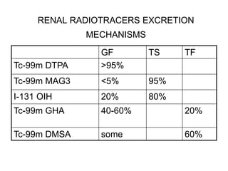
RENAL
SCINTIGRAPHY
INDICATIONS
Evaluation of:
• Renal perfusion and function
• Obstruction (Lasix renal scan)
• Renovascular HTN (Captopril renal scan)
• Infection (renal morphology scan)
• Pre-surgical quantitation (nephrectomy)
• Renal transplant
•Congenital anomalies, masses (renal morphology scan)
RENAL FUNCTION
Blood flow : 20% cardiac output to kidneys (1200
ml/min blood, 600 ml/min plasma)
 Filtration: 20% renal plasma flow filtered by
glomeruli (120 ml/min, 170 L/d)
Tubular secretion
Tubular reabsorption (1% ultrafiltrate - urine)
 Endocrine functions
RENAL RADIOTRACERS EXCRETION
MECHANISMS
TF
TS
GF
>95%
Tc-99m DTPA
95%
<5%
Tc-99m MAG3
80%
20%
I-131 OIH
20%
40-60%
Tc-99m GHA
60%
some
Tc-99m DMSA
RENAL
RADIOPHARMACEUTICALS
• Clearance
Extract. fraction
100-120 ml/min
20%
Tc-99m DTPA
~ 300 ml/minI
40-50%
Tc-99m
MAG3
500-600 ml/min
~100%
I-131 OIH
RENAL RADIOPHARMACEUTICALS
DOSIMETRY
I-131 OIH
DMSA
GHA
MAG3
DTPA
0.01
3.5
1.6
0.15
0.2
Kidney
0.3
0.3
2.7
5.1
2.8
Bladder
CHOOSING RENAL
RADIOTRACERS
Agent
Clin. Question
MAG3, DTPA, GHA
Perfusion
DMSA, GHA
Morphology
MAG3, DTPA, OIH
Obstruction
All
Relative function
I-125 iothalamate, Cr-51
EDTA , DTPA
GFR quantitation
MAG3, OIH
ERPF quantitation
BASIC RENAL SCINTIGRAPHY
PATIENT PREPARATION
Patient must be well hydrated
Give 5-10 ml/kg water (2-4 cups) 30-60 min.
pre-injection Can measure U - specific gravity (<1.015)
Void before injection
Void @ end of study
BASIC RENAL SCINTIGRAPHY
ACQUISITION
Supine position preferred
Do not inject by straight stick
Flow (angiogram) : 2-3 sec / fr x 1 min
Dynamic: 15-30 sec / frame x 20-30
min (display @ 1-3 min/frame(
BASIC RENAL SCINTIGRAPHY
PROCESSIN
Time to peak
•Best from cortical ROI
•Normal 3 - 5 min
Residual Cortical Activity (RCA 20 or 30 )
•Ratio of cts @ 20 or 30 min / peak cts
•Use cortical ROI
•Normal RCA 20 for MAG3 < 0.3
Residual Urine Volume
•(post-void cts x void. vol)
•(pre-void cts - post void cts(
RENOGRAM PHASES
I. Vascular phase (flow study):
Ao-to-Kid ~ 3”
II. Parenchymal phase (kidney-
to-bkg): Tpeak < 5’
III. Washout (excretory) phase
EVALUATION OF
HYDRONEPHROSIS
Diuretic (Lasix) Renal Scan
OBSTRUCTION
Obstruction to urine outflow leads to
obstructive uropathy (hydronephrosis,
hydroureter) and may lead to obstructive
nephropathy (loss of renal function)
DIURETIC RENAL SCAN PRINCIPLE
Hydronephrosis - tracer pooling in dilated renal
pelvis
Lasix induces increased urine flow
If obstructed >>> will not wash out
If dilated, non-obstructed >>> will wash out
Can quantitate rate of washout (T ½(
DIURETIC RENAL SCAN
INDICATIONS
Evaluate functional significance of
hydronephrosis
 Determine need for surgery
• obstructive hydronephrosis - surgical Rx
• non-obstructive hydronephrosis -
medical Rx
 Monitor effect of therapy
DIURETIC RENAL SCAN
REQUIREMENTS
Rapidly cleared tracer
 Well hydrated patient
 Good renal function
DIURETIC RENAL SCAN PROCEDURE
• Pt. preparation:
• prehydration adults - oral or 360ml/m 2 iv over 30’ peds - 10-15 ml/kg D5 0.3-
0.45%NS
• void before injection
• bladder catheterization ?
• Tracers: Tc-99m MAG3 5-10 mCi (preferred over DTPA)
• Acquisition: supine until pelvis full (can switch to sitting post- Lasix)
• Flow (angiogram) : 2-3 sec / fr x 1 min
• Dynamic: 15-30 sec / frame x 20-30 min
• Void before Lasix
• Lasix:40mg adult, 1mg/kg child iv @ ~10-20 min (when pelvis full) or @ -15min (“F-15”
method)
• Acquisition:for 30 min post Lasix
• Assess adequacy of diuresis
• Measure voided volume
• Adults produce ~200-300 ml urine post-Lasix
• Don’t give Lasix if
 Collecting system still filling
 Collecting system not full by 60 min
 Collecting system drains spontaneously
 Poor ipsilateral fct (< 20%)
DIURETIC RENAL SCAN WASHOUT
(DIURETIC RESPONSE )
T ½ : time required for 50% tracer to leave
the dilated unit i.e. time required for activity
to fall to 50% of peak
T 1/2 VALUE
 Variables influencing T1/2 value:
• Tracer
• State of hydration
• Volume of dilated pelvis
• Bladder catheterization
• Dose of Lasix
• Renal function (response to Lasix)
• ROI (kidney vs. pelvis)
• T 1/2 calculation (from inj. vs. response, curve fit)
 T ½
 Normal < 10 min
 Obstructed > 20 min
 Indeterminate 10 - 20 min
 Best to obtain own normals for each institution, depending on protocol used
DIURETIC RENAL SCAN
INTERPRETATION
• Interpret whole study, not T 1/2 alone
• Visual (dynamic images)
• Washout curve shape (concave vs.
convex)
• T 1/2
DIURETIC RENAL SCAN
PITFALLS
False positive for obstruction:
• Distended bladder
• Gross hydronephrosis T (transit time) = V
(volume)
• F (flow)
• Poorly functioning / immature kidney
• Dehydration
 False negative:
• Low grade obstruction
• Poorly functioning / immature kidney
EVALUATION OF RENOVASCULAR
HYPERTENSIONCaptopril Renal
Scan (ACEI Renography)
Renovascular hypertension is estimated to affect 3% of the unselected
hypertensive population and up to 30% of patients referred to a subspecialty
center for problematic hypertension.
Renovascular hypertension is defined as an elevated blood pressure caused by
renal hypoperfusion, usually due to anatomic renal artery stenosis and
activation of the renin-angiotensin system.
Advances in percutaneous renal angioplasty, renal artery stenting and surgical
techniques have renewed interest in developing better tests for identifying
patients with potentially correctable hypertension or renal dysfunction
secondary to renovascular disease.0
Renovascular hypertension versus renal artery stenosis.
It is important to distinguish between renovascular hypertension and the presence
of anatomic renal artery stenosis. Renal artery stenosis may be a consequence of
hypertension rather than its cause and is common even in normotensive individuals
over age 50. Renal artery stenosis may be an incidental finding in a hypertensive
patient.
Revascularization is expensive, is not without risk and may not result in any
improvement in blood pressure in as many as 30–40% of patients undergoing the
procedure. An ACE-inhibition renogram interpreted as high probability for
renovascular hypertension carries a high predictive value (90%) that renal artery
stenosis is present and that the hypertension will be ameliorated or cured by
revascularization.
Mechanism of an abnormal scan after ACE inhibition.
Renovascular hypertension is dependent on renin secretion from the juxtaglomerular apparatus of the
underperfused, stenotic kidney. Renin converts angiotensinogen to
angiotensin I, which is in turn converted to angiotensin II by ACE. Angiotensin
II causes preferential vasoconstriction of the postglomerular (efferent) arteriole.
Vasoconstriction of the efferent arteriole increases resistance to flow and can
maintain the transglomerular pressure gradient and, thereby, maintain GFR
even in the presence of a reduced perfusion pressure. Within the stenotic kidney,
ACE inhibition reduces the angiotensin II dependent constriction of the
postglomerular arteriole, decreases the resistance to flow and thereby lowers the
transcapillary pressure gradient maintaining GFR. The resulting decrease in
glomerular filtration of the stenotic kidney can be detected noninvasively with
ACE-inhibition renography by either a change in relative function or retention
of the tracer in the renal tubules due to decreased GFR and decreased flow of the
filtrate through the tubules .
*
Basic renogram followed by an ACE inhibitor (oral captopril or intravenous
enalaprilat) and a repeat renogram. This option provides the most definitive
information but it requires two renograms and requires the patient to spend 2–3 hr in the
nuclear medicine department.
The baseline renogram is omitted, and the patient receives the ACE inhibitor
followed by a renogram. If the ACE-inhibition renogram is normal, the study is
low probability for renovascular hypertension, and there is no need for the baseline renogram.
If it is abnormal, the patient needs to return another day for the baseline study to maximize
specificity; alternatively, high-risk patients with an abnormal study may be referred for
angiography. This option provides the same diagnostic information as option 1 above if the
study is normal, but it is less specific
if the study is abnormal; however, only one renogram is required, and the
patient spends only 1–2 hr in the department.
1. Hydration. The patient should be instructed to be well hydrated when he/she arrives for
the examination.
2. No food before captopril renography. If oral captopril is to be given, the
patient should be instructed not to eat any solids after midnight before the
examination. Food may delay gastric emptying and interfere with the absorption of captopril.
The instructions should clearly distinguish between avoiding solid food and the importance of
drinking water
3. Diuretics. Diuretics should be discontinued for 3 days before the study to
avoid dehydration and minimize the risk of hypotension.
4. ACE inhibitors and angiotensin II receptor blockers. Chronic ACE inhibition
and angiotensin II receptor blockade may reduce the sensitivity of the test. For optimal
sensitivity, these drugs should be discontinued for 4–7 days before the study with the longer
half-life drugs discontinued for 7 days. With the exception of diuretics, other antihypertensive
medications can be substituted for ACE inhibitors and angiotensin II receptor blockers if the
patient requires replacement therapy during the 4–7 days before the ACE-inhibition renogram.
Patient preparation
1) High probability. Significant deterioration of the renogram curve
after ACE inhibition compared with the baseline study.
2) Indeterminate (intermediate) probability. An abnormal baseline
renogram that is unchanged after ACE inhibition. The majority of
patients in this group have hypertension with azotemia or a small,
poorly functioning kidney. Depending on the level of clinical suspicion, such a
patient may be referred for an additional diagnostic test.
3) Low probability. A normal ACE-inhibition renogram is low probability for
renovascular hypertension. Even if renal artery stenosis is present, hypertension
in this group of patients is much less likely to improve with revascularization than
in patients with a positive test.
1. False-positive or false-intermediate probability studies. These can occur
because of dehydration, radiotracer extravasation, poor renal function,
obstruction or a distended patient bladder at the start of the scan. A false-
positive test also can be obtained if the patient becomes hypotensive during the
study.
2. ACE inhibitors or angiotensin II receptor blockers. The sensitivity of the test
may be reduced in patients on chronic ACE inhibition. To maximize sensitivity, ACE
inhibitors and angiotensin II receptor blockers should be withheld for 3–7 days
before the study depending on the half-life of the
particular ACE inhibitor. When
patients arrive for an ACE-inhibitor study and are found to be on chronic ACE
inhibition, most centers elect to proceed with the scan and accept a slightly lowered
sensitivity.
3. Ischemic nephropathy. A positive test result in a patient with azotemia or in a
patient with a small, poorly functioning kidney indicates a high likelihood that the
hypertension will be ameliorated by revascularization. Unlike the population of
patients with normal renal function, as many as 50% of patients in this
patient population may have an indeterminate or intermediate probability test result
(an abnormal baseline study that does not change after ACE inhibition). A false-
negative study also is more likely to occur in this patient population, possibly because
of the absence of renin-dependent hypertension. Depending on the diagnostic goals,
some clinicians will refer patients with ischemic nephropathy and a high pretest
probability for renovascular hypertension directly to angiography. Magnetic resonance
angiography or Doppler ultrasound are alternatives if there is a need to avoid
constrast.
 RENOVASCULAR DISEASE
 Renal artery stenosis (RAS)
 Ischemic nephropathy
 Renovascular hypertension
 RENOVASCULAR HYPERTENSION
 Caused by renal hypoperfusion:
 Atherosclerosis
 Fibromuscular dysplasia
 Mediated by renin - AT - aldosterone system
 Potentially curable by renal revascularization
 RENOVASCULAR HYPERTENSION Prevalence :
 <1% unselected population with HTN
 Clinical features:
 Abrupt onset HTN in child, adult < 30 or > 50y
 Severe HTN resistant to medical Rx
 Unexplained or post-ACEI impairment in ren fct
 HTN + abdominal bruits If these present - moderate risk of RVH (20-30%)
*
Gold std: angiography
 Initial non-invasive tests:
 ACEI renography
 Duplex sonography
Other tests:
 MRA - insensitive for distal / segmental RAS
 Captopril test (PRA post-C.) - low sensitivity
 Renal vein renin levels
*
In normal renal function - sens/spec ~ 90%
 In poor renal fct / ischemic nephropathy,
ACEI renography often indeterminate
>>> do MRA , Duplex US, angio
EVALUATION OF RENAL
INFECTION Renal Morphology Scan
(Renal Cortical Scintigraphy)
UTI
 VUR
• risk factor for PN,
• not all pts w PN have VUR
 PN may lead to scarring >>> ESRD, HTN
• early Dx and Rx necessary
 Clinical & laboratory Dx of renal involvement in UTI
unreliable
RENAL CORTICAL
SCINTIGRAPHY
INDICATIONS:
• Determine involvement of upper tract (kidney) in acute UTI (acute pyelonephritis) (infection)
• Detect cortical scarring (chronic pyelonephr.)
• Follow -up post Rx
• PROCEDURE:
• Tracers:
• Tc-99m DMSA
• Tc-99m GHA
IzNTERPRETATION:
 Acute PN:
• single or multiple “cold” defects
• renal contour not distorted
• diffuse decreased uptake
• diffusely enlarged kidney or focal bulging
 Chronic PN:
• volume loss, cortical thinning
• defects with sharp edges
 Differentiation of AcPN vs. ChPN unreliable
RENAL CORTICAL
SCINTIGRAPHY “COLD DEFECT “
• Acute or chronic PN
• Hydronephrosis
• Cyst
• Tumors
• Trauma (contusion, laceration,
rupture, hematoma)
• Infarct
RENAL CORTICAL SCINTIGRAPHY
CONGENITAL ANOMALIES
• Agenesis
• Ectopy
• Fusion (horseshoe, crossed fused
ectopia)
• Polycystic kidney
• Multicystic dysplastic kidney (fetal
lobulation, hypertrophic
• Pseudomasses column of Bertin)
RADIONUCLIDE
CYSTOGRAM
INDICATIONS:
• Evaluation of children with recurrent UTI
• 30-50% have VUR
• F/U after initial VCUG
• Assess ef fect of therapy / surgery
• Screening of siblings of reflux pts.
METHODS:
Direct:
 Tc-99m S.C. or TcO4
 via Foley
 can do at any age
 VUR during filling
 Catheterization
Indirect :
 Tc-99m DTPA or Tc-99m MAG3
 i.v.
 no catheter
 info on kidneys.
 need pt. cooperation
 need good renal fct
DIRECT CYSTOGRAPHY:
• 1 mCi S.C. in saline via Foley
• Fill bladder until reversal of flow
• (bladder capacity = (age+2) x 30
• Continuous imaging during filling & voiding
• Post void image
• Record
• volume instilled
• volume voided
• pre- and post- void cts
RN CYSTOGRAM VS. VCUG
Advantages:
• Lower radiation (5 vs 300 mrad to ovary)
• Smaller amount of reflux detectable
• Quantitation of post-void residual volume
Disadvantages:
• Cannot detect distal ureteral reflux
• No anatomic detail
• Grading difficult

Dynamic Renal scan lecture (2.pdf

  • 1.
  • 2.
    INDICATIONS Evaluation of: • Renalperfusion and function • Obstruction (Lasix renal scan) • Renovascular HTN (Captopril renal scan) • Infection (renal morphology scan) • Pre-surgical quantitation (nephrectomy) • Renal transplant •Congenital anomalies, masses (renal morphology scan)
  • 3.
    RENAL FUNCTION Blood flow: 20% cardiac output to kidneys (1200 ml/min blood, 600 ml/min plasma)  Filtration: 20% renal plasma flow filtered by glomeruli (120 ml/min, 170 L/d) Tubular secretion Tubular reabsorption (1% ultrafiltrate - urine)  Endocrine functions
  • 4.
    RENAL RADIOTRACERS EXCRETION MECHANISMS TF TS GF >95% Tc-99mDTPA 95% <5% Tc-99m MAG3 80% 20% I-131 OIH 20% 40-60% Tc-99m GHA 60% some Tc-99m DMSA
  • 5.
    RENAL RADIOPHARMACEUTICALS • Clearance Extract. fraction 100-120ml/min 20% Tc-99m DTPA ~ 300 ml/minI 40-50% Tc-99m MAG3 500-600 ml/min ~100% I-131 OIH
  • 6.
  • 7.
    CHOOSING RENAL RADIOTRACERS Agent Clin. Question MAG3,DTPA, GHA Perfusion DMSA, GHA Morphology MAG3, DTPA, OIH Obstruction All Relative function I-125 iothalamate, Cr-51 EDTA , DTPA GFR quantitation MAG3, OIH ERPF quantitation
  • 8.
    BASIC RENAL SCINTIGRAPHY PATIENTPREPARATION Patient must be well hydrated Give 5-10 ml/kg water (2-4 cups) 30-60 min. pre-injection Can measure U - specific gravity (<1.015) Void before injection Void @ end of study
  • 9.
    BASIC RENAL SCINTIGRAPHY ACQUISITION Supineposition preferred Do not inject by straight stick Flow (angiogram) : 2-3 sec / fr x 1 min Dynamic: 15-30 sec / frame x 20-30 min (display @ 1-3 min/frame(
  • 10.
    BASIC RENAL SCINTIGRAPHY PROCESSIN Timeto peak •Best from cortical ROI •Normal 3 - 5 min Residual Cortical Activity (RCA 20 or 30 ) •Ratio of cts @ 20 or 30 min / peak cts •Use cortical ROI •Normal RCA 20 for MAG3 < 0.3 Residual Urine Volume •(post-void cts x void. vol) •(pre-void cts - post void cts(
  • 11.
    RENOGRAM PHASES I. Vascularphase (flow study): Ao-to-Kid ~ 3” II. Parenchymal phase (kidney- to-bkg): Tpeak < 5’ III. Washout (excretory) phase
  • 12.
  • 13.
    OBSTRUCTION Obstruction to urineoutflow leads to obstructive uropathy (hydronephrosis, hydroureter) and may lead to obstructive nephropathy (loss of renal function) DIURETIC RENAL SCAN PRINCIPLE Hydronephrosis - tracer pooling in dilated renal pelvis Lasix induces increased urine flow If obstructed >>> will not wash out If dilated, non-obstructed >>> will wash out Can quantitate rate of washout (T ½(
  • 14.
    DIURETIC RENAL SCAN INDICATIONS Evaluatefunctional significance of hydronephrosis  Determine need for surgery • obstructive hydronephrosis - surgical Rx • non-obstructive hydronephrosis - medical Rx  Monitor effect of therapy
  • 15.
    DIURETIC RENAL SCAN REQUIREMENTS Rapidlycleared tracer  Well hydrated patient  Good renal function
  • 16.
    DIURETIC RENAL SCANPROCEDURE • Pt. preparation: • prehydration adults - oral or 360ml/m 2 iv over 30’ peds - 10-15 ml/kg D5 0.3- 0.45%NS • void before injection • bladder catheterization ? • Tracers: Tc-99m MAG3 5-10 mCi (preferred over DTPA) • Acquisition: supine until pelvis full (can switch to sitting post- Lasix) • Flow (angiogram) : 2-3 sec / fr x 1 min • Dynamic: 15-30 sec / frame x 20-30 min • Void before Lasix • Lasix:40mg adult, 1mg/kg child iv @ ~10-20 min (when pelvis full) or @ -15min (“F-15” method) • Acquisition:for 30 min post Lasix • Assess adequacy of diuresis • Measure voided volume • Adults produce ~200-300 ml urine post-Lasix • Don’t give Lasix if  Collecting system still filling  Collecting system not full by 60 min  Collecting system drains spontaneously  Poor ipsilateral fct (< 20%)
  • 17.
    DIURETIC RENAL SCANWASHOUT (DIURETIC RESPONSE ) T ½ : time required for 50% tracer to leave the dilated unit i.e. time required for activity to fall to 50% of peak
  • 18.
    T 1/2 VALUE Variables influencing T1/2 value: • Tracer • State of hydration • Volume of dilated pelvis • Bladder catheterization • Dose of Lasix • Renal function (response to Lasix) • ROI (kidney vs. pelvis) • T 1/2 calculation (from inj. vs. response, curve fit)  T ½  Normal < 10 min  Obstructed > 20 min  Indeterminate 10 - 20 min  Best to obtain own normals for each institution, depending on protocol used
  • 19.
    DIURETIC RENAL SCAN INTERPRETATION •Interpret whole study, not T 1/2 alone • Visual (dynamic images) • Washout curve shape (concave vs. convex) • T 1/2
  • 20.
    DIURETIC RENAL SCAN PITFALLS Falsepositive for obstruction: • Distended bladder • Gross hydronephrosis T (transit time) = V (volume) • F (flow) • Poorly functioning / immature kidney • Dehydration  False negative: • Low grade obstruction • Poorly functioning / immature kidney
  • 21.
  • 22.
    Renovascular hypertension isestimated to affect 3% of the unselected hypertensive population and up to 30% of patients referred to a subspecialty center for problematic hypertension. Renovascular hypertension is defined as an elevated blood pressure caused by renal hypoperfusion, usually due to anatomic renal artery stenosis and activation of the renin-angiotensin system. Advances in percutaneous renal angioplasty, renal artery stenting and surgical techniques have renewed interest in developing better tests for identifying patients with potentially correctable hypertension or renal dysfunction secondary to renovascular disease.0
  • 23.
    Renovascular hypertension versusrenal artery stenosis. It is important to distinguish between renovascular hypertension and the presence of anatomic renal artery stenosis. Renal artery stenosis may be a consequence of hypertension rather than its cause and is common even in normotensive individuals over age 50. Renal artery stenosis may be an incidental finding in a hypertensive patient. Revascularization is expensive, is not without risk and may not result in any improvement in blood pressure in as many as 30–40% of patients undergoing the procedure. An ACE-inhibition renogram interpreted as high probability for renovascular hypertension carries a high predictive value (90%) that renal artery stenosis is present and that the hypertension will be ameliorated or cured by revascularization.
  • 24.
    Mechanism of anabnormal scan after ACE inhibition. Renovascular hypertension is dependent on renin secretion from the juxtaglomerular apparatus of the underperfused, stenotic kidney. Renin converts angiotensinogen to angiotensin I, which is in turn converted to angiotensin II by ACE. Angiotensin II causes preferential vasoconstriction of the postglomerular (efferent) arteriole. Vasoconstriction of the efferent arteriole increases resistance to flow and can maintain the transglomerular pressure gradient and, thereby, maintain GFR even in the presence of a reduced perfusion pressure. Within the stenotic kidney, ACE inhibition reduces the angiotensin II dependent constriction of the postglomerular arteriole, decreases the resistance to flow and thereby lowers the transcapillary pressure gradient maintaining GFR. The resulting decrease in glomerular filtration of the stenotic kidney can be detected noninvasively with ACE-inhibition renography by either a change in relative function or retention of the tracer in the renal tubules due to decreased GFR and decreased flow of the filtrate through the tubules .
  • 27.
  • 28.
    Basic renogram followedby an ACE inhibitor (oral captopril or intravenous enalaprilat) and a repeat renogram. This option provides the most definitive information but it requires two renograms and requires the patient to spend 2–3 hr in the nuclear medicine department. The baseline renogram is omitted, and the patient receives the ACE inhibitor followed by a renogram. If the ACE-inhibition renogram is normal, the study is low probability for renovascular hypertension, and there is no need for the baseline renogram. If it is abnormal, the patient needs to return another day for the baseline study to maximize specificity; alternatively, high-risk patients with an abnormal study may be referred for angiography. This option provides the same diagnostic information as option 1 above if the study is normal, but it is less specific if the study is abnormal; however, only one renogram is required, and the patient spends only 1–2 hr in the department.
  • 29.
    1. Hydration. Thepatient should be instructed to be well hydrated when he/she arrives for the examination. 2. No food before captopril renography. If oral captopril is to be given, the patient should be instructed not to eat any solids after midnight before the examination. Food may delay gastric emptying and interfere with the absorption of captopril. The instructions should clearly distinguish between avoiding solid food and the importance of drinking water 3. Diuretics. Diuretics should be discontinued for 3 days before the study to avoid dehydration and minimize the risk of hypotension. 4. ACE inhibitors and angiotensin II receptor blockers. Chronic ACE inhibition and angiotensin II receptor blockade may reduce the sensitivity of the test. For optimal sensitivity, these drugs should be discontinued for 4–7 days before the study with the longer half-life drugs discontinued for 7 days. With the exception of diuretics, other antihypertensive medications can be substituted for ACE inhibitors and angiotensin II receptor blockers if the patient requires replacement therapy during the 4–7 days before the ACE-inhibition renogram. Patient preparation
  • 30.
    1) High probability.Significant deterioration of the renogram curve after ACE inhibition compared with the baseline study. 2) Indeterminate (intermediate) probability. An abnormal baseline renogram that is unchanged after ACE inhibition. The majority of patients in this group have hypertension with azotemia or a small, poorly functioning kidney. Depending on the level of clinical suspicion, such a patient may be referred for an additional diagnostic test. 3) Low probability. A normal ACE-inhibition renogram is low probability for renovascular hypertension. Even if renal artery stenosis is present, hypertension in this group of patients is much less likely to improve with revascularization than in patients with a positive test.
  • 31.
    1. False-positive orfalse-intermediate probability studies. These can occur because of dehydration, radiotracer extravasation, poor renal function, obstruction or a distended patient bladder at the start of the scan. A false- positive test also can be obtained if the patient becomes hypotensive during the study. 2. ACE inhibitors or angiotensin II receptor blockers. The sensitivity of the test may be reduced in patients on chronic ACE inhibition. To maximize sensitivity, ACE inhibitors and angiotensin II receptor blockers should be withheld for 3–7 days before the study depending on the half-life of the particular ACE inhibitor. When patients arrive for an ACE-inhibitor study and are found to be on chronic ACE inhibition, most centers elect to proceed with the scan and accept a slightly lowered sensitivity.
  • 32.
    3. Ischemic nephropathy.A positive test result in a patient with azotemia or in a patient with a small, poorly functioning kidney indicates a high likelihood that the hypertension will be ameliorated by revascularization. Unlike the population of patients with normal renal function, as many as 50% of patients in this patient population may have an indeterminate or intermediate probability test result (an abnormal baseline study that does not change after ACE inhibition). A false- negative study also is more likely to occur in this patient population, possibly because of the absence of renin-dependent hypertension. Depending on the diagnostic goals, some clinicians will refer patients with ischemic nephropathy and a high pretest probability for renovascular hypertension directly to angiography. Magnetic resonance angiography or Doppler ultrasound are alternatives if there is a need to avoid constrast.
  • 33.
     RENOVASCULAR DISEASE Renal artery stenosis (RAS)  Ischemic nephropathy  Renovascular hypertension  RENOVASCULAR HYPERTENSION  Caused by renal hypoperfusion:  Atherosclerosis  Fibromuscular dysplasia  Mediated by renin - AT - aldosterone system  Potentially curable by renal revascularization  RENOVASCULAR HYPERTENSION Prevalence :  <1% unselected population with HTN  Clinical features:  Abrupt onset HTN in child, adult < 30 or > 50y  Severe HTN resistant to medical Rx  Unexplained or post-ACEI impairment in ren fct  HTN + abdominal bruits If these present - moderate risk of RVH (20-30%)
  • 34.
    * Gold std: angiography Initial non-invasive tests:  ACEI renography  Duplex sonography Other tests:  MRA - insensitive for distal / segmental RAS  Captopril test (PRA post-C.) - low sensitivity  Renal vein renin levels
  • 35.
    * In normal renalfunction - sens/spec ~ 90%  In poor renal fct / ischemic nephropathy, ACEI renography often indeterminate >>> do MRA , Duplex US, angio
  • 36.
    EVALUATION OF RENAL INFECTIONRenal Morphology Scan (Renal Cortical Scintigraphy) UTI  VUR • risk factor for PN, • not all pts w PN have VUR  PN may lead to scarring >>> ESRD, HTN • early Dx and Rx necessary  Clinical & laboratory Dx of renal involvement in UTI unreliable
  • 37.
    RENAL CORTICAL SCINTIGRAPHY INDICATIONS: • Determineinvolvement of upper tract (kidney) in acute UTI (acute pyelonephritis) (infection) • Detect cortical scarring (chronic pyelonephr.) • Follow -up post Rx • PROCEDURE: • Tracers: • Tc-99m DMSA • Tc-99m GHA IzNTERPRETATION:  Acute PN: • single or multiple “cold” defects • renal contour not distorted • diffuse decreased uptake • diffusely enlarged kidney or focal bulging  Chronic PN: • volume loss, cortical thinning • defects with sharp edges  Differentiation of AcPN vs. ChPN unreliable
  • 38.
    RENAL CORTICAL SCINTIGRAPHY “COLDDEFECT “ • Acute or chronic PN • Hydronephrosis • Cyst • Tumors • Trauma (contusion, laceration, rupture, hematoma) • Infarct
  • 39.
    RENAL CORTICAL SCINTIGRAPHY CONGENITALANOMALIES • Agenesis • Ectopy • Fusion (horseshoe, crossed fused ectopia) • Polycystic kidney • Multicystic dysplastic kidney (fetal lobulation, hypertrophic • Pseudomasses column of Bertin)
  • 40.
  • 41.
    INDICATIONS: • Evaluation ofchildren with recurrent UTI • 30-50% have VUR • F/U after initial VCUG • Assess ef fect of therapy / surgery • Screening of siblings of reflux pts. METHODS: Direct:  Tc-99m S.C. or TcO4  via Foley  can do at any age  VUR during filling  Catheterization Indirect :  Tc-99m DTPA or Tc-99m MAG3  i.v.  no catheter  info on kidneys.  need pt. cooperation  need good renal fct
  • 42.
    DIRECT CYSTOGRAPHY: • 1mCi S.C. in saline via Foley • Fill bladder until reversal of flow • (bladder capacity = (age+2) x 30 • Continuous imaging during filling & voiding • Post void image • Record • volume instilled • volume voided • pre- and post- void cts RN CYSTOGRAM VS. VCUG Advantages: • Lower radiation (5 vs 300 mrad to ovary) • Smaller amount of reflux detectable • Quantitation of post-void residual volume Disadvantages: • Cannot detect distal ureteral reflux • No anatomic detail • Grading difficult