Downloaded 14 times
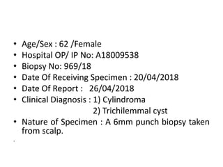

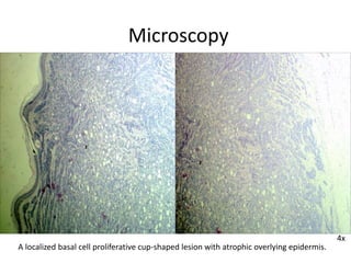

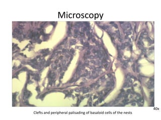


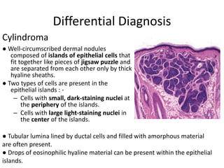
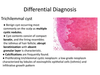


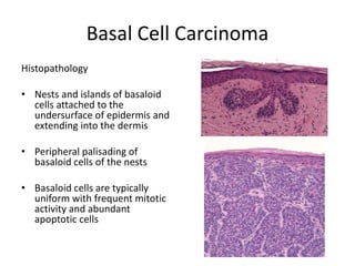
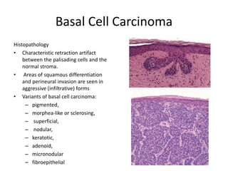
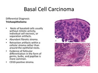

The biopsy shows features consistent with basal cell carcinoma. Microscopy revealed a localized basal cell proliferative lesion arranged in islands and cords with focal sclerosis. Basaloid cells showed peripheral palisading and clefts. The diagnosis of basal cell carcinoma was made based on these histological features. Differential considerations included cylindroma and trichilemmal cyst but were excluded.


