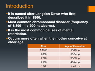
Down Syndrome
By : Najihah
Introduction
It is named after Langdon Down who first
described it in 1866.
Most common chromosomal disorder (frequency
of 1:800 – 1:1000 newborns).
It is the most common causes of mental
retardation.
Occurs more often when the mother conceive at
older age.
Risk Age of the mother
1:1550 15-29 yr
1:800 30-34 yr
1:270 35-39 yr
1:100 40-44 yr
1:50 > 45 yr
Cytogenetics
1. Meiotic non-disjunction of
chromosome 21 (94%)
- May produce an extra copy of chromosome 21
(Trisomy 21) in egg (95%) or sperm (5%).
- May be of maternal or paternal origin.
- In most cases, the origin is maternal where the
mother conceive at older age.
- Due to older age, the maternal oocyte is exposed to
harmful environmental influences for a longer period.
- Other predisposing factors are radiation, viral
infections and genetic predisposition.
- The sperm has a short lifespan and therefore has less
chances of injurious exposure.
2. Translocation (5%)
- 50% translocation are inherited from
translocation carrier parent.
- Karyotype of the parents is required.
3. Mosaicism (1%)
Clinical Features and Diagnosis
- Mental and physical
retardation.
- Flat facies.
- Upward slant of eyes.
- Epicanthic folds.
- Oblique palpebral fissure.
- Low set ears
- Small nose with flat nasal
bridges.
- Narrow short palate with
small teeth and protruding
tongue.
- Hypotonia.
- Small skull with
brachycephalic (flat occiput).
Clinical features
- Short and broad hands.
- Clinodactyly (hypoplasia
of middle phalanx of fifth
finger.
- Simian crease.
- Sandle gap toe.
Associated Abnormalities
1. Congenital heart disease
(CHD)
- 40%
- Endocardial cusion defects
(40-60%)
- Most significant factors in
determining survival.
2. Gastrointestinal
malformations
- Atresias in 12% of cases (esp
duodenal atresia)
- Increase risk of annular
pancreas and Hirschprung
disease.
3. Eye problems
- Increase risk of cataract,
nystagmus, squint, and
abnormalities of visual
acuity.
4. Hearing defects
- Prone to serious otitis
media.
Associated Abnormalies
5. Thyroid dysfunction
- 13-54% have hypothyroidism.
- Thyroid function test recommended during neonatal
period.
6. Atlanto-occipital subluxation
- Displacement of atlanto-occipital joint can cause cord
compression.
- 10-30% cases
Associated Abnormalies
7. Physical growth.
- Linear growth is retarded and tend to become obese
with age.
- Muscle tone improves with age whereas the
developmental progress slows with age.
- Regular followup for height and weight is necessary.
8. Malignancies
- Prone to develop lymphoproliferative disorders,
including acute lymphoblastic leukemia (ALL) , acute
myeloid leukemia (AML) , myelodisplasia and transient
lymphoproliferative syndrome.
Prenatal Diagnosis
 indication for screening and diagnostic tests :
- > 35 years old
- Previous child with down’s syndrome
- Translocation carrier state in parents
- History of genetic defect in family
Screening Tests
1. Antenatal ultrasonogram
- 1st trimester
Measurement of nuchal translucency (NT) -
Excess nuchal fluid accumulation is produced in
down’s syndrome foetus
- 2nd trimester
increased nuchal fold thickness
Short femur and humerus
2. Triple test (16 weeks)
- Maternal serum marker
Decreased -fetoprotein
Decreased unconjugated oestriol
Increased HCG
3. Fetal Karyotype
Chorionic villus sampling and amniocentesis
Detect non-disjunction, translocation or
mosaicism.
Management and Prognosis
Principle : early stimulation, physiotherapy
and speech therapy.
Associated problems need to be treated as
required.
Social performance is usually achieved
beyond that expected for mental age.
They behave as happy children, friendly, good
sense of rhythm and enjoy music.
Major cause of early mortality is CHD (almost
50% die in infancy)
Other common diseases :
- Chronic rhinitis
- Conjunctivitis
- Periodontal disease
Lower respiratory tract infections and
hematological malignancies are another
cause of increased mortality.
Counseling
The parents should be :
1. Informed about the disorder as early as possible after
diagnosis is confirmed.
2. Talk in simple and positive language giving hope, and
allow sufficient time to the parents to ask questions.
3. Discuss known problems and associated disorders.
4. Not talk about institutionalisation and adoption, unless
asked. Both these options should be discouraged.
5. Inform about recurrence risks and possibilities of prenatal
diagnosis.
Risk of Recurrence
Women age 35 years or less who have a child
with trisomy have a 1% risk of having another.
Risk is little increased if the mother is at risk
and 35 years or older.
For translocations inherited from the mother,
the risk is about 10% and 4-5% if inherited from
father.
QUIZ
1. Features of down syndrome :
A . Upslanting of eyes
B. Roth spots (F)
C. Single transverse palmar crease
D. Cleft lip and palate (F)
E. Flat occiput
2. The following are conditions associated with Down
Syndrome.
A. Duodenal atresia
B. Atrioventricular septal defect
C. Hypothyroidism
D. Mental retardation
E. Hypotonia
Reference
 Ghai Essential Pediatrics 7th and 8th edition
 Lippincott Clinical Pediatrics
Down syndrome

Down syndrome

  • 1.
  • 2.
    Introduction It is namedafter Langdon Down who first described it in 1866. Most common chromosomal disorder (frequency of 1:800 – 1:1000 newborns). It is the most common causes of mental retardation. Occurs more often when the mother conceive at older age. Risk Age of the mother 1:1550 15-29 yr 1:800 30-34 yr 1:270 35-39 yr 1:100 40-44 yr 1:50 > 45 yr
  • 3.
    Cytogenetics 1. Meiotic non-disjunctionof chromosome 21 (94%) - May produce an extra copy of chromosome 21 (Trisomy 21) in egg (95%) or sperm (5%). - May be of maternal or paternal origin. - In most cases, the origin is maternal where the mother conceive at older age.
  • 4.
    - Due toolder age, the maternal oocyte is exposed to harmful environmental influences for a longer period. - Other predisposing factors are radiation, viral infections and genetic predisposition. - The sperm has a short lifespan and therefore has less chances of injurious exposure.
  • 5.
    2. Translocation (5%) -50% translocation are inherited from translocation carrier parent. - Karyotype of the parents is required. 3. Mosaicism (1%)
  • 6.
    Clinical Features andDiagnosis - Mental and physical retardation. - Flat facies. - Upward slant of eyes. - Epicanthic folds. - Oblique palpebral fissure. - Low set ears - Small nose with flat nasal bridges. - Narrow short palate with small teeth and protruding tongue. - Hypotonia. - Small skull with brachycephalic (flat occiput).
  • 7.
    Clinical features - Shortand broad hands. - Clinodactyly (hypoplasia of middle phalanx of fifth finger. - Simian crease. - Sandle gap toe.
  • 8.
    Associated Abnormalities 1. Congenitalheart disease (CHD) - 40% - Endocardial cusion defects (40-60%) - Most significant factors in determining survival. 2. Gastrointestinal malformations - Atresias in 12% of cases (esp duodenal atresia) - Increase risk of annular pancreas and Hirschprung disease. 3. Eye problems - Increase risk of cataract, nystagmus, squint, and abnormalities of visual acuity. 4. Hearing defects - Prone to serious otitis media.
  • 9.
    Associated Abnormalies 5. Thyroiddysfunction - 13-54% have hypothyroidism. - Thyroid function test recommended during neonatal period. 6. Atlanto-occipital subluxation - Displacement of atlanto-occipital joint can cause cord compression. - 10-30% cases
  • 10.
    Associated Abnormalies 7. Physicalgrowth. - Linear growth is retarded and tend to become obese with age. - Muscle tone improves with age whereas the developmental progress slows with age. - Regular followup for height and weight is necessary. 8. Malignancies - Prone to develop lymphoproliferative disorders, including acute lymphoblastic leukemia (ALL) , acute myeloid leukemia (AML) , myelodisplasia and transient lymphoproliferative syndrome.
  • 11.
    Prenatal Diagnosis  indicationfor screening and diagnostic tests : - > 35 years old - Previous child with down’s syndrome - Translocation carrier state in parents - History of genetic defect in family
  • 12.
    Screening Tests 1. Antenatalultrasonogram - 1st trimester Measurement of nuchal translucency (NT) - Excess nuchal fluid accumulation is produced in down’s syndrome foetus - 2nd trimester increased nuchal fold thickness Short femur and humerus
  • 13.
    2. Triple test(16 weeks) - Maternal serum marker Decreased -fetoprotein Decreased unconjugated oestriol Increased HCG 3. Fetal Karyotype Chorionic villus sampling and amniocentesis Detect non-disjunction, translocation or mosaicism.
  • 14.
    Management and Prognosis Principle: early stimulation, physiotherapy and speech therapy. Associated problems need to be treated as required. Social performance is usually achieved beyond that expected for mental age. They behave as happy children, friendly, good sense of rhythm and enjoy music.
  • 15.
    Major cause ofearly mortality is CHD (almost 50% die in infancy) Other common diseases : - Chronic rhinitis - Conjunctivitis - Periodontal disease Lower respiratory tract infections and hematological malignancies are another cause of increased mortality.
  • 16.
    Counseling The parents shouldbe : 1. Informed about the disorder as early as possible after diagnosis is confirmed. 2. Talk in simple and positive language giving hope, and allow sufficient time to the parents to ask questions. 3. Discuss known problems and associated disorders. 4. Not talk about institutionalisation and adoption, unless asked. Both these options should be discouraged. 5. Inform about recurrence risks and possibilities of prenatal diagnosis.
  • 17.
    Risk of Recurrence Womenage 35 years or less who have a child with trisomy have a 1% risk of having another. Risk is little increased if the mother is at risk and 35 years or older. For translocations inherited from the mother, the risk is about 10% and 4-5% if inherited from father.
  • 18.
    QUIZ 1. Features ofdown syndrome : A . Upslanting of eyes B. Roth spots (F) C. Single transverse palmar crease D. Cleft lip and palate (F) E. Flat occiput 2. The following are conditions associated with Down Syndrome. A. Duodenal atresia B. Atrioventricular septal defect C. Hypothyroidism D. Mental retardation E. Hypotonia
  • 19.
    Reference  Ghai EssentialPediatrics 7th and 8th edition  Lippincott Clinical Pediatrics