Download to read offline


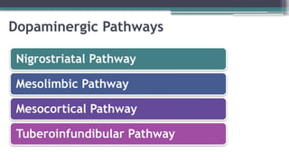


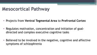
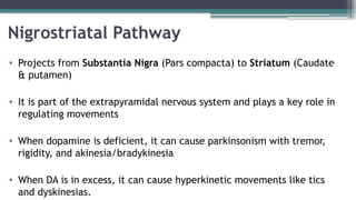

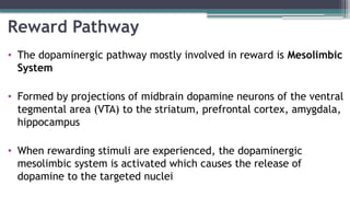


The document provides an overview of dopamine, including its synthesis, receptors, pathways, and the disorders associated with dopamine dysregulation such as Parkinson's disease and schizophrenia. Key pathways mentioned are the mesolimbic, mesocortical, and nigrostriatal pathways, with stated roles in motivation, reward, and movement regulation. It also highlights the involvement of dopamine in ADHD and how imbalances can lead to cognitive difficulties and behavioral issues.


![Neuro-humoral-Transmission-of-Dopamine[1].pptx](https://cdn.slidesharecdn.com/ss_thumbnails/neuro-humoral-transmission-of-dopamine1-241027041656-822de07d-thumbnail.jpg?width=640&height=640&fit=bounds)


