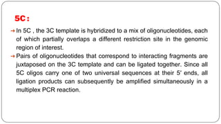
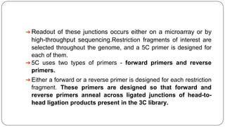
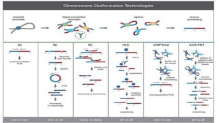
This ppt includes different 3C based techniques for study of DNA-Protein interaction. Data from given research papers are taken for education purpose only,.
This document describes optimization of the Chromosome Conformation Capture (3C) method to assay human fecal samples in order to study horizontal gene transfer between bacteria in the gut microbiome. Initial attempts to apply 3C to fecal samples produced only partially digested DNA. The authors investigated fixation methods and lysate concentrations to improve digestion efficiency. Increasing formaldehyde concentration and diluting lysate concentration enhanced digestion, likely by avoiding inhibitory factors in stool samples. With further optimization, 3C of fecal samples may help elucidate association networks between mobile elements and host bacteria in the gut microbiome.
• It is a technique that predicts the interaction between a macromolecules and a chemical molecule.
• Most of the existing efforts to identify the binding sites in protein-protein interaction are based on analyzing the differences between interface residues and non-interface residues, often through the use of machine learning or statistical methods.
• Its major application is to Identify the protein ligand binding sites is an important process in drug discovery and structure based drug design.
• Earlier, detecting protein ligand binding site is expensive and time consuming by traditional experimental method. Hence, computational approches provide many effective strategies to deal with this issue.
Recombinant DNA technology involves manipulating DNA sequences in the laboratory. DNA is isolated, cut with restriction enzymes, and joined with DNA ligase. The recombinant DNA is inserted into a cloning vector and introduced into a host cell. Cells containing the recombinant DNA are selected and amplified. This allows large quantities of identical DNA molecules to be produced for analysis, comparison, and other purposes. Common applications include understanding disease, producing therapeutic proteins, disease prevention through vaccines, diagnosis, and gene therapy.
A DNA microarray (also commonly known as DNA chip or biochip) is a collection of microscopic DNA spots attached to a solid surface.
The core principle behind microarrays is hybridization between two DNA strands, the property of complementary nucleic acid sequences to specifically pair with each other by forming hydrogen bonds between complementary nucleotide base pairs.
Molecular hybridization is the process by which two complementary strands of DNA or RNA bind together via hydrogen bonding between bases. It is used in techniques like cloning, PCR, and diagnostic tests involving nucleic acid probes. The document describes the process of hybridization, factors that affect binding strength, and techniques that utilize molecular hybridization like Southern blotting, dot/slot blotting, microarrays, and in situ hybridization.
A restriction map is a map of known restriction sites within a sequence of DNA. Restriction mapping requires the use of restriction enzymes. In molecular biology, restriction maps are used as a reference to engineer plasmids or other relatively short pieces of DNA, and sometimes for longer genomic DNA. There are other ways of mapping features on DNA for longer length DNA molecules, such as mapping by transduction (Bitner, Kuempel 1981).
Restriction mapping is a useful way to characterise a particular DNA molecule. It enables us to locate and isolate DNA fragments for further study and manipulation. The relative location of different restriction enzyme sites to each other are determined by enzymatic digest of the DNA with different restriction enzymes, alone and in various combinations.The digested DNA is separated by gel electrophoresis and the fragment sizes that have been generated are used to build the 'map' of sites of the fragment. The map lets us know 'where we are' in the linear DNA macromolecule.
This document discusses gene mapping and gene cloning. It defines gene mapping as identifying the locus and distance between genes using genetic or physical mapping techniques. Gene cloning involves inserting a fragment of DNA containing a gene into a cloning vector, which is then propagated in bacteria to make multiple copies. The document provides detailed descriptions of genetic mapping, physical mapping, gene cloning techniques like transformation, PCR cloning, and their applications and limitations.
Gene libraries, such as cDNA and genomic libraries, allow isolation of specific genes. cDNA libraries contain only exons and reflect gene expression levels, while genomic libraries contain all DNA fragments. Libraries are constructed by fragmenting DNA and cloning into vectors before transforming bacteria. They can be screened by hybridization, PCR, or immunological assays to detect gene products. Common steps include lysis, fixation, and detection to identify positive clones containing genes of interest.
Microarray and dna chips for transcriptome studyBia Khan
Microarrays and DNA chips can be used to study transcriptomes by comparing gene expression profiles. They work by immobilizing reference cDNA or oligonucleotides on a glass slide, then hybridizing labeled cDNA from the cells of interest. This allows determining which genes are expressed and their relative expression levels based on fluorescence intensities. While powerful, the method has complications like cross-hybridization of similar mRNAs and experimental errors. Normalization procedures help account for these. Yeast is commonly used as a model organism in transcriptome studies due to its stable yet responsive gene expression. Applications include stem cell research, cancer studies, and embryonic development.
This document summarizes different DNA assembly techniques. It begins by explaining that pathways of interest are selected and designed to produce chemicals of high interest. It then describes several restriction enzyme-based methods like BioBrick and Golden Gate assembly that use restriction enzymes and ligase. It also discusses sequence homology-based methods like Gibson assembly, overlap extension PCR (OE-PCR), and circular polymerase extension cloning (CPEC) that utilize overlapping DNA regions. Additionally, it covers in vivo techniques using yeast or E. coli homologous recombination as well as bridging oligo-based methods such as ligase cycling reaction (LCR). Each method is evaluated based on features like being scarless, ability to assemble multiple parts, and flexibility
After sequencing of the genome has been done, the first thing that comes to mind is "Where are the genes?". Genome annotation is the process of attaching information to the biological sequences. It is an active area of research and it would help scientists a lot to undergo with their wet lab projects once they know the coding parts of a genome.
Genome annotation is the process of analyzing genomic DNA sequences to extract biological meaning and context. It involves two main steps - structural annotation, which locates gene elements like exons and introns, and functional annotation, which predicts the functions of gene products. Computational tools are crucial given the vast amounts of sequence data. They use various approaches like identifying open reading frames, conserved sequences, statistical patterns and sequence similarities to model gene structures and infer functions. The results are then integrated into automated annotation pipelines to generate comprehensive and reliable gene annotations for genomes.
Over the past few decades, molecular biologists have developed a number of techniques that can be used to build the structure of the target DNA seed sequence more quickly so as to simplify and standardize the cloning process.
Apollo: A workshop for the Manakin Research Coordination NetworkMonica Munoz-Torres
Apollo is a web-based, collaborative genomic annotation editing platform. We need annotation editing tools to modify and refine precise location and structure of the genome elements that predictive algorithms cannot yet resolve automatically.
This presentation is an introduction to how the manual annotation process takes place using Apollo. It is addressed to the members of the Manakin Genomics research community.
This document provides an introduction and overview of manual genome annotation using the Apollo genome annotation tool. It begins with an outline of the webinar topics, which include an introduction to manual annotation and its necessity, an overview of the Apollo tool and its functionality for collaborative curation, and examples and demonstrations. The document then covers key concepts for manual annotation such as the definition of a gene, genome curation steps, transcription and translation including reading frames, splice sites, and phase. The goal of the webinar is to help participants better understand genome curation and manual annotation using Apollo to identify and modify gene models.
This presentation deals with the introduction of Recombinant DNA Technology. The role of different enzymes. Specifically Restriction endonucleases and roles of various vectors.
r-DNA technology allows the manipulation of DNA fragments through restriction endonucleases, cloning techniques, and specific probes. Restriction endonucleases cut DNA into fragments, cloning techniques amplify specific sequences, and probes identify sequences of interest. Real-time PCR and restriction fragment length polymorphism (RFLP) are techniques used to analyze DNA fragments.
This document discusses genetic methods of microbial taxonomy, focusing on nucleic acid hybridization and DNA sequencing. It provides details on hybridization probes, factors affecting hybridization, and types of hybridization including Southern, Northern, and colony hybridization. It also summarizes DNA sequencing methods such as Sanger and Maxam-Gilbert, and applications of sequencing like detecting mutations. Restriction mapping is defined as generating a map of restriction enzyme cleavage sites.
Concept: reannealing nucleic acids to identify sequence of interest.
Separates DNA/RNA in an agarose gel, then detects specific bands using probe and hybridization.
Hybridization takes advantage of the ability of a single stranded DNA or RNA molecule to find its complement, even in the presence of large amounts of unrelated DNA.
Allows detection of specific bands (DNA fragments or RNA molecules) that have complementary sequence to the probe.
Size bands and quantify abundance of molecule.
The document provides an overview of gene sequencing and DNA sequencing techniques. It discusses how DNA is composed of nucleotides containing phosphate, sugar and nitrogen bases. The order of these bases determines the genetic instructions. Each sequence of bases that codes for a protein is known as a gene. It then describes several methods for DNA sequencing, including the Maxam-Gilbert and Sanger methods. The document outlines key applications of gene sequencing such as in medicine, forensics and agriculture. Recent advances in sequencing technology including Illumina, Roche 454 and solid sequencing are also summarized.
This document discusses three biotechnology techniques: DNA microarray, gene sequencing, and SDS-PAGE. It provides details on the principles, methods, and steps for each technique. DNA microarray allows analysis of gene expression for thousands of genes using DNA spots on a solid surface. Gene sequencing determines the order of genes along a chromosome using methods like directed sequencing and shotgun libraries. SDS-PAGE separates molecules by size using polyacrylamide gel and SDS to neutralize protein charge.
The document provides an overview of recombinant DNA technology and cloning techniques. It discusses:
1) The general steps to clone DNA - isolating DNA from an organism, cutting it with restriction enzymes to create recombinant DNA, and introducing the DNA into a host.
2) Types of cloning vectors like plasmids, artificial chromosomes, and viruses that are used to clone DNA fragments. Genomic and cDNA libraries containing clones of all DNA sequences are also described.
3) Techniques for identifying recombinant clones like hybridization probes, complementation of mutations, restriction mapping, and sequencing.
Structural genomics aims to understand genome content through sequencing and mapping genomes. Genetic maps show relative gene locations based on recombination rates, while physical maps use DNA analysis to place genes by base pair distance. Whole genome sequencing involves breaking genomes into fragments that are sequenced and reassembled using overlaps. Functional genomics seeks to identify all genes, RNAs, proteins and their functions through methods like homology searches, microarrays, and mutagenesis screens.
Tracking introgressions using FISH and GISHvipulkelkar1
FISH and GISH are powerful cytogenetic techniques that allow the detection and localization of specific DNA sequences on chromosomes. FISH uses fluorescent probes to visualize DNA locations, while GISH uses total genomic DNA as probes. Both techniques have various applications, including chromosome mapping, analyzing hybrid plants and somatic variations, and detecting chromosomal abnormalities. They have improved plant breeding and furthered understanding of plant genomes, evolution, and relationships. Limitations include inability to detect small mutations and lack of commercial probes for all regions.
I. The document provides an overview of DNA sequencing methods, including a brief history and discussion of the Sanger dideoxy method, sequencing large pieces of DNA using shotgun sequencing, and progress towards achieving the "$1,000 genome".
II. It describes the Sanger dideoxy chain termination method and how primers, templates, and reagents are used. Newer methods like pyrosequencing that can sequence many DNA molecules in parallel are also covered.
III. The document discusses how sequenced DNA can be assembled and annotated, and tools for identifying genes and predicting functions like BLAST searches of databases. Reducing the cost of genome sequencing enables more widespread applications.
DNA Sequencing: History, methods and NGS4RTPCRAnand
I. The document discusses the history and methods of DNA sequencing, including the Sanger dideoxy method, sequencing large pieces of DNA using shotgun sequencing, and advances towards achieving the "$1,000 genome".
II. It describes how the Sanger method works by using DNA polymerase and dideoxynucleotides to terminate DNA strand extension at random positions, generating fragments of different lengths that can be separated by gel electrophoresis.
III. It also outlines new sequencing technologies like pyrosequencing that allow massively parallel sequencing of many DNA fragments simultaneously, enabling faster and cheaper genome sequencing.
The binding of cosmological structures by massless topological defectsSérgio Sacani
Assuming spherical symmetry and weak field, it is shown that if one solves the Poisson equation or the Einstein field
equations sourced by a topological defect, i.e. a singularity of a very specific form, the result is a localized gravitational
field capable of driving flat rotation (i.e. Keplerian circular orbits at a constant speed for all radii) of test masses on a thin
spherical shell without any underlying mass. Moreover, a large-scale structure which exploits this solution by assembling
concentrically a number of such topological defects can establish a flat stellar or galactic rotation curve, and can also deflect
light in the same manner as an equipotential (isothermal) sphere. Thus, the need for dark matter or modified gravity theory is
mitigated, at least in part.
PPT on Direct Seeded Rice presented at the three-day 'Training and Validation Workshop on Modules of Climate Smart Agriculture (CSA) Technologies in South Asia' workshop on April 22, 2024.
More Related Content
Similar to DNA-Protein interaction by 3C based method.pptx
Gene libraries, such as cDNA and genomic libraries, allow isolation of specific genes. cDNA libraries contain only exons and reflect gene expression levels, while genomic libraries contain all DNA fragments. Libraries are constructed by fragmenting DNA and cloning into vectors before transforming bacteria. They can be screened by hybridization, PCR, or immunological assays to detect gene products. Common steps include lysis, fixation, and detection to identify positive clones containing genes of interest.
Microarray and dna chips for transcriptome studyBia Khan
Microarrays and DNA chips can be used to study transcriptomes by comparing gene expression profiles. They work by immobilizing reference cDNA or oligonucleotides on a glass slide, then hybridizing labeled cDNA from the cells of interest. This allows determining which genes are expressed and their relative expression levels based on fluorescence intensities. While powerful, the method has complications like cross-hybridization of similar mRNAs and experimental errors. Normalization procedures help account for these. Yeast is commonly used as a model organism in transcriptome studies due to its stable yet responsive gene expression. Applications include stem cell research, cancer studies, and embryonic development.
This document summarizes different DNA assembly techniques. It begins by explaining that pathways of interest are selected and designed to produce chemicals of high interest. It then describes several restriction enzyme-based methods like BioBrick and Golden Gate assembly that use restriction enzymes and ligase. It also discusses sequence homology-based methods like Gibson assembly, overlap extension PCR (OE-PCR), and circular polymerase extension cloning (CPEC) that utilize overlapping DNA regions. Additionally, it covers in vivo techniques using yeast or E. coli homologous recombination as well as bridging oligo-based methods such as ligase cycling reaction (LCR). Each method is evaluated based on features like being scarless, ability to assemble multiple parts, and flexibility
After sequencing of the genome has been done, the first thing that comes to mind is "Where are the genes?". Genome annotation is the process of attaching information to the biological sequences. It is an active area of research and it would help scientists a lot to undergo with their wet lab projects once they know the coding parts of a genome.
Genome annotation is the process of analyzing genomic DNA sequences to extract biological meaning and context. It involves two main steps - structural annotation, which locates gene elements like exons and introns, and functional annotation, which predicts the functions of gene products. Computational tools are crucial given the vast amounts of sequence data. They use various approaches like identifying open reading frames, conserved sequences, statistical patterns and sequence similarities to model gene structures and infer functions. The results are then integrated into automated annotation pipelines to generate comprehensive and reliable gene annotations for genomes.
Over the past few decades, molecular biologists have developed a number of techniques that can be used to build the structure of the target DNA seed sequence more quickly so as to simplify and standardize the cloning process.
Apollo: A workshop for the Manakin Research Coordination NetworkMonica Munoz-Torres
Apollo is a web-based, collaborative genomic annotation editing platform. We need annotation editing tools to modify and refine precise location and structure of the genome elements that predictive algorithms cannot yet resolve automatically.
This presentation is an introduction to how the manual annotation process takes place using Apollo. It is addressed to the members of the Manakin Genomics research community.
This document provides an introduction and overview of manual genome annotation using the Apollo genome annotation tool. It begins with an outline of the webinar topics, which include an introduction to manual annotation and its necessity, an overview of the Apollo tool and its functionality for collaborative curation, and examples and demonstrations. The document then covers key concepts for manual annotation such as the definition of a gene, genome curation steps, transcription and translation including reading frames, splice sites, and phase. The goal of the webinar is to help participants better understand genome curation and manual annotation using Apollo to identify and modify gene models.
This presentation deals with the introduction of Recombinant DNA Technology. The role of different enzymes. Specifically Restriction endonucleases and roles of various vectors.
r-DNA technology allows the manipulation of DNA fragments through restriction endonucleases, cloning techniques, and specific probes. Restriction endonucleases cut DNA into fragments, cloning techniques amplify specific sequences, and probes identify sequences of interest. Real-time PCR and restriction fragment length polymorphism (RFLP) are techniques used to analyze DNA fragments.
This document discusses genetic methods of microbial taxonomy, focusing on nucleic acid hybridization and DNA sequencing. It provides details on hybridization probes, factors affecting hybridization, and types of hybridization including Southern, Northern, and colony hybridization. It also summarizes DNA sequencing methods such as Sanger and Maxam-Gilbert, and applications of sequencing like detecting mutations. Restriction mapping is defined as generating a map of restriction enzyme cleavage sites.
Concept: reannealing nucleic acids to identify sequence of interest.
Separates DNA/RNA in an agarose gel, then detects specific bands using probe and hybridization.
Hybridization takes advantage of the ability of a single stranded DNA or RNA molecule to find its complement, even in the presence of large amounts of unrelated DNA.
Allows detection of specific bands (DNA fragments or RNA molecules) that have complementary sequence to the probe.
Size bands and quantify abundance of molecule.
The document provides an overview of gene sequencing and DNA sequencing techniques. It discusses how DNA is composed of nucleotides containing phosphate, sugar and nitrogen bases. The order of these bases determines the genetic instructions. Each sequence of bases that codes for a protein is known as a gene. It then describes several methods for DNA sequencing, including the Maxam-Gilbert and Sanger methods. The document outlines key applications of gene sequencing such as in medicine, forensics and agriculture. Recent advances in sequencing technology including Illumina, Roche 454 and solid sequencing are also summarized.
This document discusses three biotechnology techniques: DNA microarray, gene sequencing, and SDS-PAGE. It provides details on the principles, methods, and steps for each technique. DNA microarray allows analysis of gene expression for thousands of genes using DNA spots on a solid surface. Gene sequencing determines the order of genes along a chromosome using methods like directed sequencing and shotgun libraries. SDS-PAGE separates molecules by size using polyacrylamide gel and SDS to neutralize protein charge.
The document provides an overview of recombinant DNA technology and cloning techniques. It discusses:
1) The general steps to clone DNA - isolating DNA from an organism, cutting it with restriction enzymes to create recombinant DNA, and introducing the DNA into a host.
2) Types of cloning vectors like plasmids, artificial chromosomes, and viruses that are used to clone DNA fragments. Genomic and cDNA libraries containing clones of all DNA sequences are also described.
3) Techniques for identifying recombinant clones like hybridization probes, complementation of mutations, restriction mapping, and sequencing.
Structural genomics aims to understand genome content through sequencing and mapping genomes. Genetic maps show relative gene locations based on recombination rates, while physical maps use DNA analysis to place genes by base pair distance. Whole genome sequencing involves breaking genomes into fragments that are sequenced and reassembled using overlaps. Functional genomics seeks to identify all genes, RNAs, proteins and their functions through methods like homology searches, microarrays, and mutagenesis screens.
Tracking introgressions using FISH and GISHvipulkelkar1
FISH and GISH are powerful cytogenetic techniques that allow the detection and localization of specific DNA sequences on chromosomes. FISH uses fluorescent probes to visualize DNA locations, while GISH uses total genomic DNA as probes. Both techniques have various applications, including chromosome mapping, analyzing hybrid plants and somatic variations, and detecting chromosomal abnormalities. They have improved plant breeding and furthered understanding of plant genomes, evolution, and relationships. Limitations include inability to detect small mutations and lack of commercial probes for all regions.
I. The document provides an overview of DNA sequencing methods, including a brief history and discussion of the Sanger dideoxy method, sequencing large pieces of DNA using shotgun sequencing, and progress towards achieving the "$1,000 genome".
II. It describes the Sanger dideoxy chain termination method and how primers, templates, and reagents are used. Newer methods like pyrosequencing that can sequence many DNA molecules in parallel are also covered.
III. The document discusses how sequenced DNA can be assembled and annotated, and tools for identifying genes and predicting functions like BLAST searches of databases. Reducing the cost of genome sequencing enables more widespread applications.
DNA Sequencing: History, methods and NGS4RTPCRAnand
I. The document discusses the history and methods of DNA sequencing, including the Sanger dideoxy method, sequencing large pieces of DNA using shotgun sequencing, and advances towards achieving the "$1,000 genome".
II. It describes how the Sanger method works by using DNA polymerase and dideoxynucleotides to terminate DNA strand extension at random positions, generating fragments of different lengths that can be separated by gel electrophoresis.
III. It also outlines new sequencing technologies like pyrosequencing that allow massively parallel sequencing of many DNA fragments simultaneously, enabling faster and cheaper genome sequencing.
Similar to DNA-Protein interaction by 3C based method.pptx (20)
The binding of cosmological structures by massless topological defectsSérgio Sacani
Assuming spherical symmetry and weak field, it is shown that if one solves the Poisson equation or the Einstein field
equations sourced by a topological defect, i.e. a singularity of a very specific form, the result is a localized gravitational
field capable of driving flat rotation (i.e. Keplerian circular orbits at a constant speed for all radii) of test masses on a thin
spherical shell without any underlying mass. Moreover, a large-scale structure which exploits this solution by assembling
concentrically a number of such topological defects can establish a flat stellar or galactic rotation curve, and can also deflect
light in the same manner as an equipotential (isothermal) sphere. Thus, the need for dark matter or modified gravity theory is
mitigated, at least in part.
PPT on Direct Seeded Rice presented at the three-day 'Training and Validation Workshop on Modules of Climate Smart Agriculture (CSA) Technologies in South Asia' workshop on April 22, 2024.
(June 12, 2024) Webinar: Development of PET theranostics targeting the molecu...Scintica Instrumentation
Targeting Hsp90 and its pathogen Orthologs with Tethered Inhibitors as a Diagnostic and Therapeutic Strategy for cancer and infectious diseases with Dr. Timothy Haystead.
Sexuality - Issues, Attitude and Behaviour - Applied Social Psychology - Psyc...PsychoTech Services
A proprietary approach developed by bringing together the best of learning theories from Psychology, design principles from the world of visualization, and pedagogical methods from over a decade of training experience, that enables you to: Learn better, faster!
Immersive Learning That Works: Research Grounding and Paths ForwardLeonel Morgado
We will metaverse into the essence of immersive learning, into its three dimensions and conceptual models. This approach encompasses elements from teaching methodologies to social involvement, through organizational concerns and technologies. Challenging the perception of learning as knowledge transfer, we introduce a 'Uses, Practices & Strategies' model operationalized by the 'Immersive Learning Brain' and ‘Immersion Cube’ frameworks. This approach offers a comprehensive guide through the intricacies of immersive educational experiences and spotlighting research frontiers, along the immersion dimensions of system, narrative, and agency. Our discourse extends to stakeholders beyond the academic sphere, addressing the interests of technologists, instructional designers, and policymakers. We span various contexts, from formal education to organizational transformation to the new horizon of an AI-pervasive society. This keynote aims to unite the iLRN community in a collaborative journey towards a future where immersive learning research and practice coalesce, paving the way for innovative educational research and practice landscapes.
When I was asked to give a companion lecture in support of ‘The Philosophy of Science’ (https://shorturl.at/4pUXz) I decided not to walk through the detail of the many methodologies in order of use. Instead, I chose to employ a long standing, and ongoing, scientific development as an exemplar. And so, I chose the ever evolving story of Thermodynamics as a scientific investigation at its best.
Conducted over a period of >200 years, Thermodynamics R&D, and application, benefitted from the highest levels of professionalism, collaboration, and technical thoroughness. New layers of application, methodology, and practice were made possible by the progressive advance of technology. In turn, this has seen measurement and modelling accuracy continually improved at a micro and macro level.
Perhaps most importantly, Thermodynamics rapidly became a primary tool in the advance of applied science/engineering/technology, spanning micro-tech, to aerospace and cosmology. I can think of no better a story to illustrate the breadth of scientific methodologies and applications at their best.
The cost of acquiring information by natural selectionCarl Bergstrom
This is a short talk that I gave at the Banff International Research Station workshop on Modeling and Theory in Population Biology. The idea is to try to understand how the burden of natural selection relates to the amount of information that selection puts into the genome.
It's based on the first part of this research paper:
The cost of information acquisition by natural selection
Ryan Seamus McGee, Olivia Kosterlitz, Artem Kaznatcheev, Benjamin Kerr, Carl T. Bergstrom
bioRxiv 2022.07.02.498577; doi: https://doi.org/10.1101/2022.07.02.498577
Travis Hills of MN is Making Clean Water Accessible to All Through High Flux ...Travis Hills MN
By harnessing the power of High Flux Vacuum Membrane Distillation, Travis Hills from MN envisions a future where clean and safe drinking water is accessible to all, regardless of geographical location or economic status.
Current Ms word generated power point presentation covers major details about the micronuclei test. It's significance and assays to conduct it. It is used to detect the micronuclei formation inside the cells of nearly every multicellular organism. It's formation takes place during chromosomal sepration at metaphase.
11.1 Role of physical biological in deterioration of grains.pdf
DNA-Protein interaction by 3C based method.pptx
1. By: Kashvi Jadia(MSc Biotechnology-sem-9)
Professor: Dr. Anjali Soni
Department of Biotechnology, VNSGU.
DNA PROTEIN INTERACTION
2. INTRODUCTION
➔It is well known that the chromosomes are organized in the
nucleus and this spatial arrangement of genome play a crucial
role in gene regulation and genome stability.
➔DNA- Protein interactions play very vital role in any living cell.
➔It controls replication, transcription, recombination, DNA
repair etc.
➔There are several types of proteins found in a cell.But only
those proteins interact with DNA which have the DNA
binding domains.
3. ➔Each DNA binding domain has at least one motif which is a
conserved amino acid sequence of this protein which can
potentially recognize a double stranded or a single stranded
DNA.
➔There are mainly two broad types of DNA protein interactions.
➔ Sequence specific DNA binding:
➔ Sequence non-specific DNA binding:
4. 1. Sequence specific DNA binding:
➔A DNA binding protein binds to a DNA on a site having
specific nucleotide sequence
➔Frequently involve DNA major groove.
➔Interaction maintained by Hydrogen bonds, Ionic
Interaction , Van der waals forces.
1. Sequence non-specific DNA binding:
➔The DNA binding protein can bind to a DNA in a random
position on the DNA.
➔In replication
5. ➔ Traditionally, nuclear organization is studied by microscopy, and
thus it is appropriate to start by highlighting some important
observations made under the microscope.
➔ The segregation of active and inactive chromatin inside the
nucleus raises the possibility that nuclear positioning affects gene
activity. This idea is supported by DNA fluorescence in situ
hybridization (FISH) observations that certain genes (e.g., HoxB and
uPA) loop out of their chromosome territory upon activation.
6. ➔ Some studies show inactive genes located in the interior of CTs
(chromosome territory)and active genes concentrated at the
territory periphery , but active genes can be transcribed from
inside of CTs.
➔ Moreover, regions with a high density of coordinately
expressed genes locate in loops that extend outside of CTs in
expressing cells but not in non expressing cells.
➔ Localization outside of CTs also occurs at genomic regions with
a high-density of broadly expressed genes.
➔ Hence, it has been suggested that there is a correlation
between high levels of transcriptional activity and localization
outside of CTs.
7. ➔ The power of FISH and other microscopy methods lies in their
ability to do single-cell analyses of gene positioning.
➔ However, on a genomic and cell population scale, they are
limited in throughput and resolution.
➔ It is therefore unclear whether they uncover general principles of
nuclear organization or the peculiarities of individual genes.
8. ➔ Ten years ago, Dekker et al. (2002) developed 3C technology, a
biochemical strategy to analyze contact frequencies between
selected genomic sites in cell populations. Since then, various 3C-
derived genomics methods have been developed.
➔ In comparison with microscopy, 3C-based methods enable
more systematic DNA topology studies at a higher resolution.
9. ➔ Advantage: These technologies can put observations made on
single genes in selected cells in the context of genomic behavior
in cell populations. The generated DNA contact maps start
teaching us the rules that dictate genome structure and
functioning inside the cell.
➔Morden techniques:
◆ 3C (Chromosome conformation capture)
◆ 4C (Chromosome conformation capture on chip)
◆ 5C (Chromosome conformation capture carbon copy)
◆ HiC (High-throughput Chromosome Conformation Capture)
◆ ChlAPET (Chromatin interaction analysis by paired-end tag
sequencing)
10.
11. 3C (Chromosome Conformation Capture):
➔ Chromosome folding is modulated as cells progress through the
cell cycle. During mitosis, condensins fold chromosomes into
helical loop arrays. In interphase, the cohesin complex generates
loops and topologically associating domains (TADs), while a
separate process of compartmentalization drives segregation of
active and inactive chromatin.
➔The term "3C DNA-protein interaction" refers to a laboratory
technique used to study the three-dimensional (3D) interactions
between DNA and proteins within the nucleus of a cell.
12. ➔ The strategy of 3C to discover genomic architecture is based on
quantifying the frequencies of contacts between distal DNA
segments in cell populations.
➔The 3C technique allows researchers to investigate how different
regions of the genome physically interact with each other and with
specific proteins.
➔This helps to understand the higher-order chromatin structure and
how it influences gene regulation and other genomic processes.
13. ➔The 3C technique involves several key steps:
1. Cross-Linking:
2. Cell Lysis and Restriction Enzyme
Digestion:
3. DNA Ligation:
4. Reverse Cross-Linking and Purification:
5. Quantitative Analysis:
14. 1. Cross-Linking:
➔ The initial step in 3C and 3C-derived methods is to establish a
representation of the 3D organization of the DNA. To this end, the
chromatin is fixed using a fixative agent, most often formaldehyde.
1. Cell Lysis and treatment with Restriction Enzyme:
➔ Next, the fixed chromatin is cut with a restriction enzyme
recognizing 6 base pairs (bp)—such as HindIII, BglII, SacI, BamHI,
or EcoRI—or with more frequent cutters, such as AciI or DpnII.
15.
16. 3. DNA Ligation:
➔ In the subsequent step, the sticky ends of the cross-linked DNA
fragments are religated under diluted conditions to promote
intramolecular ligations (i.e., between cross-linked fragments).
4. Reverse crosslinking and purification:
➔ DNA fragments that are far away on the linear template, but
colocalize in space(by reverse crosslinking), can, in this way, be
ligated to each other.
➔ The ligation mixture is purified. 3C yields a genome-wide ligation
product library in which each ligation product corresponds to a
specific interaction between the two corresponding loci.
17. 4. Quantitative Analysis:
➔ A template is thereby created that is, in effect, a one-
dimensional (1D) cast of the 3D nuclear structure.
➔ Conventional 3C uses polymerase chain reaction (PCR) with
specific primers to detect 3C ligation products one at a time.
The PCR primers are designed to anneal 100–150 bp
upstream of and downstream from the newly formed restriction
site of the ligation product
18. Limitation:
➔ In order to appreciate loops visualized by 3C-based
technologies, one needs to find the anchor interacting with a
distant sequence more frequently than with intervening sequences.
Therefore, 3C methods intrinsically rely on quantitative rather than
qualitative measurements.
➔ The importance of this assessment is underscored by the
following consideration:
➔ At most alleles, cross-linking will result in larger chromatin
aggregates with many DNA fragments together (“hairballs”), within
which all DNA ends compete with each other for ligation to the
anchor fragment.
19. 4C (Circular Chromosome Conformation Capture):
➔4C extends the 3C method by allowing the investigation of
interactions between a specific genomic region of interest
(viewpoint) and the entire genome.
➔In this method, DNA-protein complexes are crosslinked using
formaldehyde. The sample is fragmented, and the DNA is ligated
but with a modification that includes a biotinylated primer specific
to the viewpoint region and digested..
➔Use the biotinylated primer to perform a second round of
PCR(inversed PCR) with primers targeting the ligated DNA
fragments.
➔ Analyze the PCR products by quantitative PCR or high-throughput
sequencing to identify interacting regions with the viewpoint region.
20.
21. ❑Advantages:
1. Preferred strategy to assess the DNA contact profile
of individual genomic sites
2. Highly reproducible data
❑Disadvantages:
1. Will miss local interactions (< 50 kb) from the region
of interest
2. Large circles do not amplify efficiently
22. 5C :
➔ In 5C , the 3C template is hybridized to a mix of oligonucleotides, each
of which partially overlaps a different restriction site in the genomic
region of interest.
➔ Pairs of oligonucleotides that correspond to interacting fragments are
juxtaposed on the 3C template and can be ligated together. Since all
5C oligos carry one of two universal sequences at their 5′ ends, all
ligation products can subsequently be amplified simultaneously in a
multiplex PCR reaction.
23. ➔ Readout of these junctions occurs either on a microarray or by
high-throughput sequencing.Restriction fragments of interest are
selected throughout the genome, and a 5C primer is designed for
each of them.
➔ 5C uses two types of primers - forward primers and reverse
primers.
➔ Either a forward or a reverse primer is designed for each restriction
fragment. These primers are designed so that forward and
reverse primers anneal across ligated junctions of head-to-
head ligation products present in the 3C library.
24. ➔ 5C primers that are annealed next to each other are then ligated
with Taq ligase. This step generates a 5C library, which is amplified
with universal PCR primers that anneal to the tails of the 5C
primers.
➔ Forward and reverse 5C primers are only ligated when both are
annealed to a specific 3C ligation product. Therefore, the 3C
library determines which 5C ligation products are generated and
how frequently. As a result, the 5C library is a quantitative “carbon
copy” of a part of the 3C library, as determined by the collection of
5C primers.
25. ⚫ (A) Primers used for PCR detection
of 3C ligation product are designed
so that they anneal to the same
strand of genomic DNA and are able
to prime amplification of a head-to-
head 3C ligation product.
⚫ (B) Primers used for 5C detection
are designed so that they anneal to
the opposite strands of genomic
DNA and are able to detect a head-
to-head 3C ligation product.
Arrowheads on primers indicate the
3′ ends. The non-annealing gray
sections of the 5C primers represent
the universal tails (see the main
text). Forward primers have a 5′
universal tail, whereas reverse
primers carry a universal tail at their
3′ ends.
26. ➔5C was developed and validated by analyzing the human β-
globin Locus and a conserved gene desert region located
on human chromosome 16.
➔5C analysis also identified a looping interaction between the β-
globin Locus Control Region (LCR) and the γ–δ intergenic
region.
27. Hi-C (High-throughput Chromosome Conformation
Capture):
➔Hi-C is an advanced 3C-based method that allows genome-wide
analysis of chromatin interactions, providing a comprehensive
view of chromosomal interactions at a high resolution.
➔Similar to the classic 3C technique, Hi-C measures the
frequency (as an average over a cell population) at which two
DNA fragments physically associate in 3D space, linking
chromosomal structure directly to the genomic sequence.
➔ The general procedure of Hi-C involves generation of segments
as we did in 3C.
28. ➔After restriction enzyme digestion, the sticky ends are filled in with
biotin-labeled nucleotides followed by blunt-end ligation.
➔As a result, biotin-marked ligation junctions can be purified more
efficiently by streptavidin-coated magnetic beads, and chromatin
interaction data can be obtained by direct sequencing of the Hi-C
library.
➔only biotinylated junctions are selected for further high-throughput
sequencing and computational analysis
➔While 3C focuses on the analysis of a set of predetermined genomic
loci to offer “one-versus-some” investigations of the conformation of
the chromosome regions of interest, Hi-C enables “all-versus-all”
interaction profiling by labeling all fragmented chromatin with a
biotinylated nucleotide before ligation.
29.
30. ➔Analyses of Hi-C data not only reveal the overall genomic
structure of mammalian chromosomes but also offer insights into
the biophysical properties of chromatin as well as more specific,
long-range contacts between distant genomic elements (e.g.
between genes and regulatory elements).
➔In recent years, Hi-C has found its application in a wide variety of
biological fields, including cell growth and division,transcription
regulation, fate determination development, disease, and genome
evolution.
➔By combining Hi-C data with other datasets such as genome-wide
maps of chromatin modifications and gene expression profiles, the
functional roles of chromatin conformation in genome regulation
and stability can also be delineated.
31. ChIA-PET:
➔ The cross-linked chromatin interaction nodes bound by protein
factors are enriched by ChIP
➔ Remote DNA elements tethered together in close spatial distance in
these chromatin interaction nodes
➔ They are connected through proximity ligation with oligonucleotide
DNA linkers.
➔ Exmaple(MmeI):
➔ linker sequences are created such a way that they not only contain
MmeI restriction sites for PET extraction, but also include specific
nucleotide barcodes
32.
33. ➔ Upon MmeI digestion, the resulting PET construct contains a 20 bp
head tag, a 38 bp linker sequence, and a 20 bp tail tag, which is
the template for next generation paired-end sequencing, for
example, Illumina paired-end sequencing .
➔ When PETs are mapped to the corresponding reference genome
sequences, the genomic distance between the two mapped tags
will reveal whether a PET is derived from a self-ligation product of
a single DNA fragment (short genomic distance) or an inter-
ligation product of two DNA fragments (long genomic distance,
or inter-chromosomal)