Downloaded 559 times


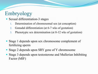


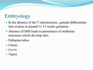


This document discusses disorders of sex development (DSD), formerly known as intersex conditions. It begins with definitions of DSD and describes when to suspect DSD based on physical examination findings in newborns. It then discusses the various types of DSD classified by chromosomal, gonadal and phenotypic sex, including 46,XX virilized females, 46,XY undervirilized males, gonadal differentiation disorders, and syndromes associated with ambiguous genitalia. The document reviews DSD embryology and hormone biosynthesis pathways. It provides guidance on the evaluation, diagnosis and management of DSD through history, physical exam, imaging, genetic and hormonal testing, and the roles of the multidisciplinary DSD team.