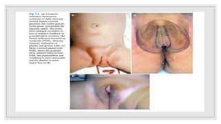
4. History
•Family history.
•History of hypospadias, amenorrhea, and infertility.
•Parental consanguinity.
•Unexplained previous infant deaths.
•Maternal ingestion of drugs (particularly progestogens,
androgens and steroids).
•Use of assisted reproductive technologies.
5. Physical examination
•General condition: ( salt wasting CAH). Usually after the first
week.
•Phallus: length (micropenis <2.5 cm), breadth, erectile tissue.
•Gonads: palpation, asymmetry.
•Scrotum: labioscrotal folding, pigmentation.
•Perineal opening: (UGS, cloaca).
•AGD: anogenital distance.
6. Anogenital distance
• AGD defined as measurement from the center of the anus
to the base of the scrotum in males and to the posterior
fourchette in females showed that mean AGD (+/- SD) at
birth was 19.8 +/- 6.1 mm in males and 9.1 +/- 2.8 mm in
females.
•Decreasing in AGD raise the suspicion of ambiguous
genitalia.
9. hCG stimulation test
•Serum testosterone, DHT, LH & FSH are measured at
baseline and then 24 hrs later after the test.
•Adequate rise indicates presence of functioning testes
• ↑ T:DHT ratio(>20:1): 5 alpha reductase deficiency.
12. • Fluoroscopy, Nonionic contrast, Metallic marker, Voiding films.
• AP, Lateral views
• Assessing: bladder position, communication with surrounding
structures, fistulae, residual MĂĽllerian or Wolffian duct structures,
vesico-ureteric reflux and possible uterine indentation on the bladder.
• Urethral length, common channel, position of the confluence.
17. •Bladder, Kidneys, Adrenals.
•Ovaries, Testis, uterus.
•Hydrometrocolpos are associated with a urogenital sinus or
cloacal malformation , vaginal atresia.
•Full bladder.
18. • In ovo-testicular DSD: the ovo-testes shows focal cystic areas among
heterogeneous tissue with the cystic areas corresponding to ovarian
tissue.
• In (CAIS): labia majora with testicles, which are often in the inguinal
region or in the inguinal canal, as well as the absence of a uterus.
• In CAH: the sonographer will be able to identify a uterus and ovaries;
however the adrenals will appear abnormal.
20. • Routine investigation in DSD ????
• Noninvasive, No radiation, Soft tissue.
• Coronal T1- and T2: wide full view including kidneys and ovaries.
• FSE T2 (sagittal): is excellent for anatomy of the uterus and vagina and
also to determine their relationship to the rectum and bladder.
• The axial plane: is used for ovaries, lower uterine segment and cervix.
• The vagina is best seen on T2 axial slices.
26. • The most common DSD at all.
• The most common enzymatic defect is 21- OH deficiency.
• Virilized female, while Boys with this enzymatic defect do not present with
genital ambiguity.
• 75% of the patients are salt-wasting CAH, Simple virilizing form.
• Ultrasound: female internal organs.
• Hormonal assay: Elevated 17OHP, androgens.
• Serum electrolytes: Hyponatremia, hyperkalemia.
27.
28. â—¦ Prenatal treatment: dexamethasone?????
â—¦ Replacement: Glucocorticoid, Mineralocorticoid.
â—¦ Bilateral adrenalectomy is not part of the standard of care and should
be considered only in cases of failed medical management.
â—¦ Gender assignment: female????
â—¦ Genitogram vs cystoscopy: length of the UGS vs level of vaginal
insertion from the bladder neck.
â—¦ Timing of surgery: early infancy or post pubertal????????
â—¦ ASTRA, PUM, TUM.
29.
30. Gonadal dysgenesis
•46 XX gonadal dysgenesis is exceedingly rare
and not seen in infancy, since there is no genital
ambiguity
31. Testicular DSD
• 80% with 46 XX testicular DSD (XX male).
• After puberty with normal male genitalia, pubic hair, small testes, and
gynecomastia.
• Infertile men due to azoospermia that demonstrate
hypergonadotropic hypogonadism.
32. â—¦ XX
â—¦ Infertile male.
â—¦ Normal male genitalia
â—¦ Small testis
â—¦ Gynecomastia
38. Disorders of gonadal development
Pure gonadal dysgenesis.
Partial gonadal dysgenesis.
Ovotesticular DSD.
Vanishing testis syndrome.
39. Partial Androgen Insensitivity Syndrome
• AR mutations, MAMLD1 mutations.
• Patients with at least one functional testicle and reasonable penile tissue
on palpation should be raised as males.
• Normal or slightly elevated androgens.
• Normal AMH level.
• Normal testicular histology.
• hCG stimulation test
• Male or female ????????
• Testosterone and DHT replacement.
40.
41.
42. Complete androgen insensitivity syndrome
• AR mutation in 80 % of cases.
• Positive family history.
• Either in infancy as inguinal hernia in phenotypically female. Or primary
amenorrhea at puberty.
• Tall female, Female external genitalia, Small nipple, pale areola, Absence pubic
hair.
• Inguinal testis, absent vas deferens and epididymis.
• Sex raring is female, Blind ending vagina.
43. • XY karyotyping.
• Ultrasound: No female internal organs, inguinal testis
• Hormonal assay: Slightly elevated testosterone level, Serum LH slightly elevated , FSH
normal.
• HCG stimulation test.
• DNA sequencing for the AR gene.
• Gonadal biopsy.
• Estrogen replacement at puberty.
• Early or delayed gonadectomy/ or no gonadectomy.
• Reconstructive surgery.
44.
45.
46. 5-Alpha-Reductase Deficiency
• High T/ DHT ratio.
• hCG stimulation test.
• Although most patients are initially raised as females, severe
virilization can take place at the time of puberty, leading to self-
reassignment to the male gender.
47. Pure gonadal dysgenesis
• Swyer’s syndrome.
• Phenotypically female with normal Mullerian structures and bilateral streak
gonads, Primary amenorrhea.
• Testosterone levels are usually low, even after stimulation.
• Gender assignment: female.
• Early gonadectomy at the time of diagnosis is advised due to the high risk of
malignancy gonadoblastoma and dysgerminoma.
49. Persistent Mullerian Duct Syndrome
â—¦Males with cryptorchidism found to harbor Mullerian
structures in the abdomen (uterus, cervix, and proximal third
of the vagina).
â—¦AR trait.
â—¦Surgical resection of the MĂĽllerian structures and repair of
any cryptorchidism.
54. Congenital hypogonadotropic hypogonadism
(CHH)
•Isolated micropenis without hypospadias + cryptorchidism.
•Mostly isolated, may be panhypopituitarism.
•Low levels of FSH, LH, and testosterone.
•Good response to an hCG stimulation test.
•Leydig cell hypoplasia in testicular histology.
•Exogenous replacement of gonadotropins and testosterone at
the puberty.
55. Testis regression syndrome
•Bilateral vanishing testis syndrome.
•Hypergonadotropic hypogonadism syndrome.
•The same picture as CHH but with no response to hCG
stimulation.
•Surgical removal Vs testosterone replacement and follow up.
58. Mixed gonadal dysgenesis
• The commonest chromosomal abnormality associated with ambiguous genitalia.
• Genital ambiguity, short stature, Asymmetrical external genitalia, Inguinal hernias.
• Gonads: streak or testicle.
• Testis: orthotopic to intra-abdominal, Normal or dysgentic.
• Testis: on one virilized side, Mullerian structures: on the other streak side.
• Uterus, hemi uterus, vaginal remnant or large utricle.
• Streak gonads should be removed.
• The more masculinized, the lower the risk of malignancy.
• If female, remove the other testis, estrogen replacement, refer to adult gynecologist.
• If male, put the testis in the scrotum and regular surveillance is VIP, growth hormone and
testosterone replacement.
59.
60. Usually raised as a male.
XO/XY.
Testis on one side and dysgentic gonad on the other side.
Male: orchiopexy and gonadectomy, remove Mullerian structure.
Female: remove testis, hormonal treatment at puberty.
61. Ovo-Testicular DSD
• Ovarian follicles and seminiferous tubules are present in the gonads of the one individual.
• Variable gonadal distribution: one pole of a gonad may consist of ovarian tissue, while the other
pole consists of testicular tissue, another variation is that one gonad is an ovary while the other is a
dysgenetic testis.
• The testicular tissue in ovo-testicular DSD undergoes atresia at a faster rate than the ovarian tissue;
while a small number of women with ovo-testicular DSD have become mothers, paternity has
never been reported.
• The karyotype in ovo-testicular DSD is most commonly 46,XX, but 45,X/46,XY and 46,XY ovo-
testicular DSD are seen.
• Mostly go as a female, usually has uterus, remove testicular tissue.
62.
63. Usually XX.
Asymmetrical gonads, testis in one side and ovary or ovotestis on
the other.
Usually raised as a female.
Gonadal biopsy.
Gonadectomy.
Hormonal replacement therapy.
64. Klinefelter Syndrome – 47,XXY
• Progressive deterioration of testicular function
• oligospermia or azoospermia, with high levels of FSH and LH.
• Males with KS tend to be taller than average.
• Verbal IQ is affected more than performance IQ.
• At puberty, gynecomastia occurs. increased risk of breast cancer.
• Increased risk of non-testicular germ cell cancers, especially in the
mediastinum.
65. Turner syndrome
• Monosomy X, 45 XO.
• webbing of the neck, lymphoedema of the hands and feet.
• Bicuspid aortic valve and coarctation of the aorta. Essential
hypertension is a risk during adolescence and adulthood.
• obstruction of their Eustachian tubes and are therefore prone to
otitis media, conductive deafness and cholesteatoma.
• High -arched palate, downturned angles of the mouth, strabismus and
epicanthic folds.
73. Case 1
â—¦ A newborn is referred to you with ambiguous genitalia. On examination, he has a
severe penoscrotal hypospadias, marked chordee, and palpable undescended
gonads. The karyotype is 46,XY.
1 Describe the normal sex development.
2 What is the differential diagnosis in this case?
3 How would you investigate this newborn?
4 He is noted to have a large prostatic utricle, which is discovered when he
has a cystoscopy. When would you operate on this? What are the various
surgical approaches?
74. Case 2
â—¦ A 3-month-old girl presents with an irreducible left inguinal hernia. At
surgical exploration, she is noted to have presence of a testis
in the hernial sac.
1 What are your surgical options?
2 What further investigations are necessary, and why?
3 What further surgical procedures may be necessary?
75. Case 3
â—¦ A 3-month-old boy is referred to you as his parents are concerned about his
penile length, which they feel is too short. He has been seen by his family
physician, who feels he may have a micropenis.
1 What is the differential diagnosis, and how can a micropenis be suspected
on clinical grounds?
2 If a micropenis is suspected, what investigations might be performed and
why?
3 What is the role of exogenous testosterone in micropenis?
4 What is the long-term outlook for males with micropenis or those that have
been gender reassigned?