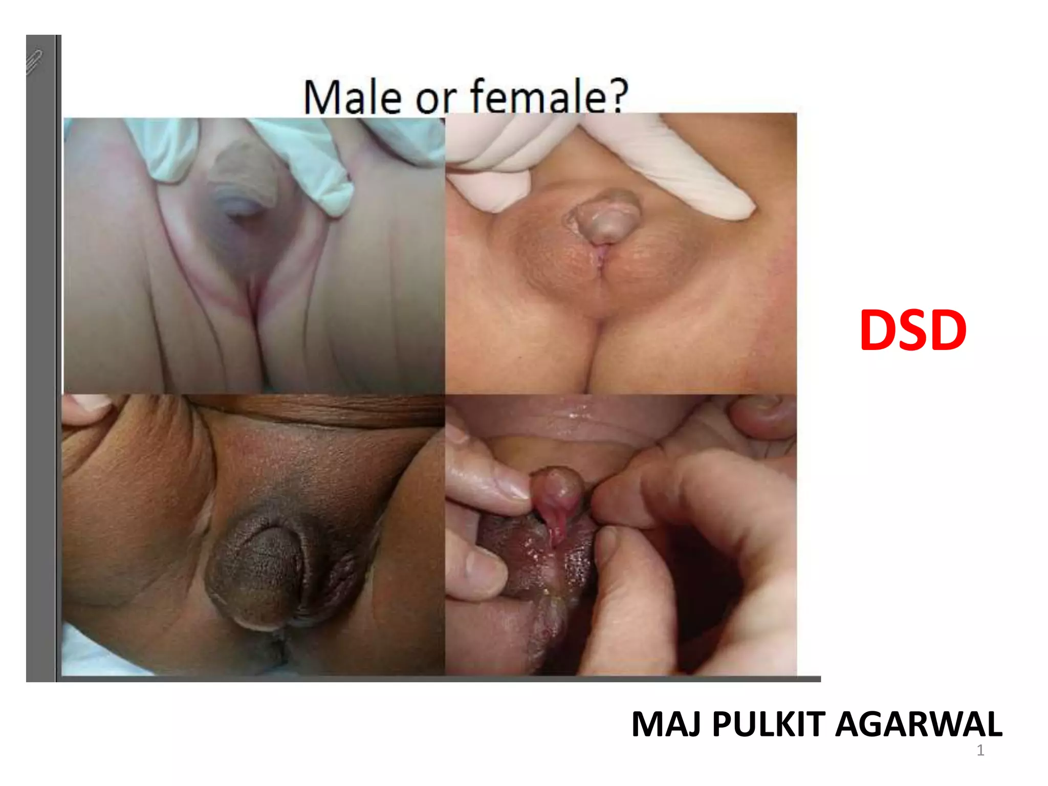
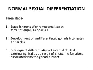
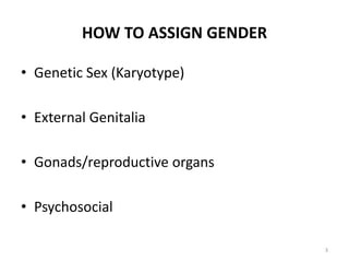
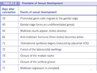
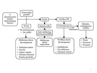
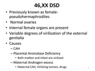
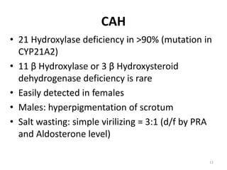
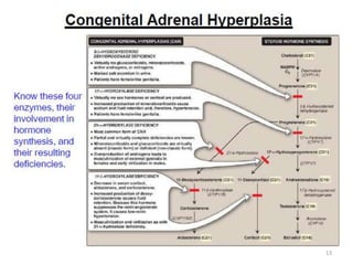
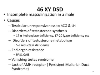
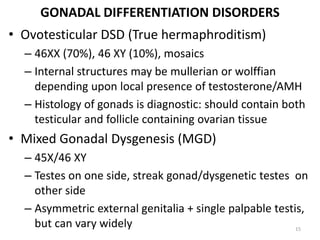
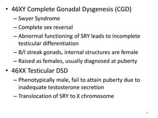
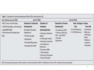
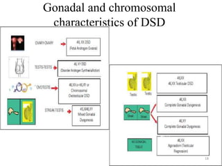
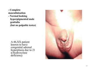
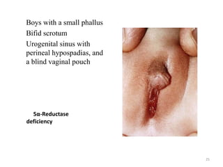
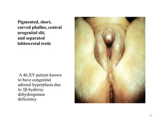
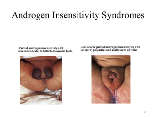
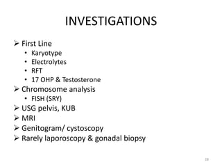
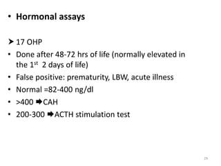
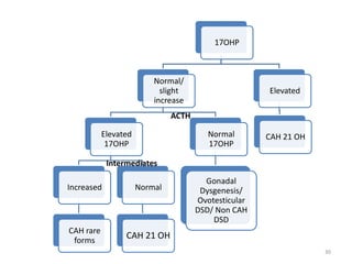
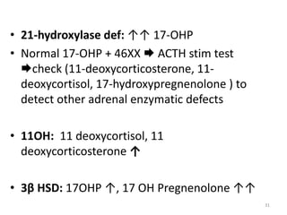
DSD
MAJ PULKIT AGARWAL1
NORMAL SEXUAL DIFFERENTIATION
Three steps-
1. Establishment of chromosomal sex at
fertilization(46,XX or 46,XY)
2. Development of undifferentiated gonads into testes
or ovaries
3. Subsequent differentiation of internal ducts &
external genitalia as a result of endocrine functions
associated with the gonad present
2
HOW TO ASSIGN GENDER
• Genetic Sex (Karyotype)
• External Genitalia
• Gonads/reproductive organs
• Psychosocial
3
4
5
7
DISORDERS OF SEXUAL DIFFERENTIATION
• When the external genitalia do not have the
typical anatomic appearance of normal male
or female genitalia.
• Infants with ambiguous genitalia have genes
of either a male or female, but with some
additional characteristics of opposite sex.
8
9
CAUSES OF DSD
• 46XX DSD
• 46XY DSD
• Disorders of Gonadal Differentiation
10
46,XX DSD
• Previously known as female-
pseudohermaphrodites
• Normal ovaries
• Internal female organs are present
• Variable degrees of virilisation of the external
genitalia
• Causes
– CAH
– Placental Aromatase Deficiency
• Both mother and infant are virilised
– Maternal Androgen excess
• Maternal CAH, Virilizing tumors, drugs 11
CAH
• 21 Hydroxylase deficiency in >90% (mutation in
CYP21A2)
• 11 β Hydroxylase or 3 β Hydroxysteroid
dehydrogenase deficiency is rare
• Easily detected in females
• Males: hyperpigmentation of scrotum
• Salt wasting: simple virilizing = 3:1 (d/f by PRA
and Aldosterone level)
12
13
46 XY DSD
• Incomplete masculinization in a male
• Causes
– Testicular unresponsiveness to hCG & LH
– Disorders of testosterone synthesis
• 17 α hydroxylase deficiency, 17-20 lyase deficiency etc
– Disorders of testosterone metabolism
• 5 α reductase deficiency
– End organ resistance
• PAIS, CAIS
– Vanishing testes syndrome
– Lack of AMH receptor ( Persistent Mullerian Duct
Syndrome)
14
GONADAL DIFFERENTIATION DISORDERS
• Ovotesticular DSD (True hermaphroditism)
– 46XX (70%), 46 XY (10%), mosaics
– Internal structures may be mullerian or wolffian
depending upon local presence of testosterone/AMH
– Histology of gonads is diagnostic: should contain both
testicular and follicle containing ovarian tissue
• Mixed Gonadal Dysgenesis (MGD)
– 45X/46 XY
– Testes on one side, streak gonad/dysgenetic testes on
other side
– Asymmetric external genitalia + single palpable testis,
but can vary widely 15
• 46XY Complete Gonadal Dysgenesis (CGD)
– Swyer Syndrome
– Complete sex reversal
– Abnormal functioning of SRY leads to incomplete
testicular differentiation
– B/l streak gonads, internal structures are female
– Raised as females, usually diagnosed at puberty
• 46XX Testicular DSD
– Phenotypically male, fail to attain puberty due to
inadequate testosterone secretion
– Translocation of SRY to X chromosome
16
17
Gonadal and chromosomal
characteristics of DSD
18
EVALUATION
19
HISTORY
Family history
• CAH:Neonatal death (male baby with
vomiting/dehydration)
• Hypospadias/ cryptorchidism
• Infertility/Pubertal delay
• Consanguinity
Pregnancy history
-Placental aromatase deficiency allows fetal adrenal
androgens to virilize both mother & fetus
-Androgen secreting tumors
– Medication: Androgens, antiandrogens (finesteride,
spironolactone), estrogens
20
PHYSICAL EXAMINATION
General Examination
– Dysmorphic features
– Evidence of salt wasting skin turgor, poor tone,
dehydration, low BP, vomiting, poor feeding
– Hyper pigmentation
– In adolescent evidence of hirsutism/ virilization
Tanner staging
21
External Genitalia
• Phallus
– Stretched length (>2.5 cm in male term infant/ < 1cm
clitoris in female term infant)
– Chordee
– Position of orifice
• Labioscrotal folds
– Separated or fused fusion is an androgen effect
– Skin texture rugosity suggests exposure to androgens
– Color of the skin ↑↑ pigmentation may be evidence
for CAH
– Vaginal opening 22
Gonadal Examination
• Palpate labioscrotal tissue & inguinal canal for
presence of gonads
• Note No. of gonads, size, symmetry, position.
• Palpable gonads below the inguinal canal are almost
always testicles
• Ovotestis may be present as inguinal hernia
Rectal exam
• To palpate for presence or absence of the uterus
23
- Complete
masculinisation
- Normal looking
hyperpigmented male
genitalia
(but no palpable testes)
A 46,XX patient
known to have
congenital adrenal
hyperplasia due to 21
α-hydroxylase
deficiency
24
5α-Reductase
deficiency
Boys with a small phallus
Bifid scrotum
Urogenital sinus with
perineal hypospadias, and
a blind vaginal pouch
25
Pigmented, short,
curved phallus, central
urogenital slit,
and separated
labioscrotal testis
A 46,XY patient known
to have congenital
adrenal hyperplasia due
to 3β-hydroxy
dehydrogenase
deficiency
26
Androgen Insensitivity Syndromes
Partial androgen insensitivity with
descended testes in bifid labioscrotal folds
Less severe partial androgen insensitivity with
severe hypospadias and maldescent of testes
27
INVESTIGATIONS
 First Line
• Karyotype
• Electrolytes
• RFT
• 17 OHP & Testosterone
 Chromosome analysis
• FISH (SRY)
 USG pelvis, KUB
 MRI
 Genitogram/ cystoscopy
 Rarely laporoscopy & gonadal biopsy
28
• Hormonal assays
 17 OHP
• Done after 48-72 hrs of life (normally elevated in
the 1st 2 days of life)
• False positive: prematurity, LBW, acute illness
• Normal =82-400 ng/dl
• >400 CAH
• 200-300 ACTH stimulation test
29
17OHP
Normal/
slight
increase
Elevated
17OHP
Increased
CAH rare
forms
Normal
CAH 21 OH
Normal
17OHP
Gonadal
Dysgenesis/
Ovotesticular
DSD/ Non CAH
DSD
Elevated
CAH 21 OH
ACTH
Intermediates
30
• 21-hydroxylase def: ↑↑ 17-OHP
• Normal 17-OHP + 46XX  ACTH stim test
check (11-deoxycorticosterone, 11-
deoxycortisol, 17-hydroxypregnenolone ) to
detect other adrenal enzymatic defects
• 11OH: 11 deoxycortisol, 11
deoxycorticosterone ↑
• 3β HSD: 17OHP ↑, 17 OH Pregnenolone ↑↑
31
Testosterone
Normal/ Slight
Increase
No increase,
normal T:DHT
?PAIS
Phallus increase
<2+/- 0.6
Very high
CAIS
Low
No change
Gonadal
dysgenesis/LH
receptor mutation/
Gest loss of
testes/defect of
Testosterone synth
T:DHT >20:1
5 α reductase def
T:DHT<0.8:1
17 β HSD
deficiency
HCG stimulation
HCG stimulation
Monthly
testosterone
x 3 months
32
33
34
THANKS
35

Approach to DSD (Ambiguous genitalia)

  • 1.
  • 2.
    NORMAL SEXUAL DIFFERENTIATION Threesteps- 1. Establishment of chromosomal sex at fertilization(46,XX or 46,XY) 2. Development of undifferentiated gonads into testes or ovaries 3. Subsequent differentiation of internal ducts & external genitalia as a result of endocrine functions associated with the gonad present 2
  • 3.
    HOW TO ASSIGNGENDER • Genetic Sex (Karyotype) • External Genitalia • Gonads/reproductive organs • Psychosocial 3
  • 4.
  • 5.
  • 6.
  • 7.
    DISORDERS OF SEXUALDIFFERENTIATION • When the external genitalia do not have the typical anatomic appearance of normal male or female genitalia. • Infants with ambiguous genitalia have genes of either a male or female, but with some additional characteristics of opposite sex. 8
  • 8.
  • 9.
    CAUSES OF DSD •46XX DSD • 46XY DSD • Disorders of Gonadal Differentiation 10
  • 10.
    46,XX DSD • Previouslyknown as female- pseudohermaphrodites • Normal ovaries • Internal female organs are present • Variable degrees of virilisation of the external genitalia • Causes – CAH – Placental Aromatase Deficiency • Both mother and infant are virilised – Maternal Androgen excess • Maternal CAH, Virilizing tumors, drugs 11
  • 11.
    CAH • 21 Hydroxylasedeficiency in >90% (mutation in CYP21A2) • 11 β Hydroxylase or 3 β Hydroxysteroid dehydrogenase deficiency is rare • Easily detected in females • Males: hyperpigmentation of scrotum • Salt wasting: simple virilizing = 3:1 (d/f by PRA and Aldosterone level) 12
  • 12.
  • 13.
    46 XY DSD •Incomplete masculinization in a male • Causes – Testicular unresponsiveness to hCG & LH – Disorders of testosterone synthesis • 17 α hydroxylase deficiency, 17-20 lyase deficiency etc – Disorders of testosterone metabolism • 5 α reductase deficiency – End organ resistance • PAIS, CAIS – Vanishing testes syndrome – Lack of AMH receptor ( Persistent Mullerian Duct Syndrome) 14
  • 14.
    GONADAL DIFFERENTIATION DISORDERS •Ovotesticular DSD (True hermaphroditism) – 46XX (70%), 46 XY (10%), mosaics – Internal structures may be mullerian or wolffian depending upon local presence of testosterone/AMH – Histology of gonads is diagnostic: should contain both testicular and follicle containing ovarian tissue • Mixed Gonadal Dysgenesis (MGD) – 45X/46 XY – Testes on one side, streak gonad/dysgenetic testes on other side – Asymmetric external genitalia + single palpable testis, but can vary widely 15
  • 15.
    • 46XY CompleteGonadal Dysgenesis (CGD) – Swyer Syndrome – Complete sex reversal – Abnormal functioning of SRY leads to incomplete testicular differentiation – B/l streak gonads, internal structures are female – Raised as females, usually diagnosed at puberty • 46XX Testicular DSD – Phenotypically male, fail to attain puberty due to inadequate testosterone secretion – Translocation of SRY to X chromosome 16
  • 16.
  • 17.
  • 18.
  • 19.
    HISTORY Family history • CAH:Neonataldeath (male baby with vomiting/dehydration) • Hypospadias/ cryptorchidism • Infertility/Pubertal delay • Consanguinity Pregnancy history -Placental aromatase deficiency allows fetal adrenal androgens to virilize both mother & fetus -Androgen secreting tumors – Medication: Androgens, antiandrogens (finesteride, spironolactone), estrogens 20
  • 20.
    PHYSICAL EXAMINATION General Examination –Dysmorphic features – Evidence of salt wasting skin turgor, poor tone, dehydration, low BP, vomiting, poor feeding – Hyper pigmentation – In adolescent evidence of hirsutism/ virilization Tanner staging 21
  • 21.
    External Genitalia • Phallus –Stretched length (>2.5 cm in male term infant/ < 1cm clitoris in female term infant) – Chordee – Position of orifice • Labioscrotal folds – Separated or fused fusion is an androgen effect – Skin texture rugosity suggests exposure to androgens – Color of the skin ↑↑ pigmentation may be evidence for CAH – Vaginal opening 22
  • 22.
    Gonadal Examination • Palpatelabioscrotal tissue & inguinal canal for presence of gonads • Note No. of gonads, size, symmetry, position. • Palpable gonads below the inguinal canal are almost always testicles • Ovotestis may be present as inguinal hernia Rectal exam • To palpate for presence or absence of the uterus 23
  • 23.
    - Complete masculinisation - Normallooking hyperpigmented male genitalia (but no palpable testes) A 46,XX patient known to have congenital adrenal hyperplasia due to 21 α-hydroxylase deficiency 24
  • 24.
    5α-Reductase deficiency Boys with asmall phallus Bifid scrotum Urogenital sinus with perineal hypospadias, and a blind vaginal pouch 25
  • 25.
    Pigmented, short, curved phallus,central urogenital slit, and separated labioscrotal testis A 46,XY patient known to have congenital adrenal hyperplasia due to 3β-hydroxy dehydrogenase deficiency 26
  • 26.
    Androgen Insensitivity Syndromes Partialandrogen insensitivity with descended testes in bifid labioscrotal folds Less severe partial androgen insensitivity with severe hypospadias and maldescent of testes 27
  • 27.
    INVESTIGATIONS  First Line •Karyotype • Electrolytes • RFT • 17 OHP & Testosterone  Chromosome analysis • FISH (SRY)  USG pelvis, KUB  MRI  Genitogram/ cystoscopy  Rarely laporoscopy & gonadal biopsy 28
  • 28.
    • Hormonal assays 17 OHP • Done after 48-72 hrs of life (normally elevated in the 1st 2 days of life) • False positive: prematurity, LBW, acute illness • Normal =82-400 ng/dl • >400 CAH • 200-300 ACTH stimulation test 29
  • 29.
    17OHP Normal/ slight increase Elevated 17OHP Increased CAH rare forms Normal CAH 21OH Normal 17OHP Gonadal Dysgenesis/ Ovotesticular DSD/ Non CAH DSD Elevated CAH 21 OH ACTH Intermediates 30
  • 30.
    • 21-hydroxylase def:↑↑ 17-OHP • Normal 17-OHP + 46XX  ACTH stim test check (11-deoxycorticosterone, 11- deoxycortisol, 17-hydroxypregnenolone ) to detect other adrenal enzymatic defects • 11OH: 11 deoxycortisol, 11 deoxycorticosterone ↑ • 3β HSD: 17OHP ↑, 17 OH Pregnenolone ↑↑ 31
  • 31.
    Testosterone Normal/ Slight Increase No increase, normalT:DHT ?PAIS Phallus increase <2+/- 0.6 Very high CAIS Low No change Gonadal dysgenesis/LH receptor mutation/ Gest loss of testes/defect of Testosterone synth T:DHT >20:1 5 α reductase def T:DHT<0.8:1 17 β HSD deficiency HCG stimulation HCG stimulation Monthly testosterone x 3 months 32
  • 32.
  • 33.
  • 34.