Downloaded 122 times
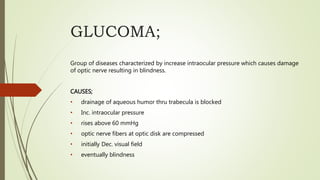
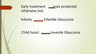
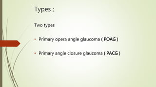
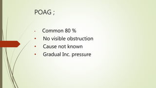
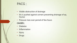

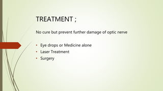
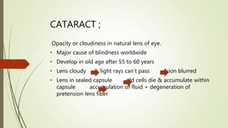
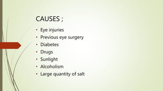
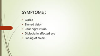
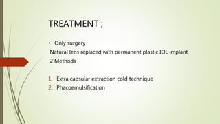


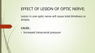
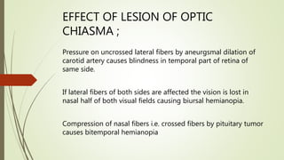
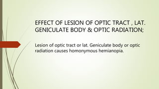
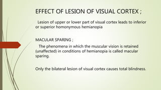
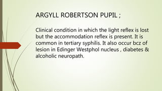
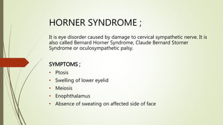

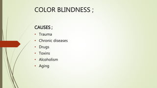
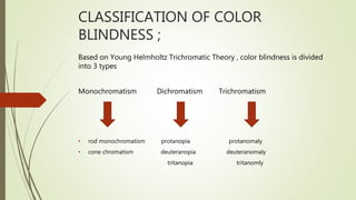


The document discusses various eye diseases, primarily glaucoma, cataracts, and their effects on vision, highlighting causes, symptoms, and treatment options. Glaucoma is characterized by increased intraocular pressure leading to optic nerve damage, while cataracts cause lens opacity resulting in blurred vision, and other conditions like night blindness and color blindness are also detailed. It also describes the effects of lesions on different parts of the visual pathway and the impact on visual fields.


![CTEV [ clubfoot] DR ARUN LAL ,DR MOHAMED ASHRAF travancore medical college k...](https://cdn.slidesharecdn.com/ss_thumbnails/ctevclubfootdrarunlaldrmohamedashraftravancoremedicalcollegekollamkeralaindia-260208063247-18fc466c-thumbnail.jpg?width=640&height=640&fit=bounds)


