Human have a special digestive system than the other animals. We have to know how our body digest our food after having a meal. So, let's check it out.
The Interaction of the internal and external organs that are responsible for the digestion process.
Grade 8 Power Point Presentation
Prepared by: Edsyl Berongoy Penas, LPT
Austin Digestive System is an open access, peer reviewed, scholarly journal dedicated to publish articles covering all areas of Digestive System.
The journal aims to promote latest information and provide a forum for doctors, researchers, physicians, and healthcare professionals to find most recent advances in the areas of Digestive System. Austin Digestive System accepts research articles, reviews, mini reviews, case reports and rapid communication covering all aspects of Digestive System.
Austin Digestive System strongly supports the scientific up gradation and fortification in related scientific research community by enhancing access to peer reviewed scientific literary works. Austin Publishing Group also brings universally peer reviewed journals under one roof thereby promoting knowledge sharing.
Anatomy of GI Tract with special reference to anatomy and functions of stomach,
( Acid production in the stomach, regulation of acid production through
parasympathetic nervous system, pepsin role in protein digestion) small intestine
and large intestine, anatomy and functions of salivary glands, pancreas and liver,
movements of GIT, digestion and absorption of nutrients and disorders of GIT.
753 Learning OutcomesAfter reading this chapter, .docxpoulterbarbara
75
3
Learning Outcomes
After reading this chapter, you will be able to:
3.1 Describe the processes and organs involved in
digestion.
3.2 Explain how food is propelled through the gas-
trointestinal tract.
3.3 Identify the role of enzymes and other secre-
tions in chemical digestion.
3.4 Describe how digested nutrients are absorbed.
3.5 Explain how hormones and the nervous sys-
tem regulate digestion.
3.6 Explain how absorbed nutrients are trans-
ported throughout the body.
3.7 Discuss the most common digestive disorders.
True or False?
1. Saliva can alter the taste of food. T/F
2. Without mucus, the stomach would digest itself. T/F
3. The major function of bile is to emulsify fats. T/F
4. Acid reflux is caused by gas in the stom-ach. T/F
5. The primary function of the large intes-tine is to absorb water. T/F
6. Feces contain a high amount of bacteria. T/F
7. The lymphatic system transports all nutrients through the body once they’ve been absorbed. T/F
8. Hormones play an important role in digestion. T/F
9. Diarrhea is always caused by bacterial infection. T/F
10. Irritable bowel syndrome is caused by an allergy to gluten. T/F
See page 110 for the answers.
Digestion,
Absorption,
and Transport
M03_BLAK8260_04_SE_C03.indd 75 12/1/17 11:28 PM
76 Chapter 3 | Digestion, Absorption, and Transport
The digestion of food begins even before you take that first bite. Just the sight and smell of homemade apple pie stimulates the release of saliva in
the mouth. The secretion of saliva and other digestive juices starts a cascade of
events that prepares the body for digestion, the chemical and mechanical
processes by which the body breaks food down into individual nutrient
molecules ready for absorption. Food components that aren’t absorbed are
excreted as waste (feces) by elimination. Although these are complex
processes, they go largely unnoticed. You consciously chew and swallow the
pie, but you don’t feel the release of chemicals or the muscular contractions
that cause it to be digested or the absorption of nutrient molecules through
the intestinal lining cells. In fact, you may be unaware of the entire process
until about 48 hours after eating, when the body is ready to eliminate waste.
In this chapter, we explore the processes of digestion, absorption, and
elimination, the organs involved, and the other biological mechanisms that
regulate our bodies’ processing of food and nutrients. We also discuss the causes
and treatments of some common gastrointestinal conditions and disorders.
What Are the Processes and Organs
Involved in Digestion?
LO 3.1 Describe the processes and organs involved in digestion.
Digestion, absorption, and elimination occur in the gastrointestinal (GI) tract, a mus-
cular tube approximately 20–24 feet long in an adult. Stretched vertically, the tube would
be about as high as a two-story building. It provides a barrier between the food within the
lumen (the hollow .
Introduction to digestive system
Organs of digestive tract
Mouth and their different enzymes and actions
salivary glands
Oesophagus
Stomach
Small Intestine and funcions
Large Intestine and functions
Anus
Assessary Organs
Liver
Pancreas
Digestive system Physiology
Ingestion
Digestion
Absorption
Assimilation.
Excretion
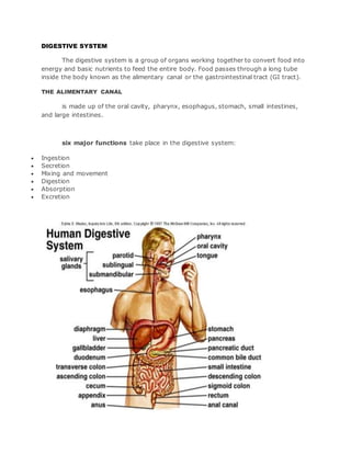
1. DIGESTIVE SYSTEM
The digestive system is a group of organs working together to convert food into
energy and basic nutrients to feed the entire body. Food passes through a long tube
inside the body known as the alimentary canal or the gastrointestinal tract (GI tract).
THE ALIMENTARY CANAL
is made up of the oral cavity, pharynx, esophagus, stomach, small intestines,
and large intestines.
six major functions take place in the digestive system:
Ingestion
Secretion
Mixing and movement
Digestion
Absorption
Excretion
2. Digestive System Anatomy
Mouth
Food begins its journey through the digestive system in the mouth, also known as the
oral cavity.
Teeth
The teeth are 32 small, hard organs found along the anterior and lateral
edges of the mouth.
Tongue
The tongue is located on the inferior portion of the mouth just posterior
and medial to the teeth.
Salivary Glands
Surrounding the mouth are 3 sets of salivary glands. The salivary glands
are accessory organs that produce a watery secretion known as saliva.
Saliva helps to moisten food and begins the digestion of carbohydrates.
The body also uses saliva to lubricate food as it passes through the
mouth, pharynx, and esophagus.
Pharynx
The pharynx, or throat, is a funnel-shaped tube connected to the posterior end of the
mouth.
Esophagus
The esophagus is a muscular tube connecting the pharynx to the stomach that is
part of the upper gastrointestinal tract.
Stomach
The stomach is a muscular sac that is located on the left side of the abdominal
cavity, just inferior to the diaphragm.
Small Intestine
The small intestine is a long, thin tube about 1 inch in diameter and about 10 feet
long that is part of the lower gastrointestinal tract.
Liver and Gallbladder
The liver is a roughly triangular accessory organ of the digestive system located to
the right of the stomach, just inferior to the diaphragm and superior to the small
intestine.
Pancreas
The pancreas is a large gland located just inferior and posterior to the stomach.
3. Large Intestine
The large intestine is a long, thick tube about 2 ½ inches in diameter and about 5
feet long.
Digestive System Physiology
The six primary processes of the digestive system include:
1. Ingestion of food
2. Secretion of fluids and digestive enzymes
3. Mixing and movement of food and wastes through the body
4. Digestion of food into smaller pieces
5. Absorption of nutrients
6. Excretion of wastes
Ingestion
The first function of the digestive system is ingestion, or the intake of food.
Secretion
In the course of a day, the digestive system secretes around 7 liters of fluids. These
fluids include saliva, mucus, hydrochloric acid, enzymes, and bile.
Mixing and Movement
The digestive system uses 3 main processes to move and mix food:
Swallowing
is the process of using smooth and skeletal muscles in the mouth,
tongue, and pharynx to push food out of the mouth, through the pharynx,
and into the esophagus.
Peristalsis
is a muscular wave that travels the length of the GI tract, moving
partially digested food a short distance down the tract. It takes many
waves of peristalsis for food to travel from the esophagus, through the
stomach andintestines, and reach the end of the GI tract.
Segmentation
occurs only in the small intestine as short segments of intestine contract
like hands squeezing a toothpaste tube. Segmentation helps to increase
the absorption of nutrients by mixing food and increasing its contact with
the walls of the intestine.
4. Digestion
is the process of turning large pieces of food into its component chemicals.
Absorption
Once food has been reduced to its building blocks, it is ready for the body to
absorb.
Excretion
The final function of the digestive system is the excretion of waste in a process
known as defecation.
5. The Digestive System of a Chicken
the digestive system of a chicken mechanically and chemically breaks down food
and allows nutrients to be absorbed ready for use in the body. It is important to
understand how the digestive system works in order to get a better idea of digestive
system problems and to know what is normal (like a bulging crop or the caecal
discharge). The main problems that we encounter as hobby poultry keepers in the
digestive system are worms and crop problems such as impacted crop / sour crop.
The basic function of the digestive system is described here:
The beak moistens food with Saliva. Food is not chewed.
The oesophagus takes the food down to the crop to be stored. After a chicken has eaten, the
crop will feel full and bulge.
Food from the crop slowly passes down to the proventriculus.
The proventriculus mixes the food with acids and digestive enzymes.
Food is then passed through to the gizzard where insoluble (flint) grit has accumulated.
Food is ground down by strong muscular action in the gizzard.
From the gizzard, food is passed through to the small intestine and is reduced further with
enzymes from the pancreas.
Bile produced by the liver and stored in the gall bladder helps to break down fat.
The intestines digest the food, taking nutrients from it.
Water and the remaining undigested food is absorbed in the large intestine.
The caeca are a pair of tubes that allow fermentation of undigested food to take place. This is
emptied every 24 hours or so and is a light brown (mustard colour) froth. This can often be
confused as diarrhoea by the novice.
The cloaca / vent passes a combination of faeces and urine, together with eggs from the oviduct.
6.
7. Digestive System of the Pig: Anatomy and Function
The digestive system of a pig is well suited for complete concentrate based rations that are
typically fed. The entire digestive tract is relatively simple in terms of the organs involved,
which are connected in a continuous musculo-membanous tube from mouth to anus. Yet this
multi-faceted system involves many complex interactive functions.
Mouth
The mouth serves a valuable role not only for the consumption of food but it
also provides for the initial partial size reduction though grinding. While teeth
serve the main role in grinding to reduce food size and increase surface area,
the first action to begin the chemical breakdown of food occurs when feed is
mixed with saliva.
Stomach
The stomach is a muscular organ responsible for storage, initiating the
breakdown of nutrients, and passing the digesta into the small intestine.
8. Small Intestine, Pancreas and Liver
The small intestine is the major site of nutrient absorption, and is divided into
three sections. The first section is the duodenum. The duodenum is
approximately 12 inches long and is the portion of the small intestine that
ducts from the pancreas and the liver (gall bladder). The pancreas is involved
with both exocrine and endocrine excretions. This means the pancreas is
responsible for secretion of insulin and glucagon in response to high or low
glucose levels in the body. In addition, it has exocrine functions of secreting
digestive enzymes and sodium bicarbonate.
Large Intestine
The large intestine or hindgut encompasses four main sections. First, digesta
from the small intestine passes into the caecum. The caecum has two
sections, first a section that has a blind end, where material can not pass
though. The caecum has a second portion where it connects to the colon,
where digesta is passed to the rectum and anus where the remaining digesta
is excreted.
The main function of the large intestine is the absorption of water. The chyme
that passes through the small intestine and into the large intestine initially is very
fluid. The large intestine epithelium has a large capacity for water absorption.
9. Digestive System of a Frog
The digestive system of frog consists of digestive tract and the accessory organs which help to
process the food consumed into small molecules (nutrients) which then can be easily absorbed and
then utilized by the cells of the body.
Parts of the digestive system of a frog includes:
Mouth
Pharynx
Oesophagus
Stomach
Small intestine
Large intestine
Cloaca
Accessory organs
Mechanism of Digestion in Digestive System of a Frog
The digestive system of a frog starts with the mouth. Mouth helps in consumption of food. This
process is known as ingestion. Frog feeds on flies or insects. As the teeth’s present in frog is very
week they are not useful to catch the agile prey. Frogs catch their food (such as insects and flies)
with the help of its stick tongue and mixes it with the saliva. The teeth’s present in the upper jaw are
called the maxillary teeth, it helps in grinding the ingested food before it is swallowed.
The saliva produced and secreted by the salivary glands helps in conversion of starch to sugar and
adds liquid to the ingested food.
The food mixed with saliva then moves from mouth into the pharynx, and then into the oesophagus.
It pushes the food further into the sac like structure stomach. This movement is food into the
stomach is known as deglutition or swallowing.
Food particles in the stomach mix thoroughly with enzymes and other fluids due to contraction of
smooth muscles present in the stomach. Peristaltic movement propels the food particles into the
digestive tract and the pyloric sphincter valve is involved in preventing the movement of food
backward from the stomach.
10. The food which is partially digested in stomach then proceeds in to the small intestine, where most
part of the digestion occurs. It is divisible into duodenum and ileum. Pancreatic juice is secreted from
the pancreas and bile through the gallbladder from the liver to the small intestine, which helps in
completion of digestion.
Absorption of the digested nutrients in digestive system of a frog occurs in small intestine.
Absorption unabsorbed nutrients and reabsorption of water takes place in the large intestine. Liquid
wastes are in frog is passed to the urinary bladder, while solids are routed to the cloaca. Both liquid
and solid wastes in frog are expelled out through cloaca which is a slit that opens out finally in
digestive system of a frog.
11. Digestive System of the Dog
The pictures in this section are reprinted with permission by the copyright owner, Hill's Pet Nutrition,
from the Atlas of Veterinary Clinical Anatomy. These illustrations should not be downloaded, printed or
copied except for personal, non-commercial use.
The digestive system includes the:
mouth
teeth
salivary glands
esophagus
stomach
small intestine
large intestine
pancreas
liver and gall bladder
12. The digestive system absorbs and digests food and eliminates solid wastes from the body.
mouth
19. The Digestive Tract
As with all animals digestion in fish involves the breakdown of eaten food into its
smaller component parts, amino acids, vitamins, fatty acids etc. which can then be
used to build up new fish body. The breaking apart or breaking down of the eaten
material is called anabolism, the building up of new material is called catabolism and
these two together make up the whole of metabolism. Grammatically it follows from
this that the respective adjectives are anabolic, catabolic and metabolic.
Mouth - Pharynx - Oesophagus - Gizzard - Stomach - Intestines - Rectum.
However not all fish have all these parts, some, like many of the Cyprinids and
Cyprinidonts, lack a stomach, while a gizzard is only found in a relatively few species.
The Mouth
Food is brought into the body via the mouth, and the jaws of modern teleost fish are a
mechanical wonder, and the way the many bones work together is quite inspiring.
However there is, as always a large variety in fish as a whole and the mouths of a
Basking Shark, a Yellowfin Tuna and a Seahorse are quite different in both form and
function. Lips are rare in fish, most species have a hard edge to their mouth. Some
suction feeders that take in small prey items have small protractible lips that help give
the mouth the form of a tube with a circular opening.
The Pharynx
Immediately behind the mouth is the pharynx which is the continuation of the tube
started at the mouth and in which the are found the gill clefts, through which water
20. flows out of the alimentary canal and into the gills. It is short which leads to the
oesophagus. It is lined with squamous epithelium. As I mentioned above the pharynx
may possess teeth, both upper and lower and as many as 4 rows of them. These
pharyngeal teeth may be specialised forgrinding like molars, comb-like forbreaking
up fine materials of sharp and pointed forpiercing prey, in some species they are
even hinged so that they fold up to allow foodto pass and hang down again
afterwards to prevent its escape. For the most part however pharyngeal teeth seem
to have evolved in order to assist in the act of swallowing food.
The Oesophagus
After the pharynx comes the oesophagus, a muscular tube that leads to the stomach.
It is constructed of two layers of non-striated muscle, one of which is longitudinal
and the other circular, strangely in some species of fish the longitudinal muscle is the
inner layer while in others the circular muscle is the inner layer. With so many
species generalisations only apply to the majority, there are always exceptions. The
Tench (Tinca tinca) forinstance is unusual in having striated muscle all through the
oesophagus and stomach ant into the intestines. The walls of the anterior portion of
the Oesophagus are lined squamous epithelium while those of the posterior section
are lined with columnar epithelium, the whole contains many mucus cells, the mucus
keeps the foodlubricated and helps it to move along the tube.
The Gizzard
The gizzard is really a highly muscular modificationof the first part of the stomach.
Its main purpose is to grind up coarse fooditems into smaller pieces thus facilitating
their later digestion. In those fish which have a gizzard, such as Shad, it is the place
where digestion begins because as well as its muscular activity the gizzard also
secretes digestive enzymes into the food.
The Stomach
The stomach of fish is less well delineated than it is in the higher vertebrates, and in
some cases it is considered to be absent. Where a true stomach is found to exist it is
a muscular bag, or tube with a highly acidic internal environment.
Pyloric Caeca
At th hind end of the stomach, before, just at the beginning of the intestines many
fish have some thin blind tubes called Pyloric Caeca.
21. Intestines
The intestine is a long thin tube with a thin double layer musculature, the outer layer
being longitudinal and the inner layer being circular.
The Rectum
The rectum is the end of the intestines and through it faeces pass out of the fish's
body and into the surrounding water.
The Pancreas
The Pancreas is well developed in the lungfish, sharks and rays and most juvenile
fish, however in many teleosts it becomes quite reduced and diffuse in the adults. In
sharks and rays it is quite distinct from the liver, but in those teleosts wherein it is
found it is oftenpartially embedded in the liver. The pancreas secretes enzymes such
as trypsin (attacks proteins), amylases (attack carbohydrates) and lipases (attack fats)
into the intestines either through sharing one of the hepatic ducts (those belonging
to the liver), or through its own pancreatic duct.
The Liver
Is a large organ that play various roles in the fishes body, it is the site of glycogen
storage, it produces a variety of substances, including enzymes that help with the
digestion and it is a major chemical factory producing various hormones as well as
numerous other important molecules. The liver has no specific shape in fish and
generally molds itself into the space around the stomachand the heart however it
has a tendancy to reflect the fish's body shape, being long and tin in eels and wide in
rays and skates.
22. Muscularsystem
The muscular system is an organ system consisting of skeletal, smooth and cardiac muscles.
It permits movement of the body, maintains posture, and circulates blood throughout the
body. The muscular system in vertebrates is controlled through the nervous system,
although some muscles (such as the cardiac muscle) can be completely autonomous.
Together with the skeletal system it forms the musculoskeletal system, which is responsible
for movement of the human body.
Muscles
Muscles provide strength, balance, posture, movement and heat for the body to keep warm.
There are three distinct types of muscles:
skeletal muscles
cardiac or heart muscles
and smooth (non-striated) muscles
Aerobic and anaerobic muscle activity
At rest, the body produces the majority of its ATP aerobically in
the mitochondria[1]
without producing lactic acid or other fatiguing byproducts.[2]
During
exercise, the method of ATP production varies depending on the fitness of the individual
as well as the duration, and intensity of exercise.
Cardiac muscle
Heart muscles are distinct from skeletal muscles because the muscle fibers are laterally
connected to each other. Furthermore, just as with smooth muscles, they are not
controlling themselves. Heart muscles are controlled by the sinus node influenced by
the autonomic nervous system.
Smooth muscle
Smooth muscles are controlled directly by the autonomic nervous system and are
involuntary, meaning that they are incapable of being moved by conscious thought.
Functions such as heart beat and lungs (which are capable of being willingly controlled,
be it to a limited extent) are involuntary muscles but are not smooth muscles.
Skeletal muscle
There are approximately 639 skeletal muscles in the human body.
23. Control of muscle contraction
Neuromuscular junctions are the focal point where a motor neuron attaches to a muscle.
Acetylcholine, (a neurotransmitter used in skeletal muscle contraction) is released from the axon
terminal of the nerve cell when an action potential reaches the microscopic junction, called
a synapse. A group of chemical messengers cross the synapse and stimulate the formation of
electrical changes, which are produced in the muscle cell when the acetylcholine binds to receptors
on its surface. Calcium is released from its storage area in the cell's sarcoplasmic reticulum. An
impulse from a nerve cell causes calcium release and brings about a single, short muscle
contraction called a muscle twitch. If there is a problem at the neuromuscular junction, a very
prolonged contraction may occur, tetanus. Also, a loss of function at the junction can
produce paralysis.
Skeletal muscles are organized into hundreds of motor units, each of which involves a motor
neuron, attached by a series of thin finger-like structures called axon terminals. These attach to and
control discrete bundles of muscle fibers. A coordinated and fine tuned response to a specific
circumstance will involve controlling the precise number of motor units used. While individual muscle
units contract as a unit, the entire muscle can contract on a predetermined basis due to the structure
of the motor unit. Motor unit coordination, balance, and control frequently come under the direction
of the cerebellum of the brain. This allows for complex muscular coordination with little conscious
effort, such as when one drives a car without thinking about the process.
24.
25. The muscular system
is responsible for the movement of the human body. Attached to the bones of
the skeletal system are about 700 named muscles that make up roughly half of a
person’s body weight. Each of these muscles is a discrete organ constructed of skeletal
muscle tissue, blood vessels, tendons, and nerves. Muscle tissue is also found inside of
the heart, digestive organs, and blood vessels. In these organs, muscles serve to move
substances throughout the body....
Muscle Types
There are three types of muscle tissue: Visceral, cardiac, and skeletal;
1. Visceral Muscle. Visceral muscle is found inside of organs like the stomach, intestines,
and blood vessels. The weakest of all muscle tissues, visceral muscle makes organs
contract to move substances through the organ. Because visceral muscle is controlled
by the unconscious part of the brain, it is known as involuntary muscle—it cannot be
directly controlled by the conscious mind. The term “smooth muscle” is often used to
describe visceral muscle because it has a very smooth, uniform appearance when
26. viewed under a microscope. This smooth appearance starkly contrasts with the banded
appearance of cardiac and skeletal muscles.
2. Cardiac Muscle. Found only in the heart, cardiac muscle is responsible for pumping
blood throughout the body. Cardiac muscle tissue cannot be controlled consciously, so
it is an involuntary muscle. While hormones and signals from thebrain adjust the rate
of contraction, cardiac muscle stimulates itself to contract. The natural pacemaker of
the heart is made of cardiac muscle tissue that stimulates other cardiac muscle cells to
contract. Because of its self-stimulation, cardiac muscle is considered to be
autorhythmic or intrinsically controlled.
The cells of cardiac muscle tissue are striated—that is, they appear to have light and
dark stripes when viewed under a light microscope. The arrangement of protein fibers
inside of the cells causes these light and dark bands. Striations indicate that a muscle
cell is very strong, unlike visceral muscles.
The cells of cardiac muscle are branched X or Y shaped cells tightly connected together
by special junctions called intercalated disks. Intercalated disks are made up of
fingerlike projections from two neighboring cells that interlock and provide a strong
bond between the cells. The branched structure and intercalated disks allow the muscle
cells to resist high blood pressures and the strain of pumping blood throughout a
lifetime. These features also help to spread electrochemical signals quickly from cell to
cell so that the heart can beat as a unit.
3. Skeletal Muscle. Skeletal muscle is the only voluntary muscle tissue in the human
body—it is controlled consciously. Every physical action that a person consciously
performs (e.g. speaking, walking, or writing) requires skeletal muscle. The function of
skeletal muscle is to contract to move parts of the body closer to the bone that the
muscle is attached to. Most skeletal muscles are attached to two bones across a joint,
so the muscle serves to move parts of those bones closer to each other.
Skeletal muscle cells form when many smaller progenitor cells lump themselves
together to form long, straight, multinucleated fibers. Striated just like cardiac muscle,
these skeletal muscle fibers are very strong. Skeletal muscle derives its name from the
fact that these muscles always connect to the skeleton in at least one place.
Gross Anatomy of a Skeletal Muscle
Most skeletal muscles are attached to two bones through tendons. Tendons are tough
bands of dense regular connective tissue whose strong collagen fibers firmly attach
muscles to bones. Tendons are under extreme stress when muscles pull on them, so
they are very strong and are woven into the coverings of both muscles and bones.
Muscles move by shortening their length, pulling on tendons, and moving bones closer
to each other. One of the bones is pulled towards the other bone, which remains
stationary. The place on the stationary bone that is connected via tendons to the
muscle is called the origin. The place on the moving bone that is connected to the
muscle via tendons is called the insertion. The belly of the muscle is the fleshy part of
the muscle in between the tendons that does the actual contraction.
27. Names of Skeletal Muscles
Skeletal muscles are named based on many different factors, including their location,
origin and insertion, number of origins, shape, size, direction, and function.
Location. Many muscles derive their names from their anatomical region. The rectus
abdominis and transverse abdominis, for example, are found in theabdominal
region. Some muscles, like the tibialis anterior, are named after the part of the
bone (the anterior portion of the tibia) that they are attached to. Other muscles use a
hybrid of these two, like the brachioradialis, which is named after a region (brachial)
and a bone (radius).
Origin and Insertion. Some muscles are named based upon their connection to a
stationary bone (origin) and a moving bone (insertion). These muscles become very
easy to identify once you know the names of the bones that they are attached to.
Examples of this type of muscle include the sternocleidomastoid (connecting
thesternum and clavicle to the mastoid process of the skull) and the occipitofrontalis
(connecting the occipital bone to the frontal bone).
Number of Origins. Some muscles connect to more than one bone or to more than
one place on a bone, and therefore have more than one origin. A muscle with two
origins is called a biceps. A muscle with three origins is a triceps muscle. Finally, a
muscle with four origins is a quadriceps muscle.
Shape, Size, and Direction. We also classify muscles by their shapes. For example,
the deltoids have a delta or triangular shape. The serratus muscles feature a
serrated or saw-like shape. The rhomboid major is a rhombus or diamond shape. The
size of the muscle can be used to distinguish between two muscles found in the same
region. The gluteal region contains three muscles differentiated by size—the gluteus
maximus (large), gluteus medius (medium), and gluteus minimus (smallest). Finally,
the direction in which the muscle fibers run can be used to identify a muscle. In the
abdominal region, there are several sets of wide, flat muscles. The muscles whose
fibers run straight up and down are the rectus abdominis, the ones running
transversely (left to right) are the transverse abdominis, and the ones running at an
angle are the obliques.
Function. Muscles are sometimes classified by the type of function that they perform.
Most of the muscles of the forearms are named based on their function because they
are located in the same region and have similar shapes and sizes. For example, the
flexor group of the forearm flexes the wrist and the fingers. Thesupinator is a muscle
that supinates the wrist by rolling it over to face palm up. In the leg, there are
muscles called adductors whose role is to adduct (pull together) the legs.
28. Groups Action in Skeletal Muscle
Skeletal muscles rarely work by themselves to achieve movements in the body. More
often they work in groups to produce precise movements. The muscle that produces
any particular movement of the body is known as an agonist or prime mover. The
agonist always pairs with an antagonist muscle that produces the opposite effect on the
same bones. For example, the biceps brachii muscle flexes the arm at the elbow. As
the antagonist for this motion, the triceps brachii muscle extends the arm at the elbow.
When the triceps is extending the arm, the biceps would be considered the antagonist.
In addition to the agonist/antagonist pairing, other muscles work to support the
movements of the agonist. Synergists are muscles that help to stabilize a movement
and reduce extraneous movements. They are usually found in regions near the agonist
and often connect to the same bones. Because skeletal muscles move the insertion
closer to the immobile origin, fixator muscles assist in movement by holding the origin
stable. If you lift something heavy with your arms, fixators in the trunk region hold
your body upright and immobile so that you maintain your balance while lifting.
Skeletal Muscle Histology
Skeletal muscle fibers differ dramatically from other tissues of the body due to their
highly specialized functions. Many of the organelles that make up muscle fibers are
unique to this type of cell.
The sarcolemma is the cell membrane of muscle fibers. The sarcolemma acts as a
conductor for electrochemical signals that stimulate muscle cells. Connected to the
sarcolemma are transverse tubules (T-tubules) that help carry these electrochemical
signals into the middle of the muscle fiber. The sarcoplasmic reticulum serves as a
storage facility for calcium ions (Ca2+) that are vital to muscle contraction.
Mitochondria, the “power houses” of the cell, are abundant in muscle cells to break
down sugars and provide energy in the form of ATP to active muscles. Most of the
muscle fiber’s structure is made up of myofibrils, which are the contractile structures of
the cell. Myofibrils are made up of many proteins fibers arranged into repeating
subunits called sarcomeres. The sarcomere is the functional unit of muscle fibers.
(See Macronutrients for more information about the roles of sugars and proteins.)
Sarcomere Structure
Sarcomeres are made of two types of protein fibers: thick filaments and thin filaments.
Thick filaments. Thick filaments are made of many bonded units of the protein
myosin. Myosin is the protein that causes muscles to contract.
Thin filaments. Thin filaments are made of three proteins:
29. 1. Actin. Actin forms a helical structure that makes up the bulk of the thin filament mass.
Actin contains myosin-binding sites that allow myosin to connect to and move actin
during muscle contraction.
2. Tropomyosin. Tropomyosin is a long protein fiber that wraps around actin and covers
the myosin binding sites on actin.
3. Troponin. Bound very tightly to tropomyosin, troponin moves tropomyosin away from
myosin binding sites during muscle contraction.
Function of Muscle Tissue
The main function of the muscular system is movement. Muscles are the only tissue in
the body that has the ability to contract and therefore move the other parts of the
body.
Related to the function of movement is the muscular system’s second function: the
maintenance of posture and body position. Muscles often contract to hold the body still
or in a particular position rather than to cause movement. The muscles responsible for
the body’s posture have the greatest endurance of all muscles in the body—they hold
up the body throughout the day without becoming tired.
Another function related to movement is the movement of substances inside the body.
The cardiac and visceral muscles are primarily responsible for transporting substances
like blood or food from one part of the body to another.
The final function of muscle tissue is the generation of body heat. As a result of the
high metabolic rate of contracting muscle, our muscular system produces a great deal
of waste heat. Many small muscle contractions within the body produce our natural
body heat. When we exert ourselves more than normal, the extra muscle contractions
lead to a rise in body temperature and eventually to sweating.
Skeletal Muscles as Levers
Skeletal muscles work together with bones and joints to form lever systems. The
muscle acts as the effort force; the joint acts as the fulcrum; the bone that the muscle
moves acts as the lever; and the object being moved acts as the load.
There are three classes of levers, but the vast majority of the levers in the body are
third class levers. A third class lever is a system in which the fulcrum is at the end of
the lever and the effort is between the fulcrum and the load at the other end of the
lever. The third class levers in the body serve to increase the distance moved by the
load compared to the distance that the muscle contracts.
The tradeoff for this increase in distance is that the force required to move the load
must be greater than the mass of the load. For example, the biceps brachia of the arm
pulls on the radius of the forearm, causing flexion at the elbow joint in a third class
lever system. A very slight change in the length of the biceps causes a much larger
30. movement of the forearm and hand, but the force applied by the biceps must be higher
than the load moved by the muscle.
Motor Units
Nerve cells called motor neurons control the skeletal muscles. Each motor neuron
controls several muscle cells in a group known as a motor unit. When a motor neuron
receives a signal from the brain, it stimulates all of the muscles cells in its motor unit at
the same time.
The size of motor units varies throughout the body, depending on the function of a
muscle. Muscles that perform fine movements—like those of the eyes or fingers—have
very few muscle fibers in each motor unit to improve the precision of the brain’s control
over these structures. Muscles that need a lot of strength to perform their function—like
leg or arm muscles—have many muscle cells in each motor unit. One of the ways that
the body can control the strength of each muscle is by determining how many motor
units to activate for a given function. This explains why the same muscles that are used
to pick up a pencil are also used to pick up a bowling ball.
Contraction Cycle
Muscles contract when stimulated by signals from their motor neurons. Motor neurons
contact muscle cells at a point called the Neuromuscular Junction (NMJ). Motor neurons
release neurotransmitter chemicals at the NMJ that bond to a special part of the
sarcolemma known as the motor end plate. The motor end plate contains many ion
channels that open in response to neurotransmitters and allow positive ions to enter the
muscle fiber. The positive ions form an electrochemical gradient to form inside of the
cell, which spreads throughout the sarcolemma and the T-tubules by opening even
more ion channels.
When the positive ions reach the sarcoplasmic reticulum, Ca2+ ions are released and
allowed to flow into the myofibrils. Ca2+ ions bind to troponin, which causes the
troponin molecule to change shape and move nearby molecules of tropomyosin.
Tropomyosin is moved away from myosin binding sites on actin molecules, allowing
actin and myosin to bind together.
ATP molecules power myosin proteins in the thick filaments to bend and pull on actin
molecules in the thin filaments. Myosin proteins act like oars on a boat, pulling the thin
filaments closer to the center of a sarcomere. As the thin filaments are pulled together,
the sarcomere shortens and contracts. Myofibrils of muscle fibers are made of many
sarcomeres in a row, so that when all of the sarcomeres contract, the muscle cells
shortens with a great force relative to its size.
Muscles continue contraction as long as they are stimulated by a neurotransmitter.
When a motor neuron stops the release of the neurotransmitter, the process of
contraction reverses itself. Calcium returns to the sarcoplasmic reticulum; troponin and
tropomyosin return to their resting positions; and actin and myosin are prevented from
31. binding. Sarcomeres return to their elongated resting state once the force of myosin
pulling on actin has stopped.
Types of Muscle Contraction
The strength of a muscle’s contraction can be controlled by two factors: the number of
motor units involved in contraction and the amount of stimulus from the nervous
system. A single nerve impulse of a motor neuron will cause a motor unit to contract
briefly before relaxing. This small contraction is known as a twitch contraction. If the
motor neuron provides several signals within a short period of time, the strength and
duration of the muscle contraction increases. This phenomenon is known as temporal
summation. If the motor neuron provides many nerve impulses in rapid succession, the
muscle may enter the state of tetanus, or complete and lasting contraction. A muscle
will remain in tetanus until the nerve signal rate slows or until the muscle becomes too
fatigued to maintain the tetanus.
Not all muscle contractions produce movement. Isometric contractions are light
contractions that increase the tension in the muscle without exerting enough force to
move a body part. When people tense their bodies due to stress, they are performing
an isometric contraction. Holding an object still and maintaining posture are also the
result of isometric contractions. A contraction that does produce movement is an
isotonic contraction. Isotonic contractions are required to develop muscle mass through
weight lifting.
Muscle tone is a natural condition in which a skeletal muscle stays partially contracted
at all times. Muscle tone provides a slight tension on the muscle to prevent damage to
the muscle and joints from sudden movements, and also helps to maintain the body’s
posture. All muscles maintain some amount of muscle tone at all times, unless the
muscle has been disconnected from the central nervous system due to nerve damage.
Functional Types of Skeletal Muscle Fibers
Skeletal muscle fibers can be divided into two types based on how they produce and
use energy: Type I and Type II.
1. Type I fibers are very slow and deliberate in their contractions. They are very
resistant to fatigue because they use aerobic respiration to produce energy from
sugar. We find Type I fibers in muscles throughout the body for stamina and posture.
Near the spine and neck regions, very high concentrations of Type I fibers hold the
body up throughout the day.
2. Type II fibers are broken down into two subgroups: Type II A and Type II B.
Type II A fibers are faster and stronger than Type I fibers, but do not have as much
endurance. Type II A fibers are found throughout the body, but especially in the legs
where they work to support your body throughout a long day of walking and
32. standing.
Type II B fibers are even faster and stronger than Type II A, but have even less
endurance. Type II B fibers are also much lighter in color than Type I and Type II A
due to their lack of myoglobin, an oxygen-storing pigment. We find Type II B fibers
throughout the body, but particularly in the upper body where they give speed and
strength to the arms and chest at the expense of stamina.
Muscle Metabolism and Fatigue
Muscles get their energy from different sources depending on the situation that the
muscle is working in. Muscles use aerobic respiration when we call on them to produce
a low to moderate level of force. Aerobic respiration requires oxygen to produce about
36-38 ATP molecules from a molecule of glucose. Aerobic respiration is very efficient,
and can continue as long as a muscle receives adequate amounts of oxygen and
glucose to keep contracting. When we use muscles to produce a high level of force,
they become so tightly contracted that oxygen carrying blood cannot enter the muscle.
This condition causes the muscle to create energy using lactic acid fermentation, a form
of anaerobic respiration. Anaerobic respiration is much less efficient than aerobic
respiration—only 2 ATP are produced for each molecule of glucose. Muscles quickly tire
as they burn through their energy reserves under anaerobic respiration.
To keep muscles working for a longer period of time, muscle fibers contain several
important energy molecules. Myoglobin, a red pigment found in muscles, contains iron
and stores oxygen in a manner similar to hemoglobin in the blood. The oxygen from
myoglobin allows muscles to continue aerobic respiration in the absence of oxygen.
Another chemical that helps to keep muscles working is creatine phosphate. Muscles
use energy in the form of ATP, converting ATP to ADP to release its energy. Creatine
phosphate donates its phosphate group to ADP to turn it back into ATP in order to
provide extra energy to the muscle. Finally, muscle fibers contain energy-storing
glycogen, a large macromolecule made of many linked glucoses. Active muscles break
glucoses off of glycogen molecules to provide an internal fuel supply.
When muscles run out of energy during either aerobic or anaerobic respiration, the
muscle quickly tires and loses its ability to contract. This condition is known as muscle
fatigue. A fatigued muscle contains very little or no oxygen, glucose or ATP, but instead
has many waste products from respiration, like lactic acid and ADP. The body must take
in extra oxygen after exertion to replace the oxygen that was stored in myoglobin in
the muscle fiber as well as to power the aerobic respiration that will rebuild the energy
supplies inside of the cell. Oxygen debt (or recovery oxygen uptake) is the name for the
extra oxygen that the body must take in to restore the muscle cells to their resting
state. This explains why you feel out of breath for a few minutes after a strenuous
activity—your body is trying to restore itself to its normal state.