Downloaded 340 times


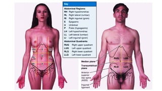

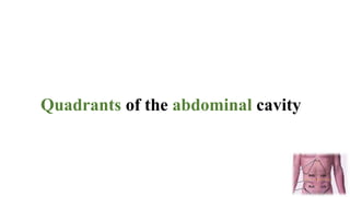
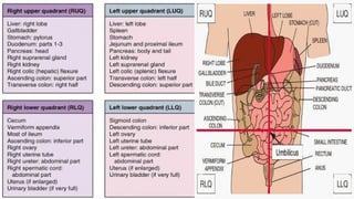
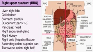
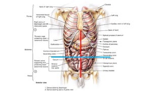
![Four quadrants of the abdominal cavity (right and left
upper and lower quadrants) are defined by two readily
defined planes:
• (1) The transverse transumbilical plane passing
through the umbilicus (and the intervertebral [IV] disc
between the L3 and L4 vertebrae), dividing it into upper
and lower halves,
• (2) The vertical median plane passing longitudinally
through the body, dividing it into right and left halves](https://image.slidesharecdn.com/anteriorabdomenalwall-171229132729/85/Anterior-abdominal-wall-29-320.jpg)


![External oblique muscle
• Origin : external surfaces of 5th
to 12th
ribs.
• Insertion: linea and alba, pubic tubercle, and
anterior half of iliac crest.
• Nerve supply: thoracoabdominal nerves
(inferior 5 [T7 to T11] thoracic nerves) and
subcostal nerve.
• Action: compress and support abdominal
viscera, flex and rotate trunk.](https://image.slidesharecdn.com/anteriorabdomenalwall-171229132729/85/Anterior-abdominal-wall-50-320.jpg)
![Muscle origin insertion Nerve supply action
Rectus abdominis Pubic symphysis and
pubic crest
Xiphoid process and 5th
to
7th costal cartilages
Thoracoabdominal
nerves (anterior rami of
inferior 6 thoracic
nerves)
Flexes tru
Compresses ab
viscera; stabili
controls tilt of
(antilordo
Transverse
abdominal
Internal surfaces of 7th
12th costal cartilages,
thoracolumbar fascia,
iliac crest, and lateral
third of inguinal
ligament
Linea alba with
aponeurosis of internal
oblique, pubic crest, and
pecten pubis via conjoint
tendon
Thoracoabdominal
nerves (anterior rami of
inferior 6 thoracic
nerves) and first lumbar
nerves
Compresses and
abdominal v
Internal oblique Thoracolumbar fascia,
anterior two-thirds of
iliac crest, and lateral
half of inguinal
ligament
Inferior borders of
10th–12th ribs, linea
alba, and pecten pubis via
conjoint tendon
Compress and
abdominal viscer
rotate tru
External oblique External surfaces of
5th–12th ribs
Linea and alba, pubic
tubercle, and anterior
half of iliac crest
Thoracoabdominal
nerves (inferior 5
[T7–T11] thoracic
nerves) and subcostal](https://image.slidesharecdn.com/anteriorabdomenalwall-171229132729/85/Anterior-abdominal-wall-54-320.jpg)


The document provides an overview of the anatomy of the anterior abdominal wall, including its regions, quadrants, and the muscles involved. It describes the structure and boundaries of the abdominal wall and details the innervation and blood supply to this area. Additionally, it touches on surgical considerations such as incision types and lymphatic drainage patterns.


