
Recommended
More Related Content
Similar to FEDHA GIT HISTO (1).pptx
Similar to FEDHA GIT HISTO (1).pptx (20)
Recently uploaded
Recently uploaded (20)
FEDHA GIT HISTO (1).pptx
- 3. Introduction GIT function-break down food for absorption into the body The process occurs in five main phases; 1. ingestion, 2. fragmentation, 3. digestion, (Food enzyme molecules) 4. absorption 5. elimination of waste products
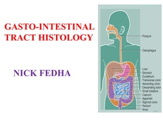
- 4. The digestive system consists of the digestive tract: oral cavity (lip and tongue) Esophagus Stomach small large intestines Rectum anus and its associated glands: Salivary glands Liver Pancreas.
- 5. The tongue • A mass of striated muscle covered by a mucous membrane whose structure varies according to the region. • The mucous membrane is smooth on the lower (ventral) surface but has papillae on the dorsal part. • The surface of the tongue has two types of small lymphoid aggregations, lymphoid nodules and the lingual tonsils, the lymphoid nodules aggregate around invaginations (crypts).
- 6. Histological components Epithelium - What type of epithelium covers the tongue? Keratinised stratified squamous epithelium. - On which surface of the tongue is the epithelium most keratinised and why? On the dorsal surface. There is most contact on this surface. Fat (adipose tissue) Salivary glands Blood vessels and nerves Skeletal (striated) muscle- seen in longitudinal, oblique and transverse orientations. Papillae.
- 7. Skeletal muscle and adipose tissue adipocytes B : LS section A : TS section
- 8. Blood vessels and nerves N N A A V nerve artery vein
- 9. Papillae Filiform Papillae Filiform papillae have an elongated conical shape; they are quite numerous and are present over the entire surface of the tongue. Their epithelium, has no taste buds, but is keratinized. Fungiform Papillae Fungiform papillae resemble mushrooms in that they have a narrow stalk and a smooth-surfaced, dilated upper part. These papillae, which contain scattered taste buds on their upper surfaces, are irregularly interspersed among the filiform papillae.
- 10. Foliate Papillae Foliate papillae are poorly developed in humans. They consist of two or more parallel ridges and furrows on the dorso-lateral surface of the tongue and contain many taste buds. Circumvallate Papillae Circumvallate papillae are 7–12 extremely large circular papillae whose flattened surfaces extend above the other papillae. They are distributed in the posterior portion of the tongue.
- 11. Identify the main papilla in this section. Circumvallate (vallate) papilla (arrowed). Decide whether : glands : Yes taste buds : Yes lymphatic tissue : Yes can be associated with this papilla
- 12. Identify the main papillae in this section. Foliate papillae form a series of parallel folds.
- 13. Decide whether : glands : Yes taste buds : Yes lymphatic tissue : Some (seen here labelled L) can be associated with this papilla. L : Some lymphocytes
- 14. Identify the main papillae in this section. Filiform papillae (arrowed). Decide whether : glands : No taste buds : No lymphatic tissue : No can be associated with these papillae.
- 15. Salivary glands
- 16. Taste buds
- 17. Lymphatic tissue : found deeper into the papillae.
- 18. ESOPHAGUS It is a muscular tube whose function is to transport foodstuffs from the mouth to the stomach and to prevent the retrograde flow of gastric contents. Transport is achieved by peristaltic contractions and relaxation of the esophageal sphincters (upper and lower), usually controlled by reflexes and by the autonomic nervous system. In humans the esophagus is covered by nonkeratinized stratified squamous epithelium
- 19. Name the four layers: 1: mucosa. 2: submucosa. 3: tunica muscularis. 4: serosa or adventitia.
- 20. How is the tunica muscularis (skeletal subdivided further? Tunica muscularis consists of : 1. outer longitudinal muscle layer. 2. inner circular muscle layer.
- 21. How is the mucosal layer subdivided further? The mucosal layer consists of : 1. epithelium. 2. lamina propria. 3. lamina muscularis. epithelium lamina propria lamina muscularis
- 22. NOTE: In the submucosa are groups of small mucus- secreting glands, the esophageal glands, whose secretion facilitates the transport of foodstuffs and protects the mucosa. In the lamina propria of the region near the stomach are groups of glands, the esophageal cardiac glands, that also secrete mucus.
- 23. Gastro-oesophageal junction What is the most obvious change at this junction? Epithelium changes from stratified squamous nonkeratinized in the oesophagus to simple columnar in the stomach. Glands from few or no mucous glands in oesophagus to gastric glands in stomach. Thickening of tunica muscularis in the stomach (cardiac sphincter muscle).
- 24. Lumen of stomach Lumen of oesophagus Stratified squamous nonkeratinized Simple columnar
- 25. gastric glands columnar epithelium stratified squamous epithelium
- 26. Stomach
- 27. Food starts to be digested and absorbed in the stomach, although absorption is mostly limited to water, alcohol and some drugs. The stomach is an expandable, muscular bag, and it keeps swallowed food inside it by contracting the muscular pyloric sphincter. Food can stay in the stomach for 2 hours or more. Food is broken down chemically, by gastric juice, and mechanically, by contraction of the three layers of smooth muscle in the muscular externa layer. The broken up food at the end of this process is called chyme.
- 28. Gastric juice is secreted by gastric mucosal glands, and contains hydrochloric acid, mucus, and proteolytic enzymes pepsin (which breaks down proteins), and lipase (which breaks down fats). When the stomach is empty, and not distended, the lining is thrown up into folds called rugae. After eating, these folds flatten, and the stomach is able to distend greatly.
- 29. The stomach has three anatomical regions: 1. cardiac, which contains mucous secreting glands (called cardiac glands) and is closest to the oesophagus. 2. fundus, the body or largest part of the stomach which contain the gastric (fundic) glands. 3. pyloric, which secretes mucus, and the hormone gastrin.
- 30. Layers of the stomach. Mucosa Submucosa Muscularis externa Serosa
- 31. The mucosa is made up of: Epethelium (simple columnar). Full of gastric glands and pits. Laminar propria (loose connective tissue). • The lamina propria, located just below the epithelium, is rich in macrophages and lymphoid cells(lymphocytes), some of which actively produce antibodies mainly IgA against viral & bacterial invasion. • viral and bacterial invasion. Prominent layer of smooth muscle - the muscularis mucosae (contraction of this muscle helps to expel the contents of the gastric glands).
- 35. Pyloric region of glandular stomach Identify : deep gastric pits coiled glands in lamina propria opening into gastric pits lamina muscularis Gastric pits Coiled glands Lamina muscularis
- 36. Gastric pits and glands opening of gastric pit to stomach Tubular glands
- 37. The submucosa is composed of dense connective tissue containing blood and lymph vessels; it is infiltrated by lymphoid cells, macrophages, and mast cells. The muscularis externa layer has three layers of muscle. An innner oblique layer , a middle circular and an external longitudinal layer. The contraction of these muscle layers help to break up the food mechanically. The stomach is covered by a thin serosa.
- 38. N/B: Gastric Glands in the fundus (body) of the stomach The epithelium of the mucosa of the fundus and body of the stomach forms invaginations called gastric pits. The lamina propria contains gastric glands, which open into the bases of the gastric pits. These glands are responsible for the synthesis and secretion of the gastric juice. The lining epithelium of the stomach, and gastric pits is entirely made up of mucous columnar cells. These cells produce a thick coating of mucus, that protects the gastric mucosa from acid and enzymes in the lumen. Even so, these cells have to be replaced after 4-6 days.
- 39. Small Intestine Site of terminal food digestion, nutrient absorption, and endocrine secretion. The processes of digestion are completed here. is relatively long—approximately 5 m—and consists of three segments: the duodenum, jejunum, and ileum.
- 43. Columnar epithelial cells with the brush border (arrowhead) interspersed with mucus-secreting goblet cells.
- 44. DUODENUM