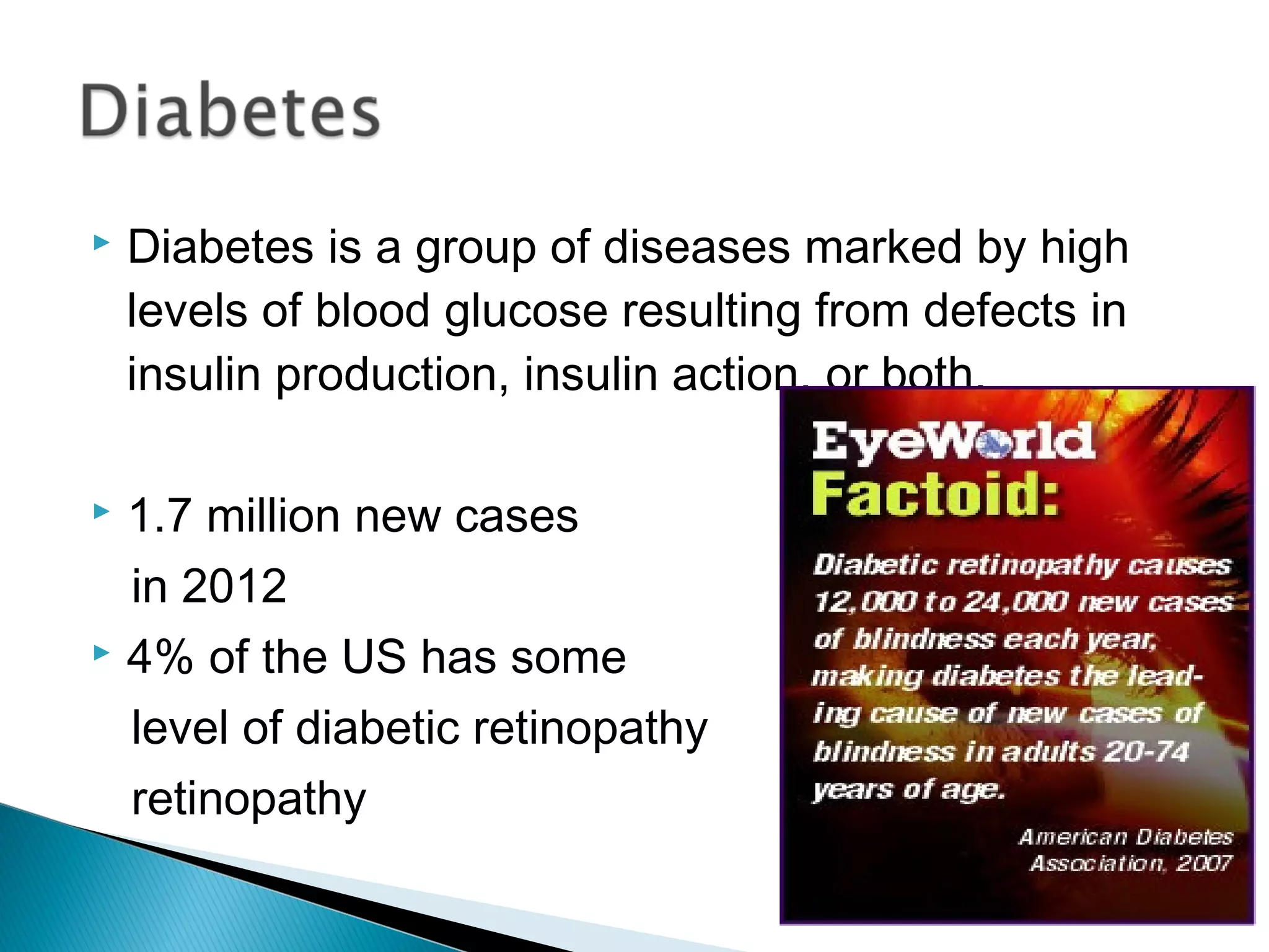
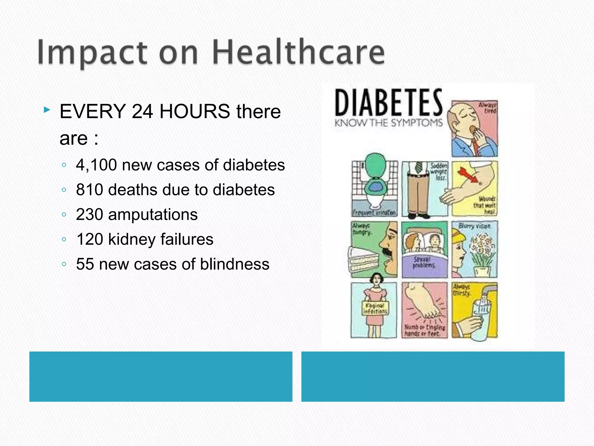
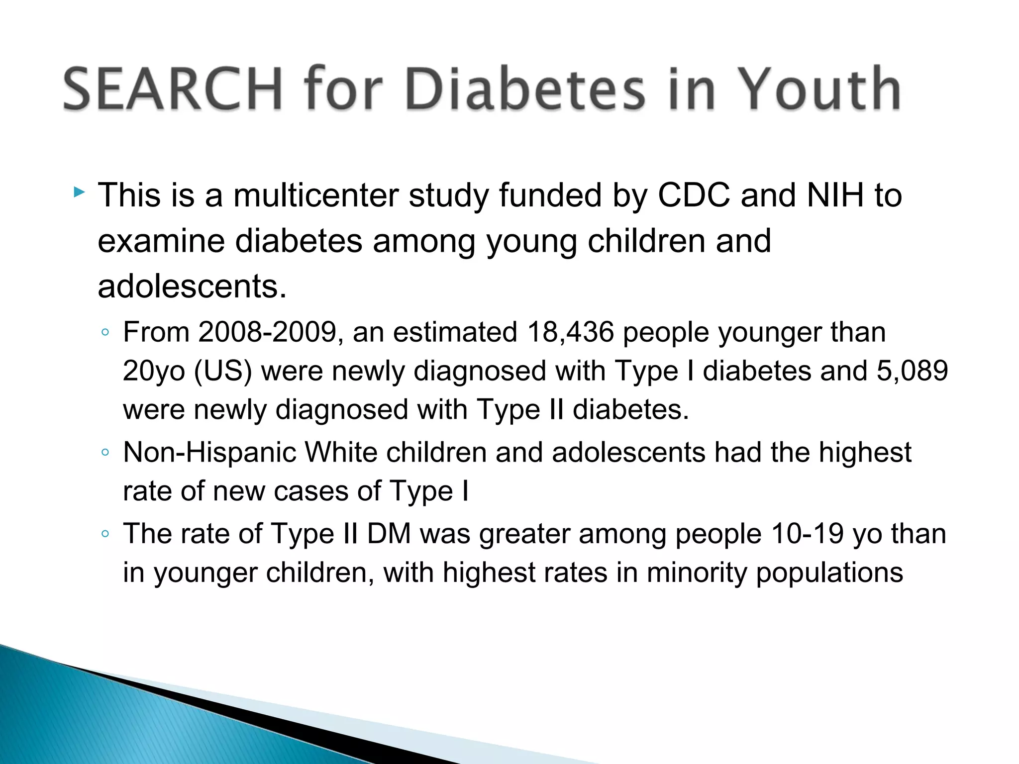
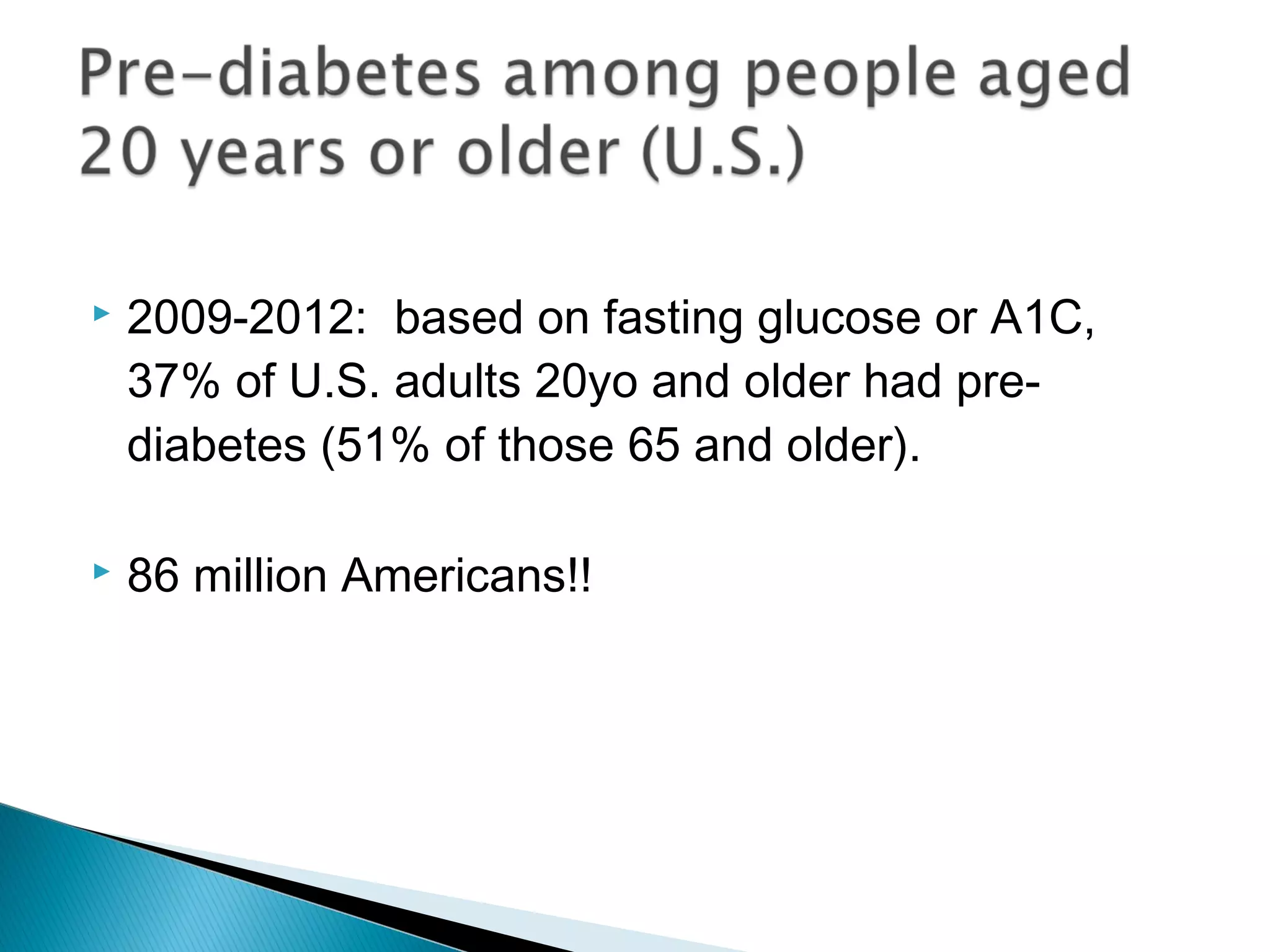
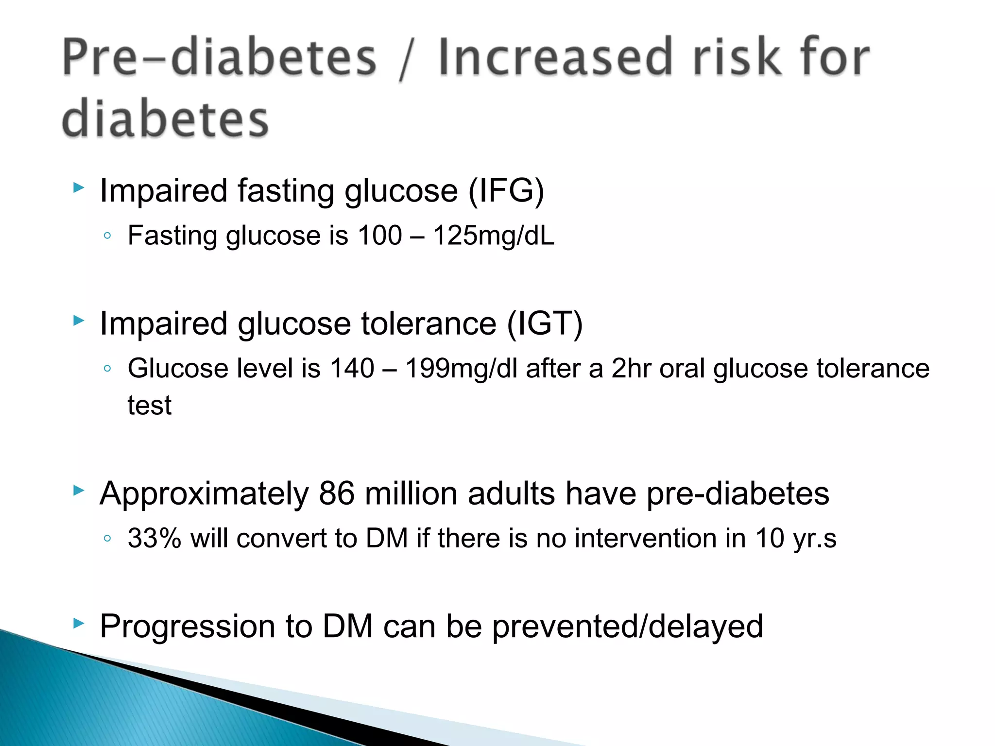
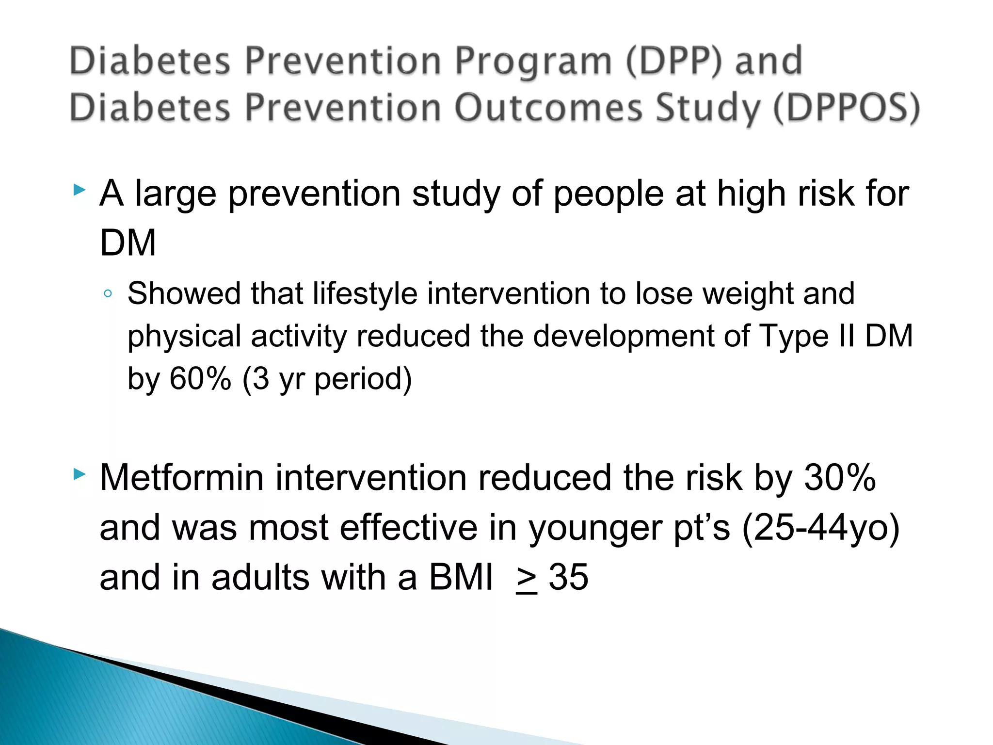
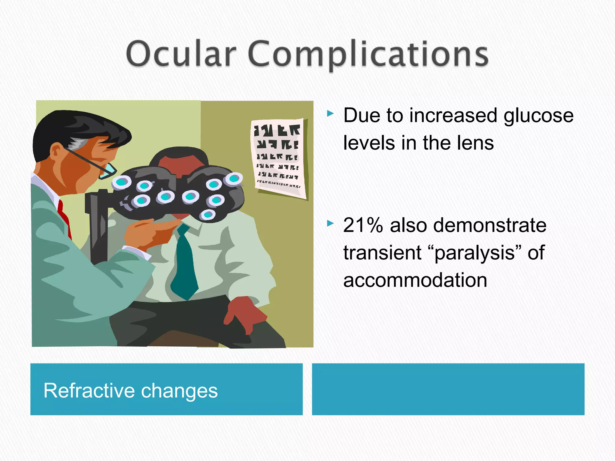
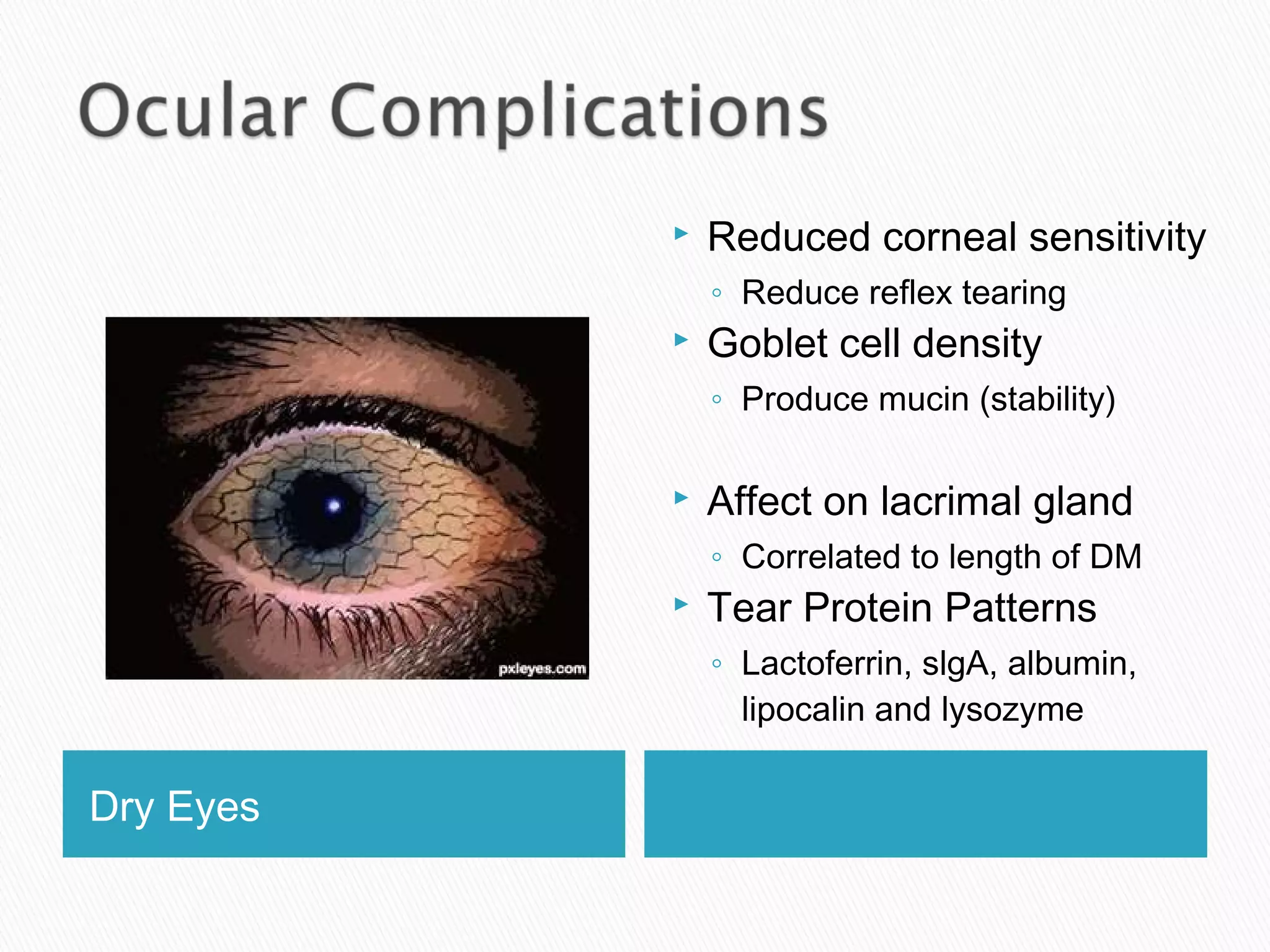
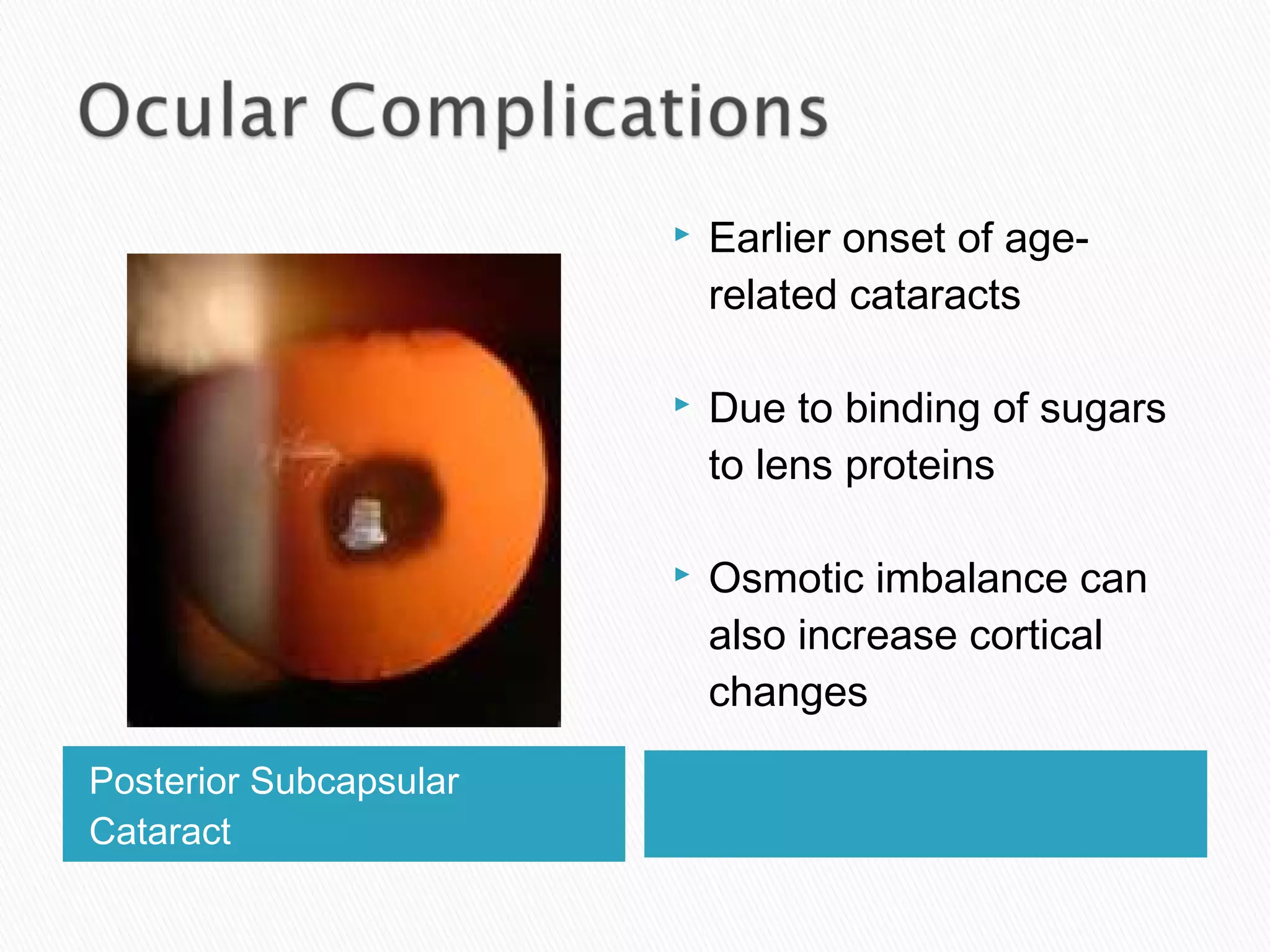
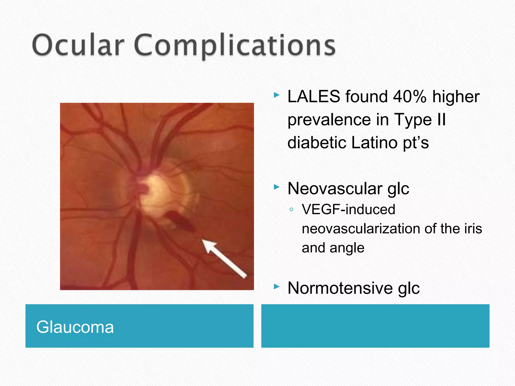
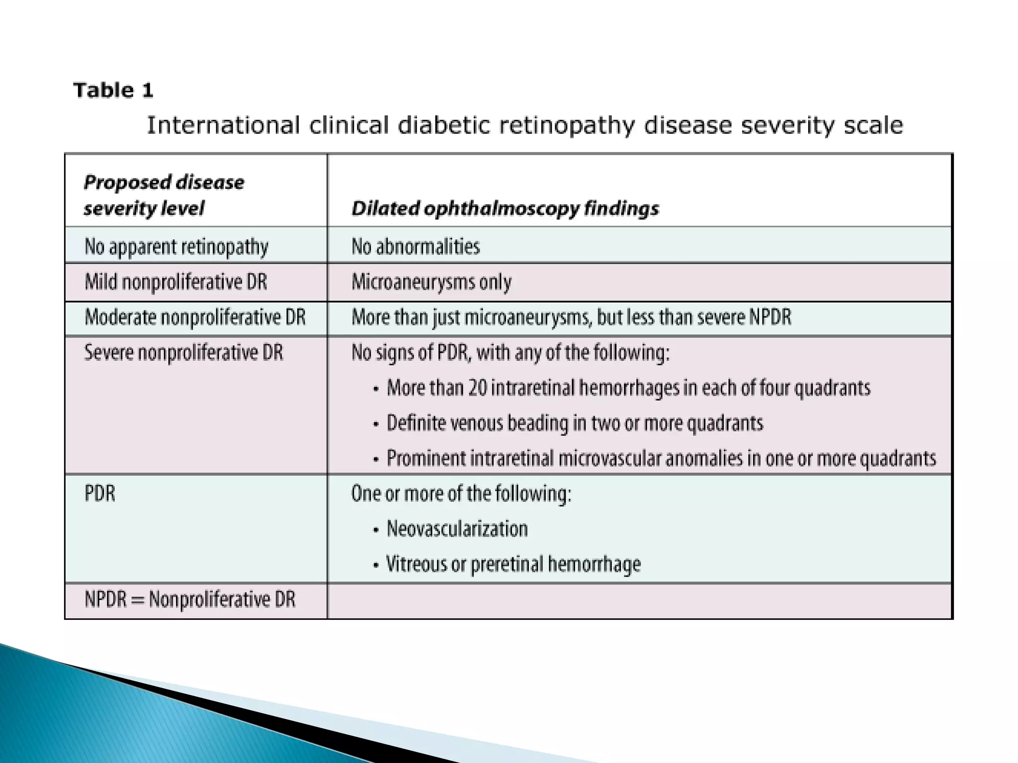
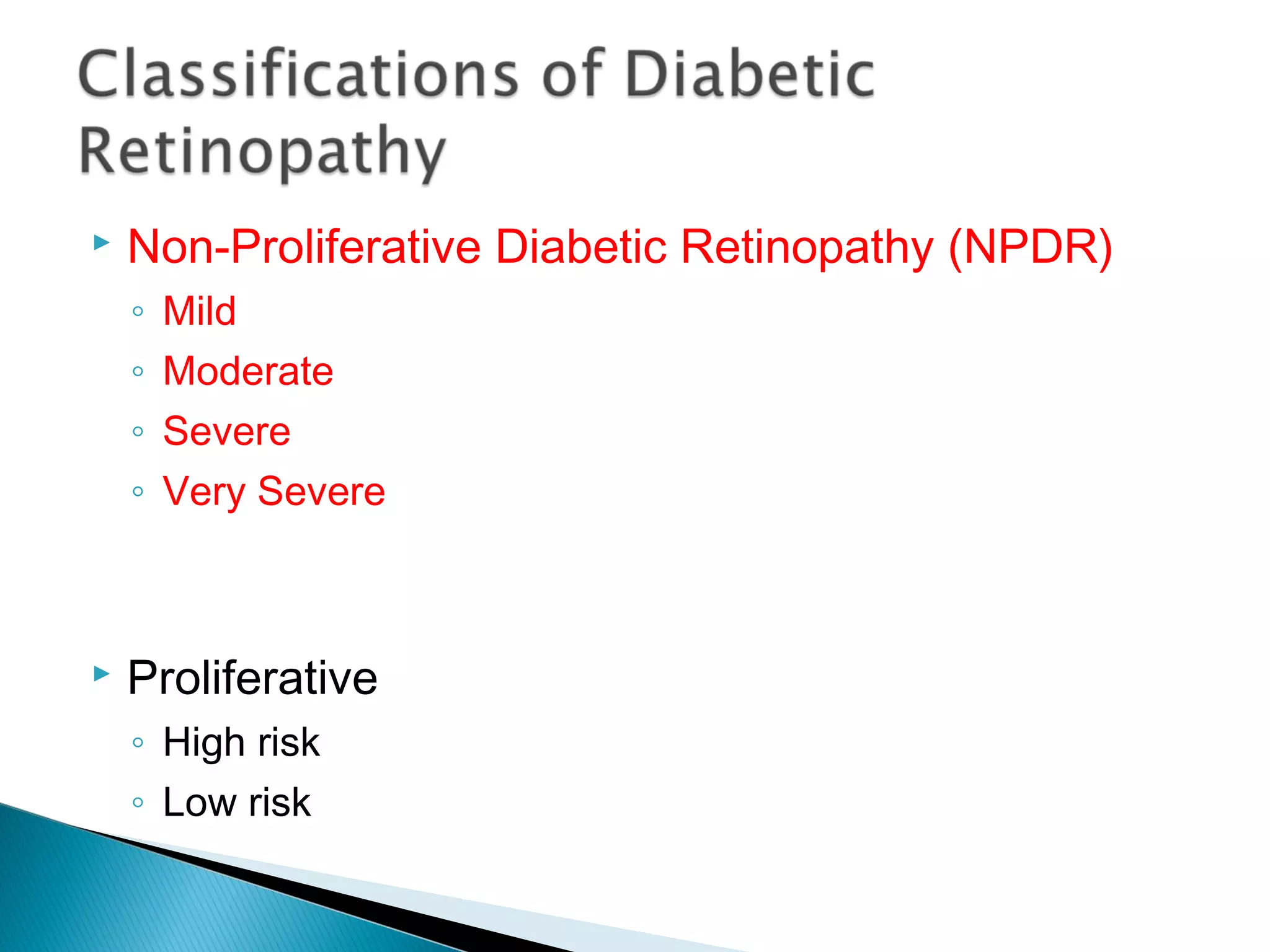
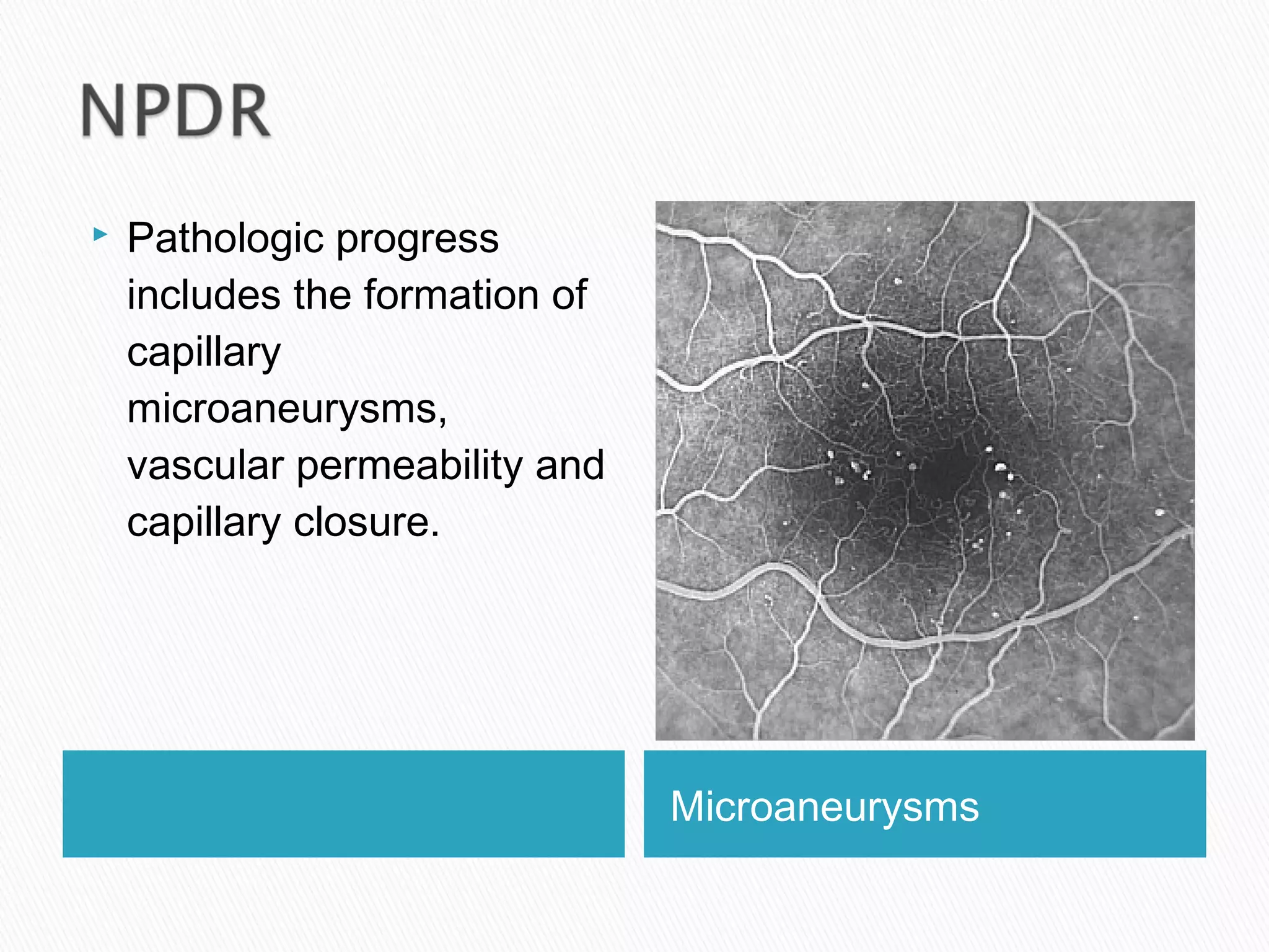
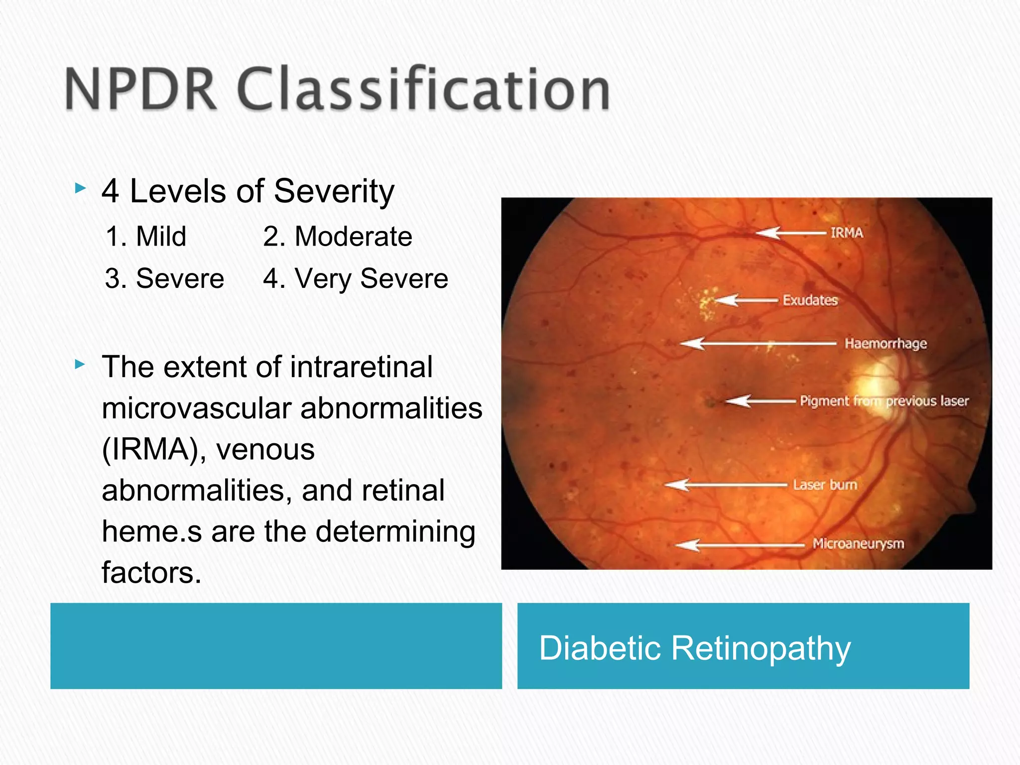
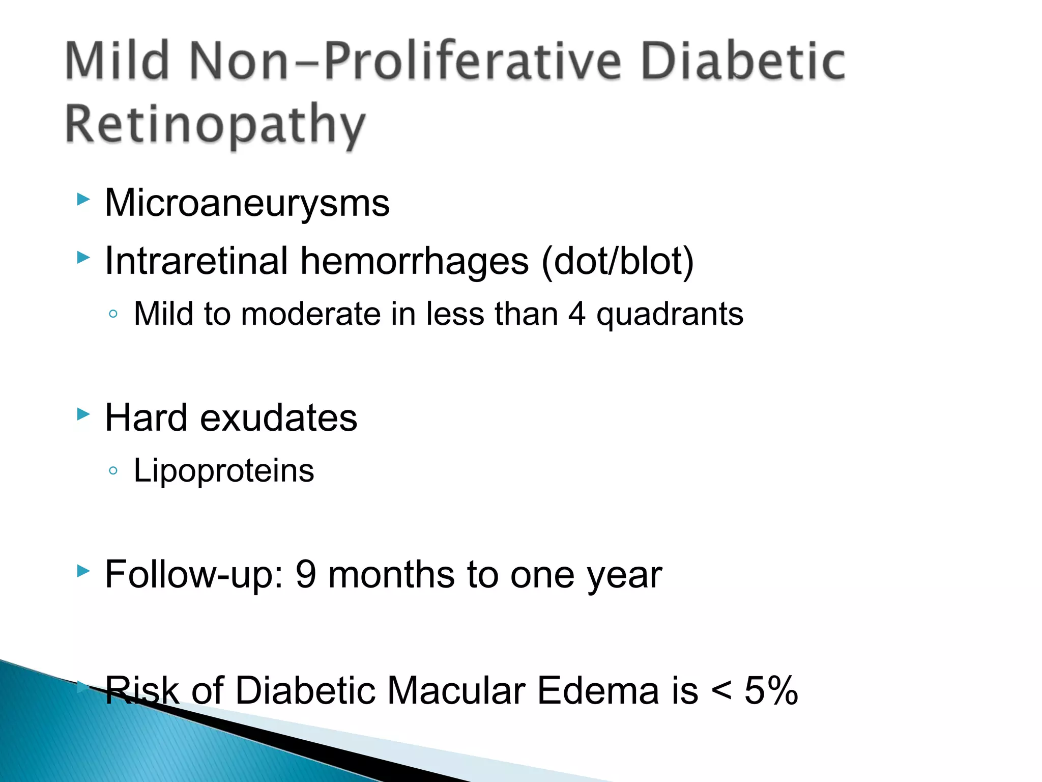
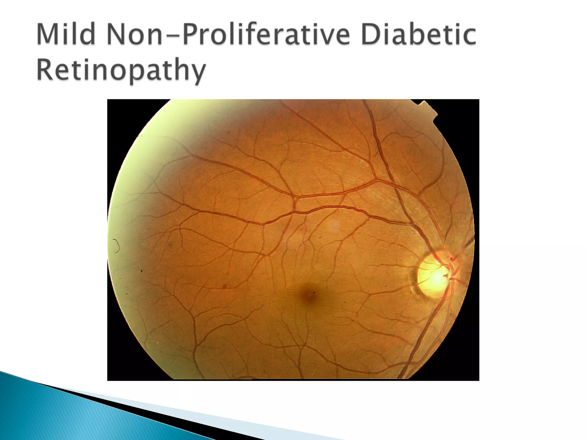
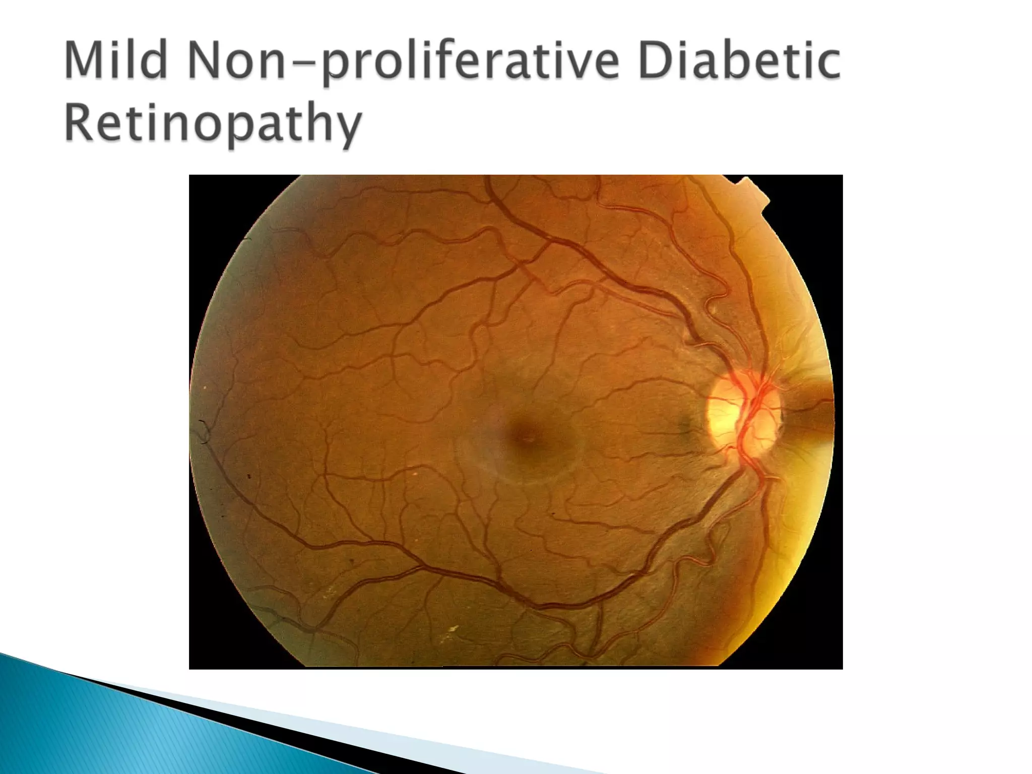
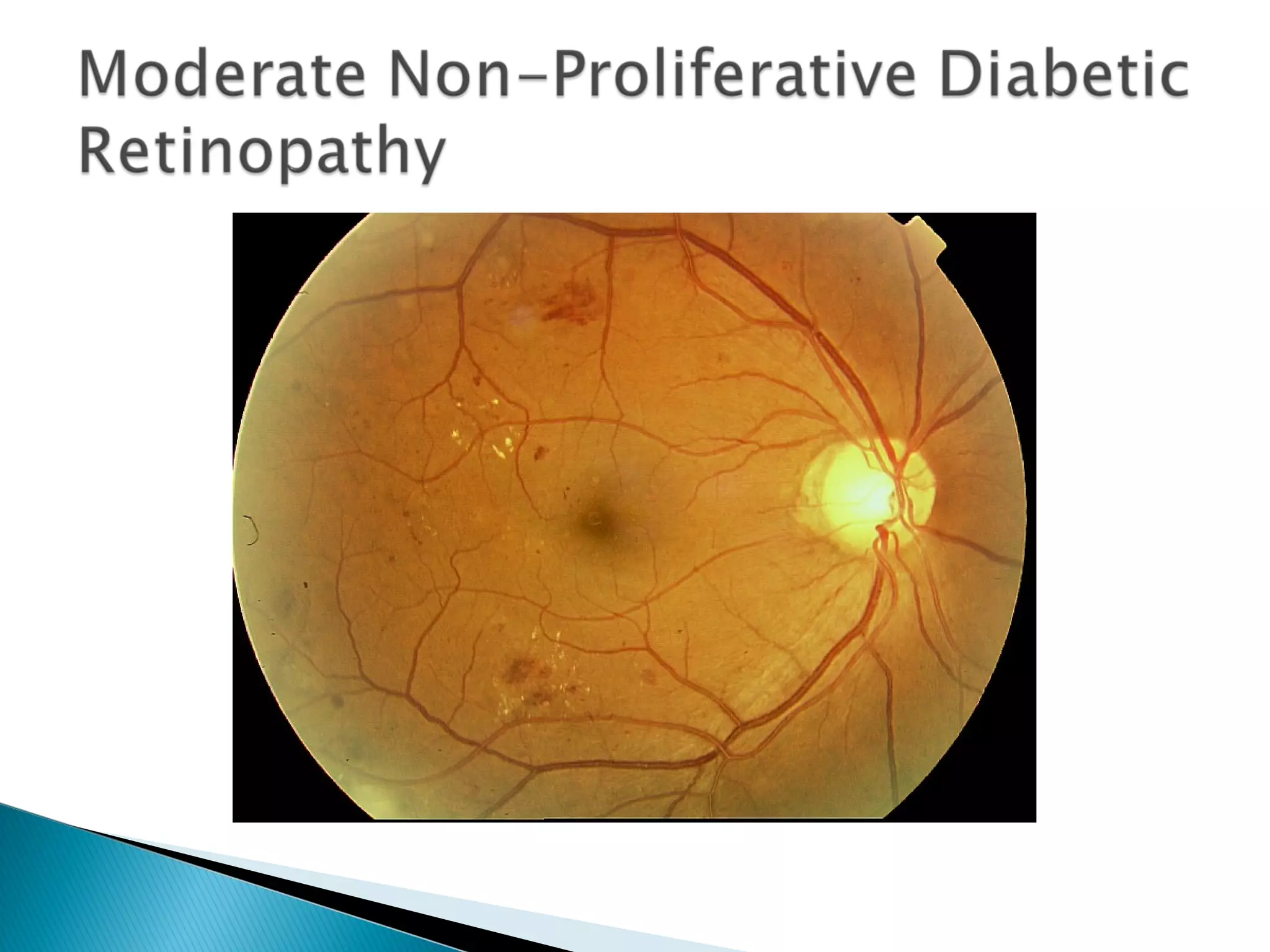
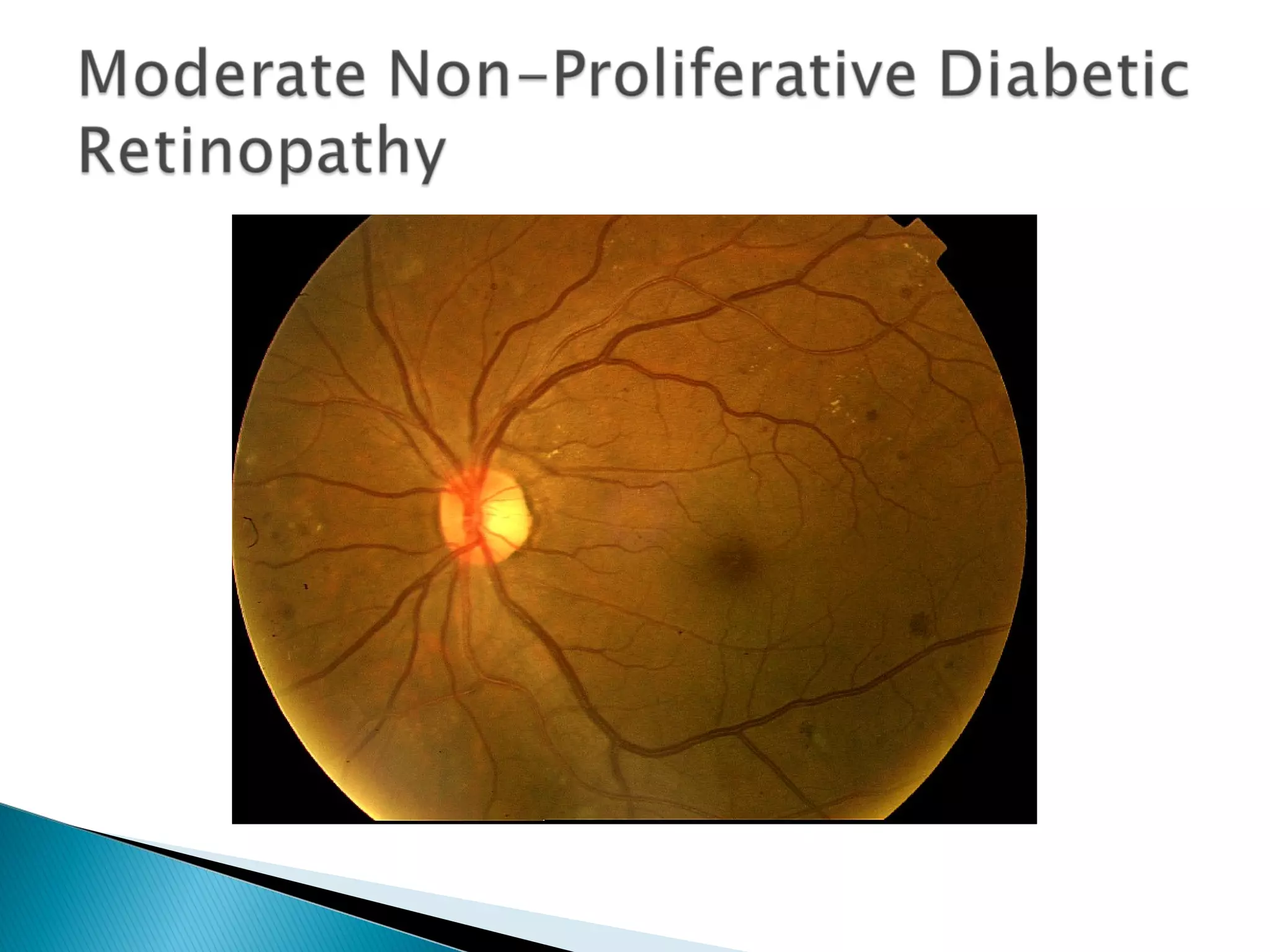
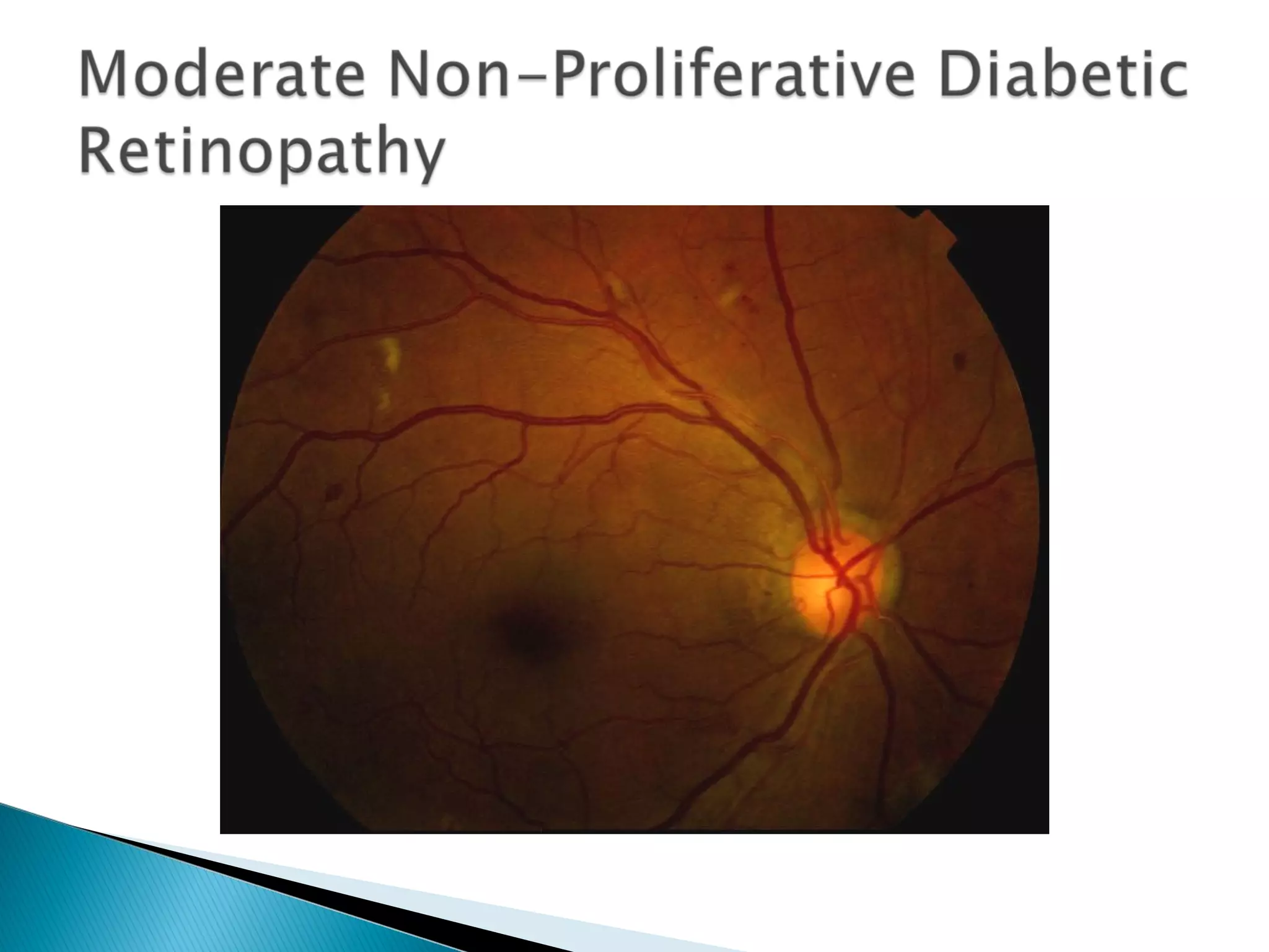
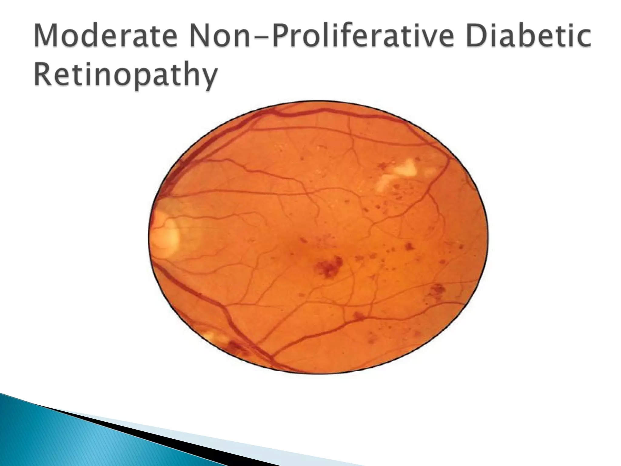
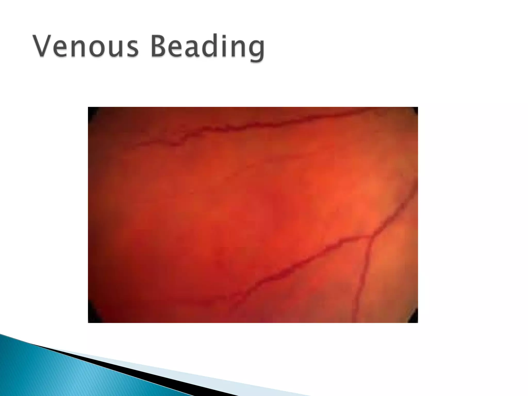
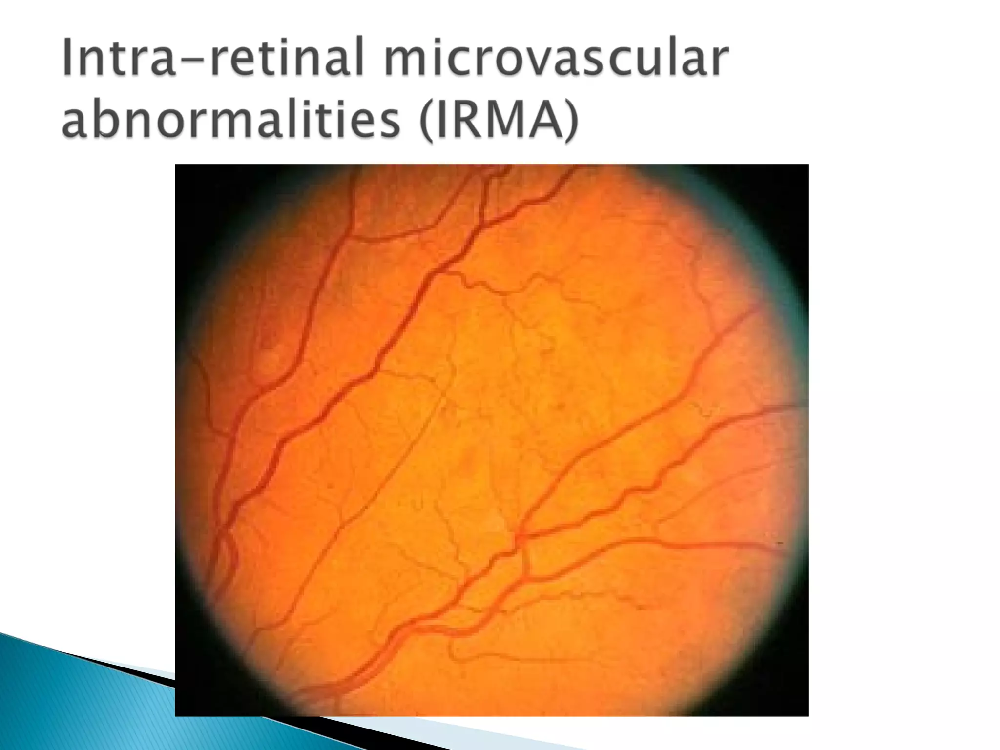
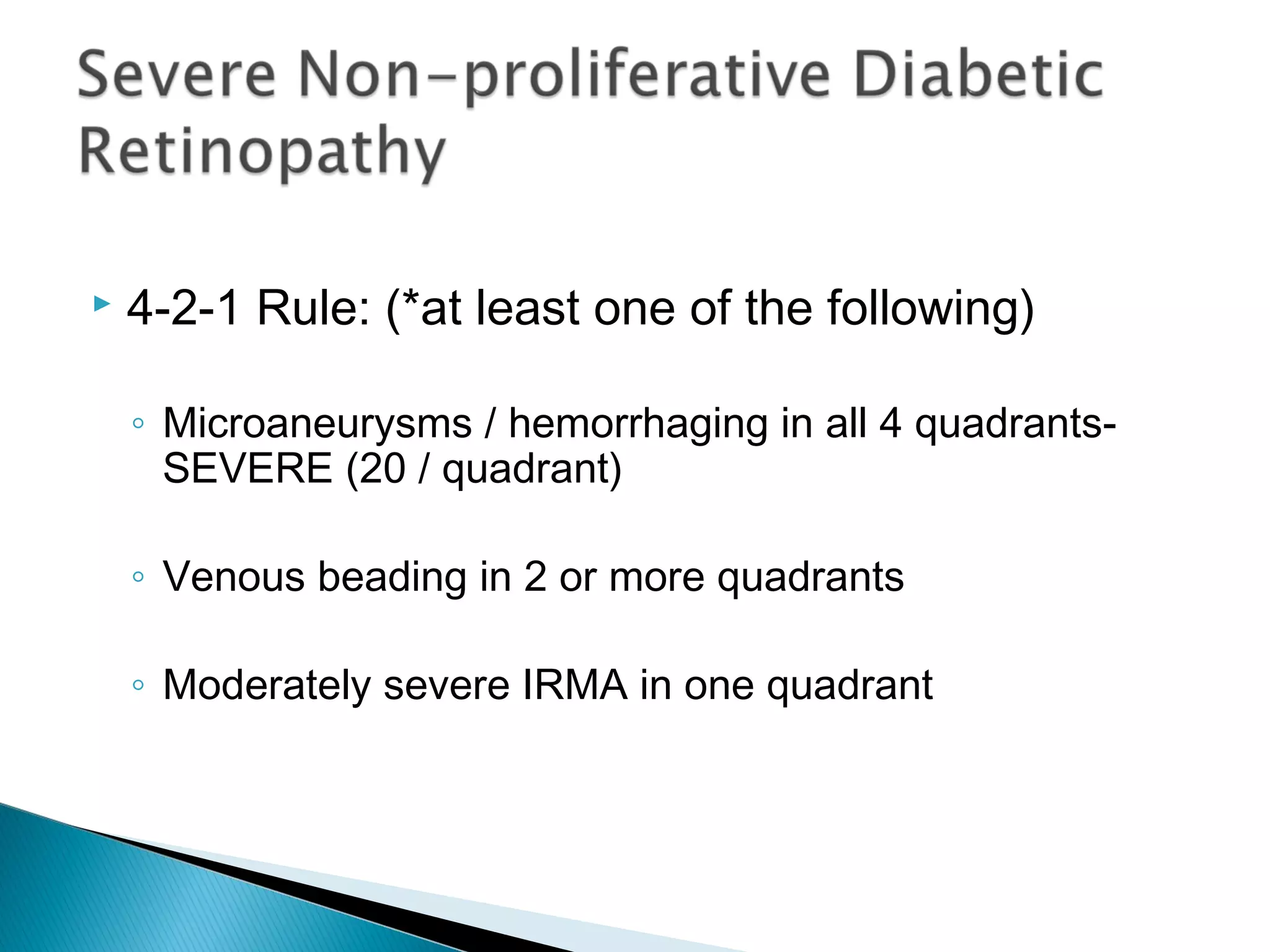
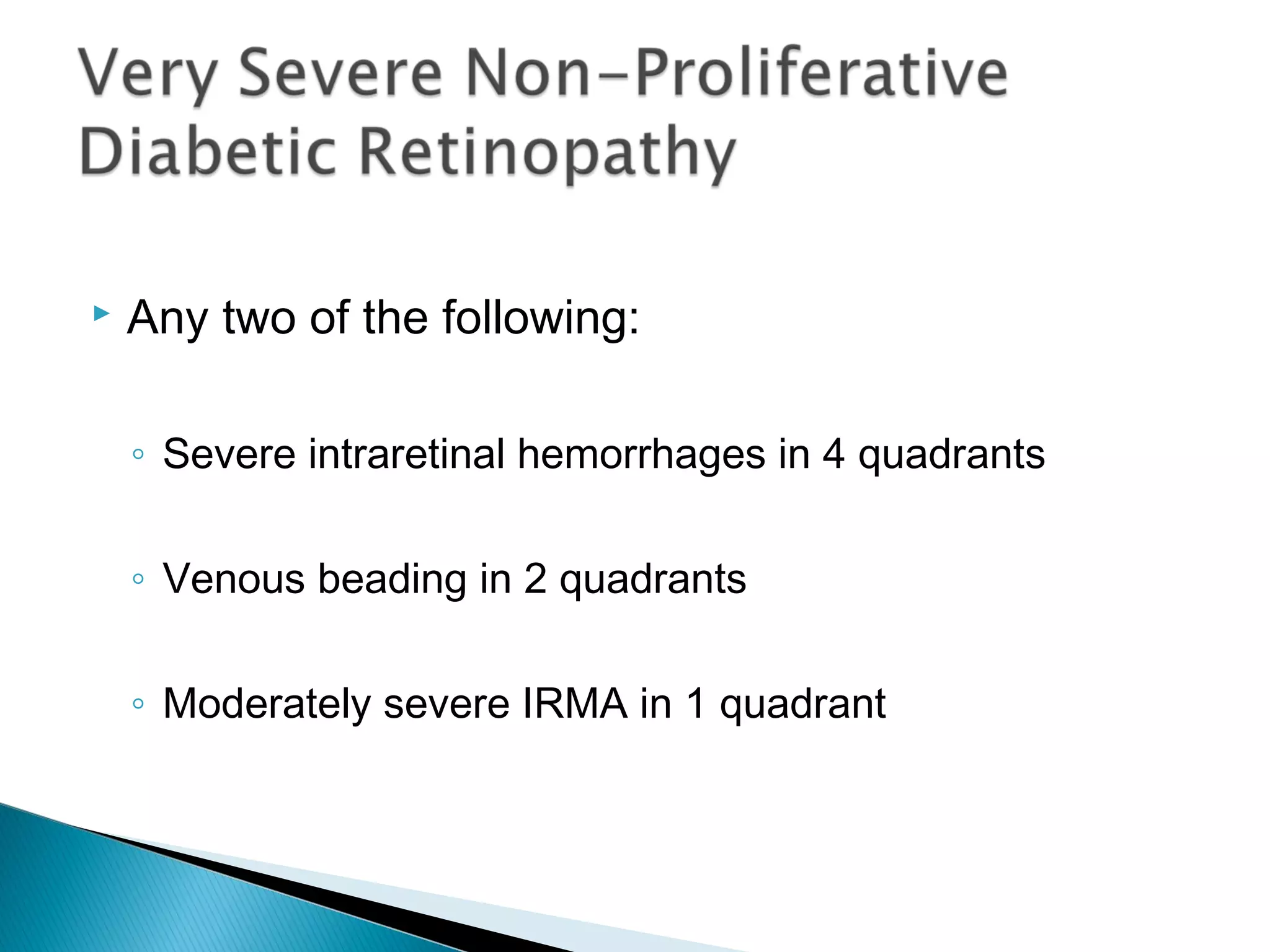
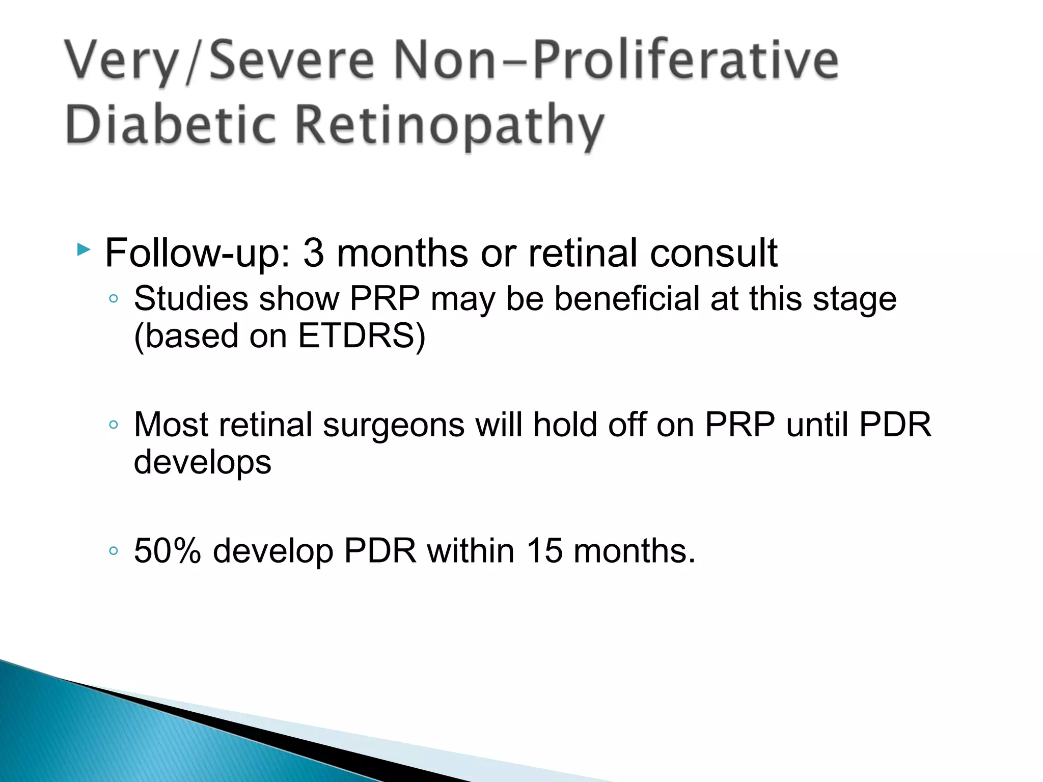
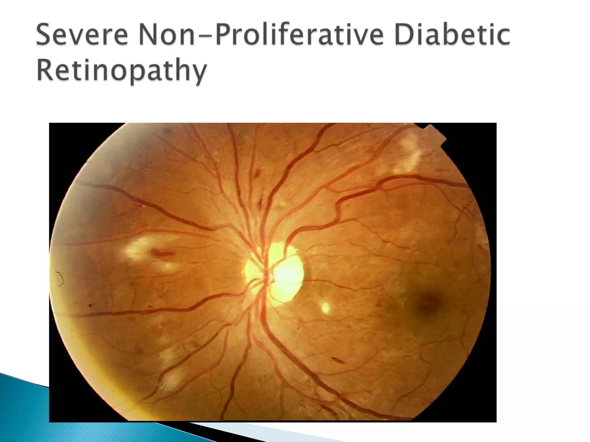
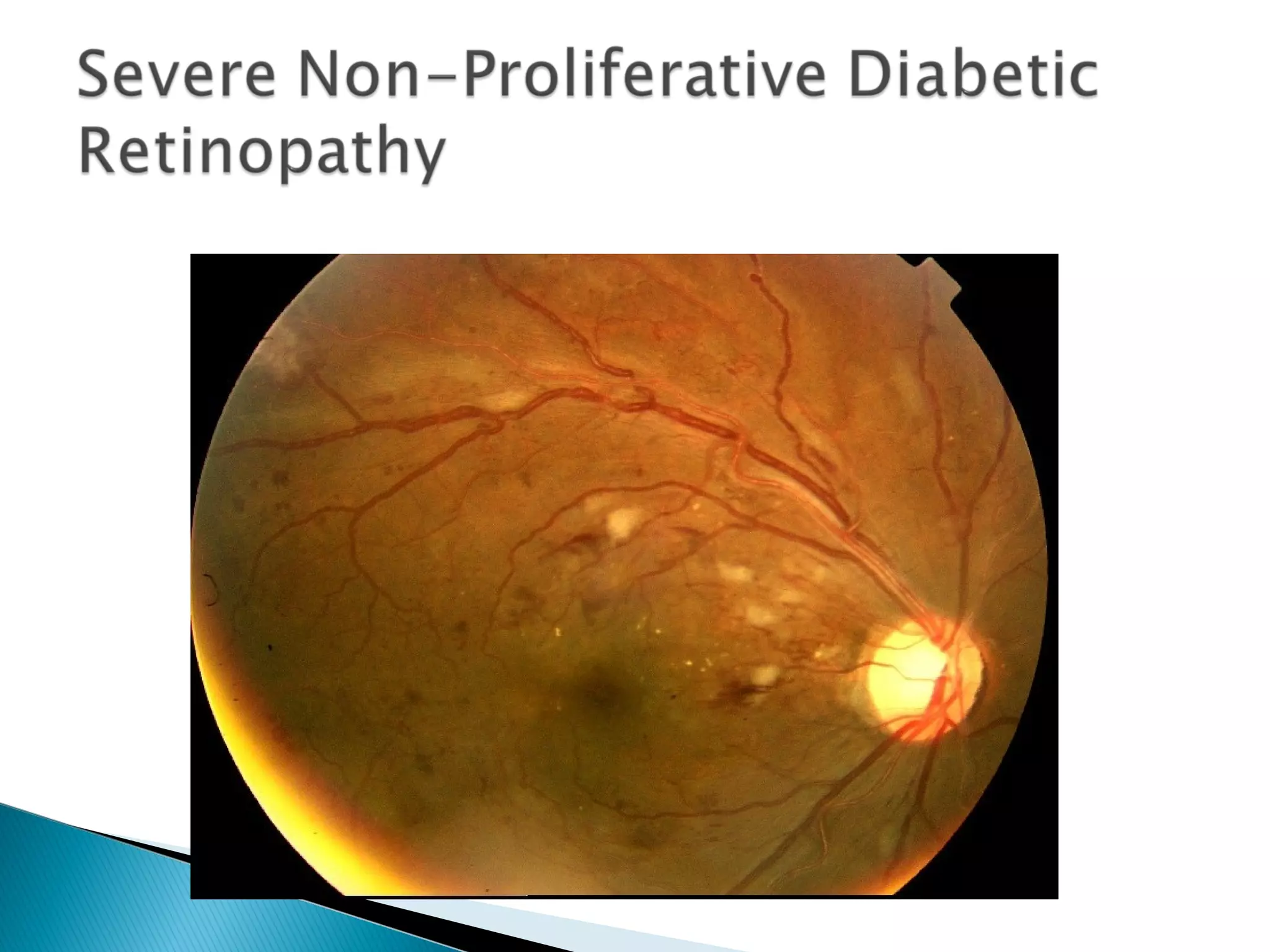
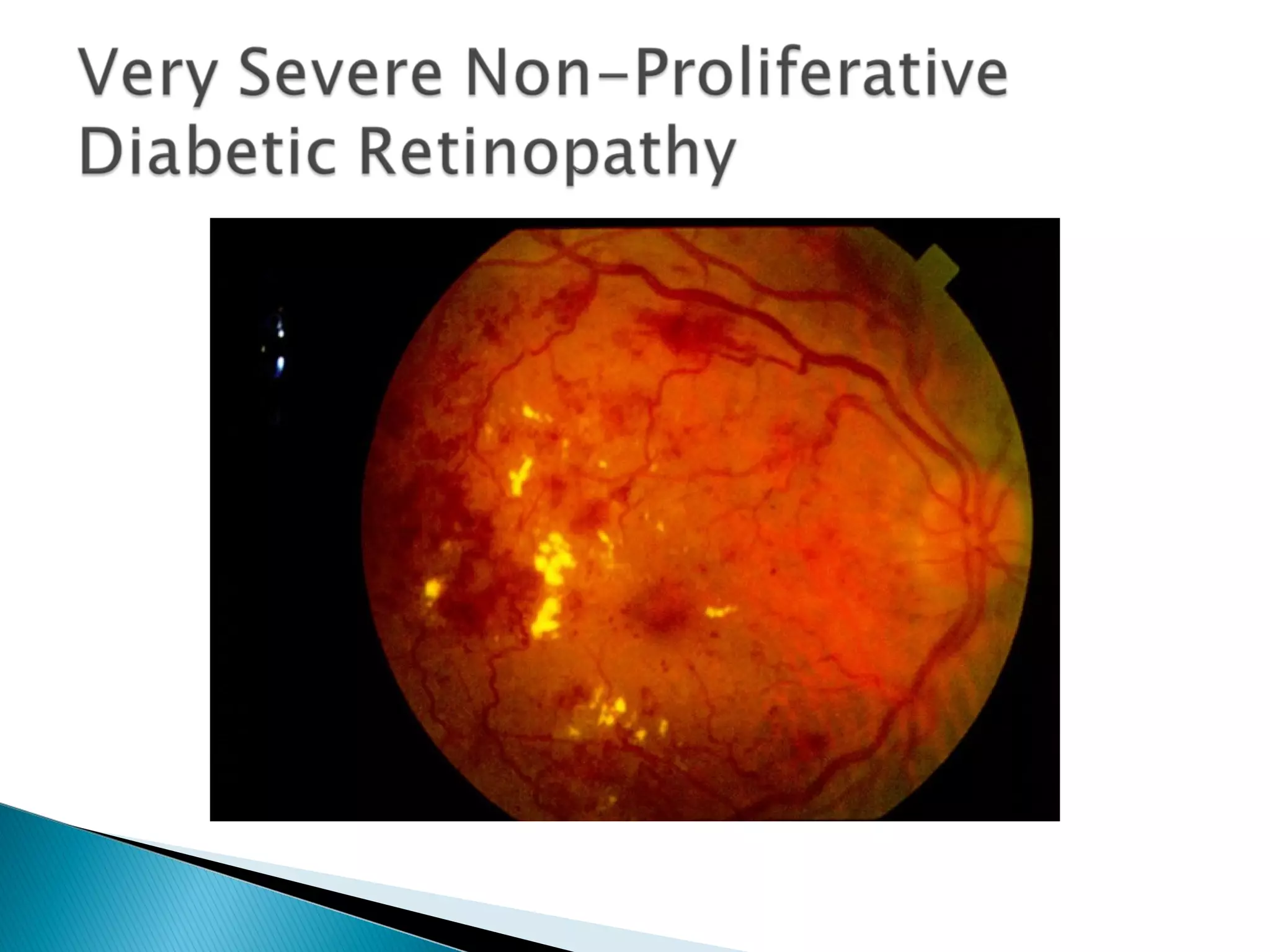
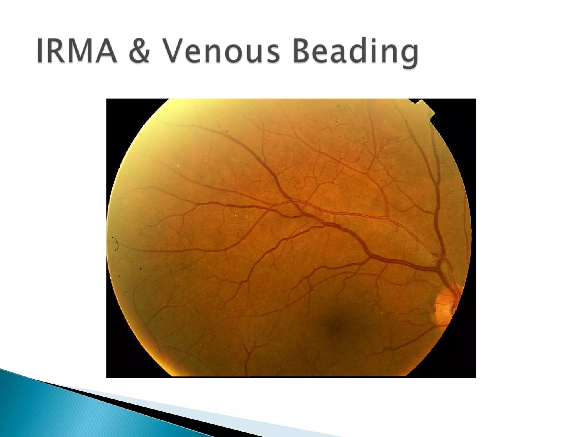
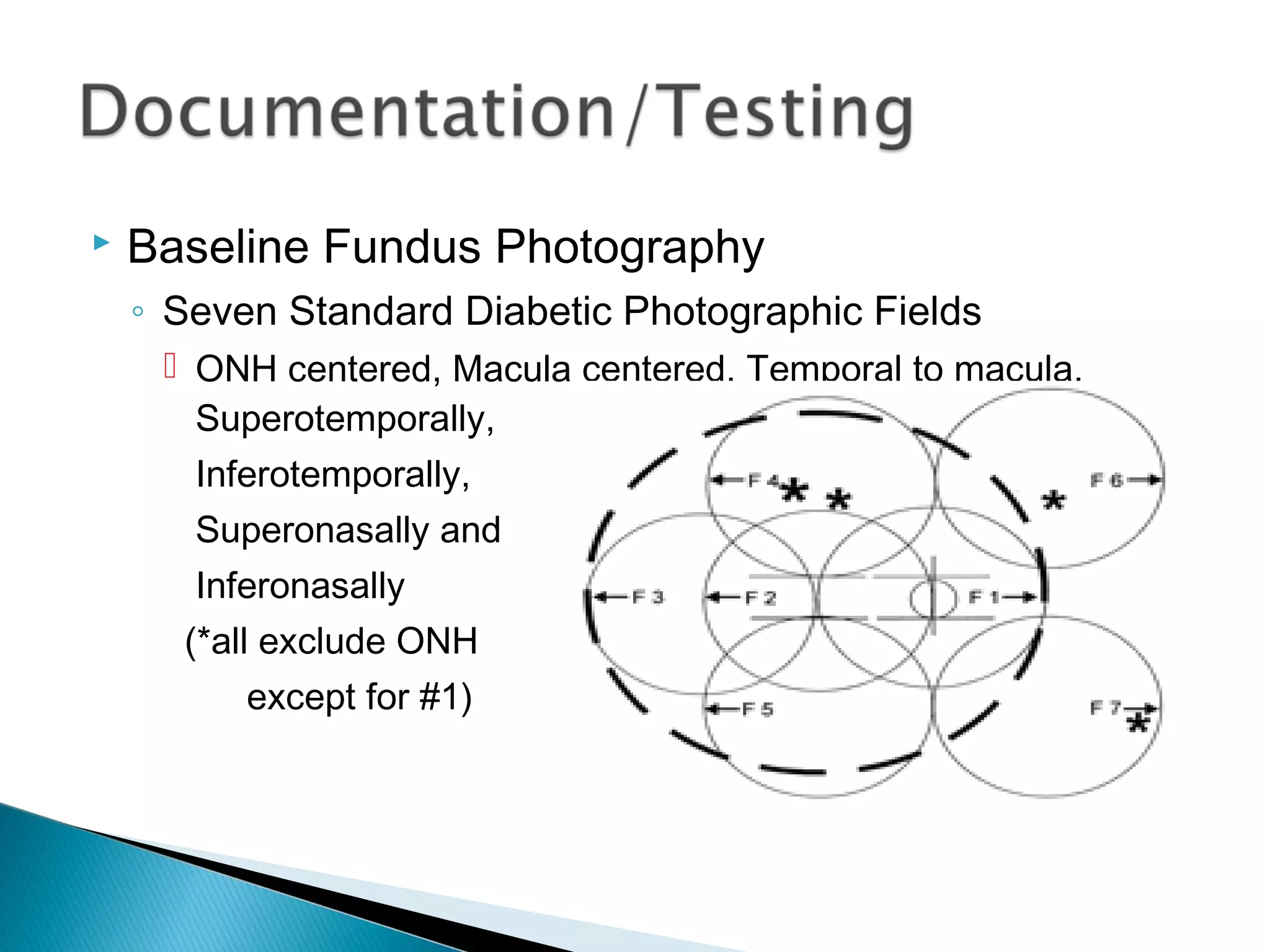
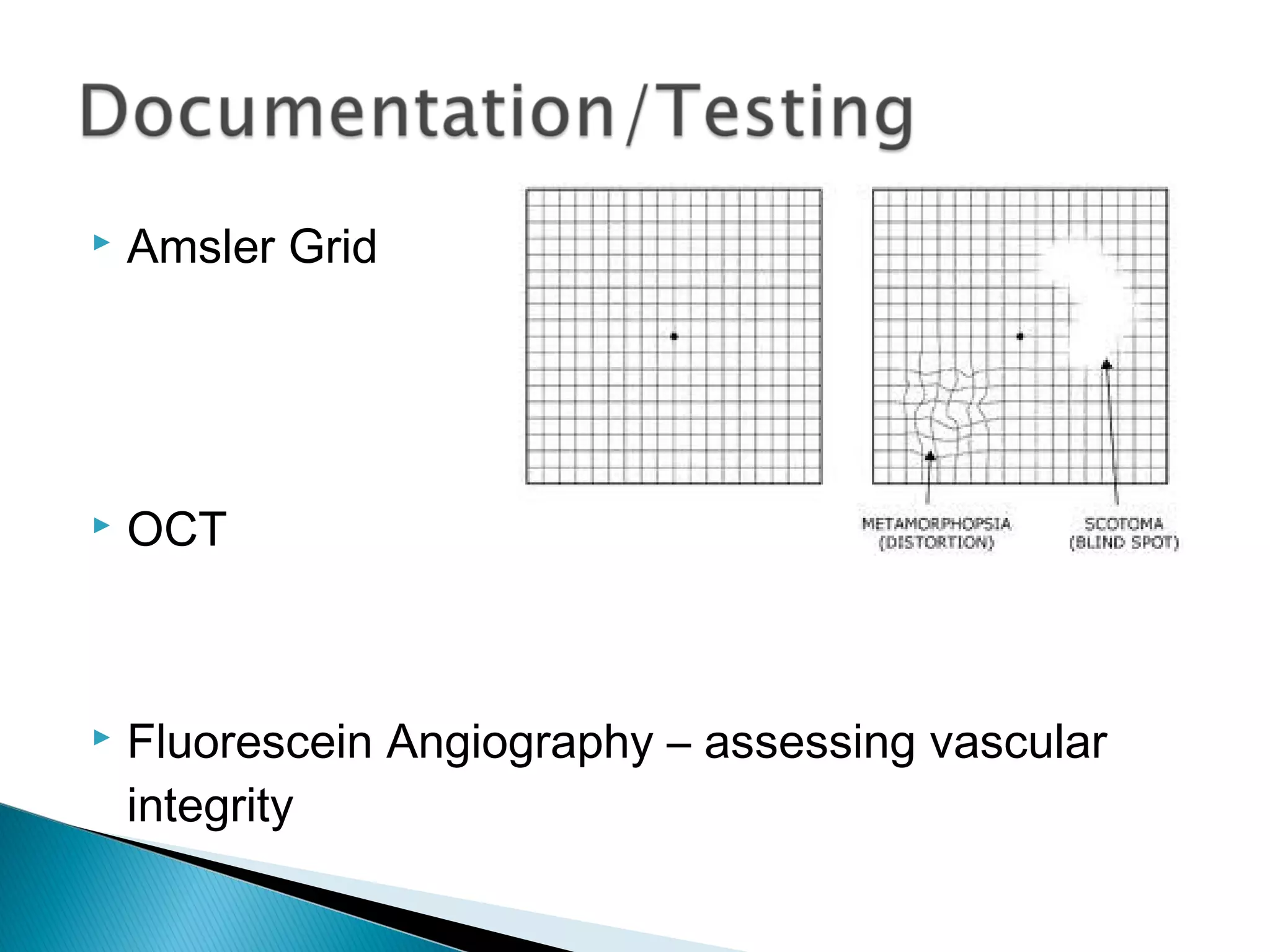
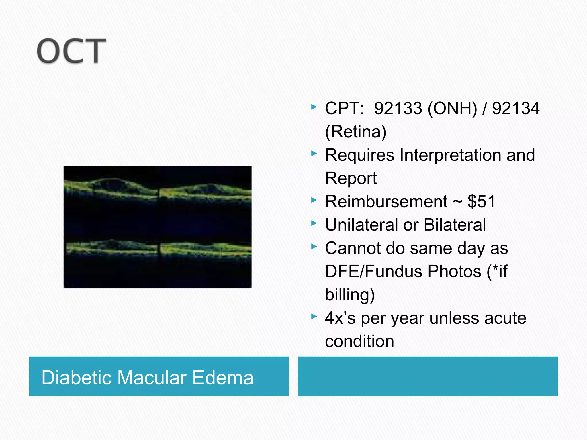
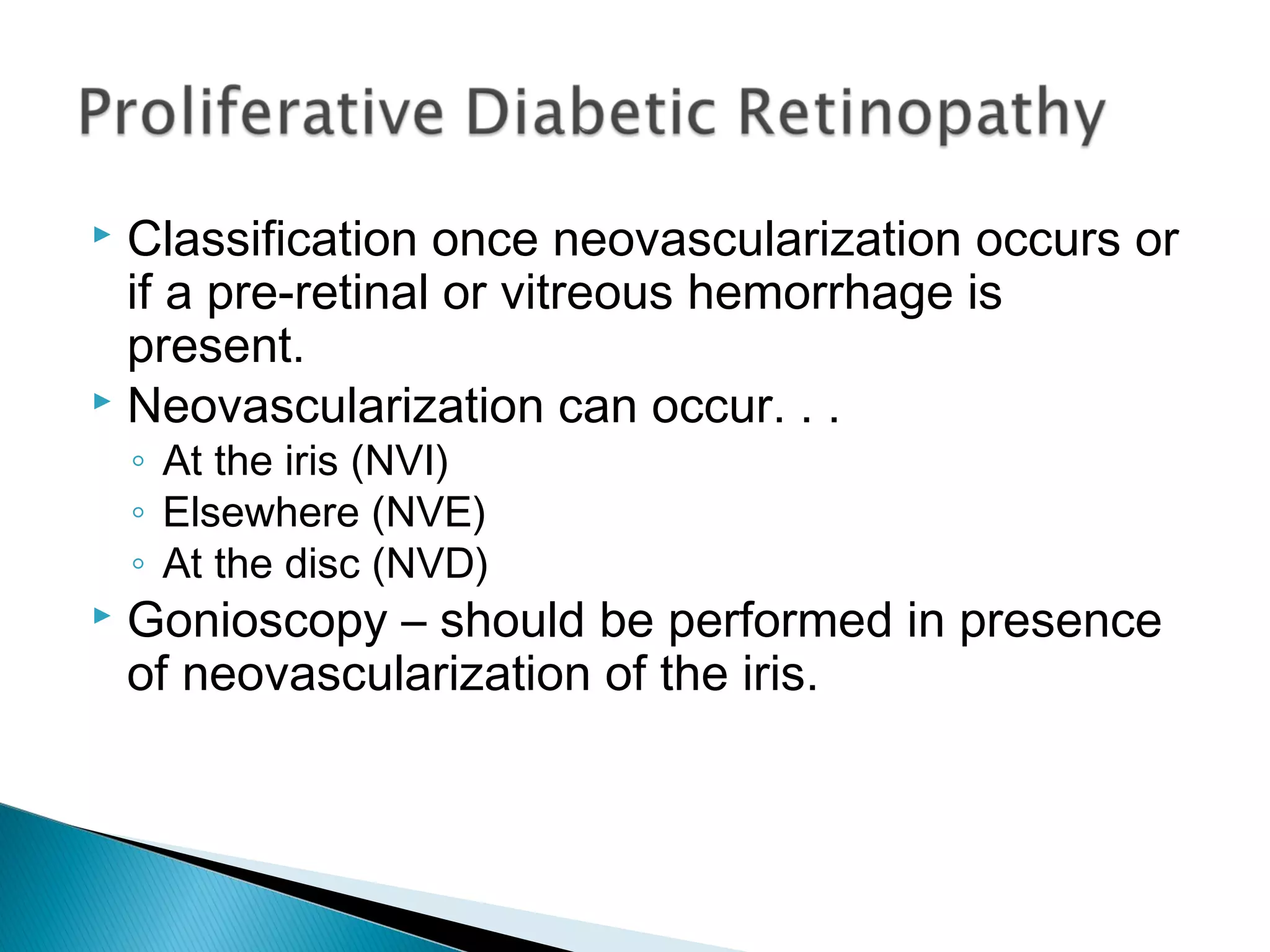
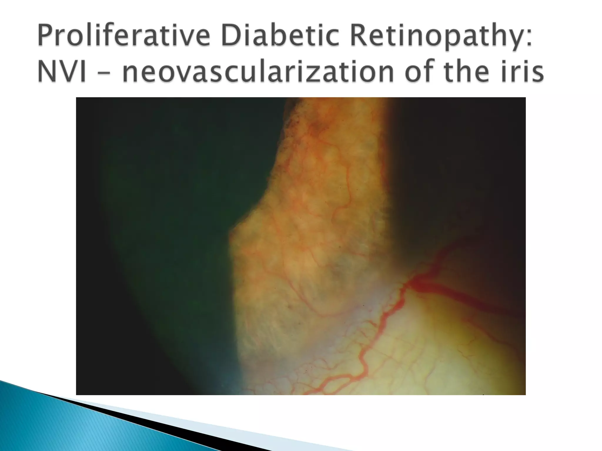
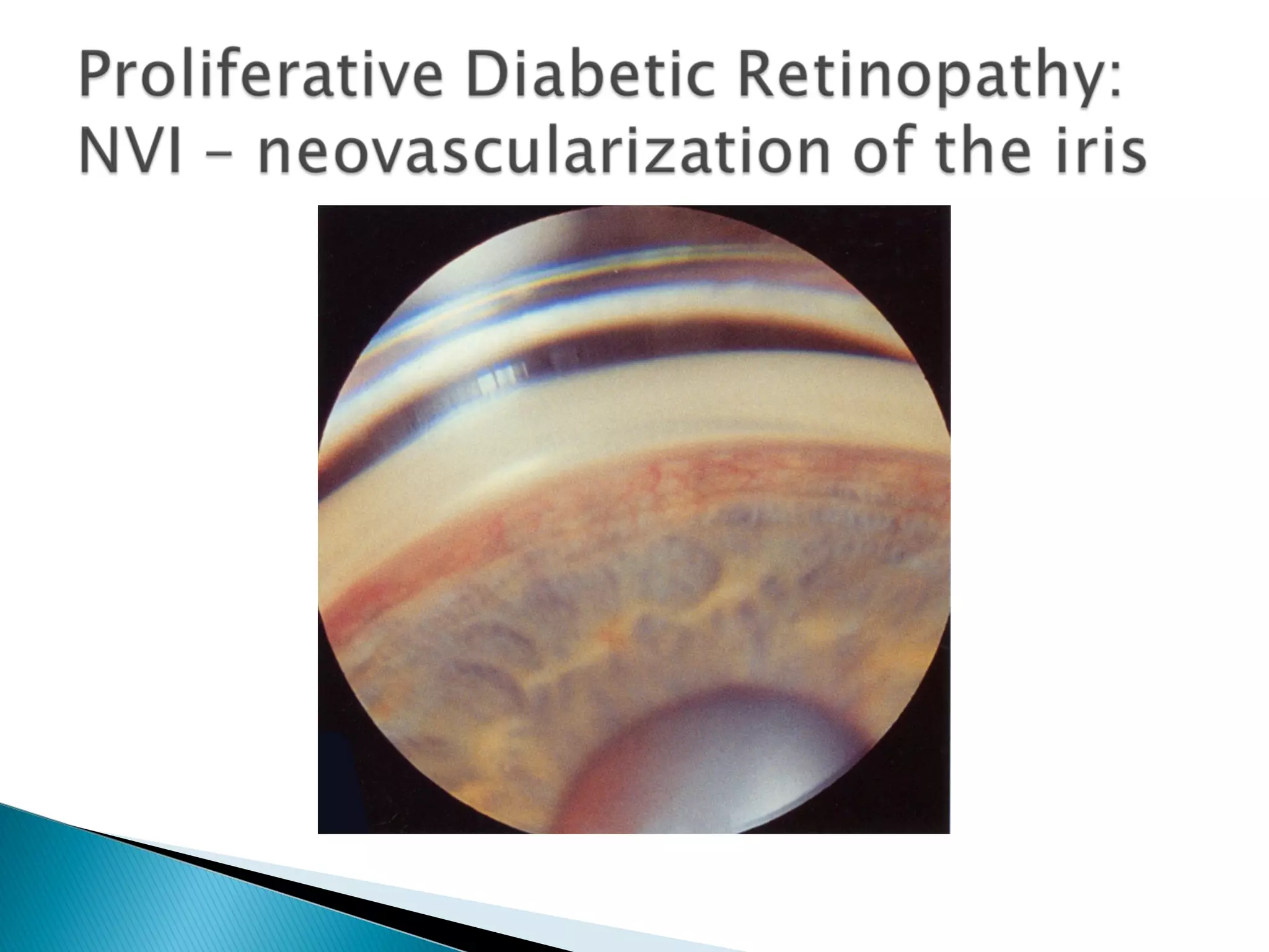
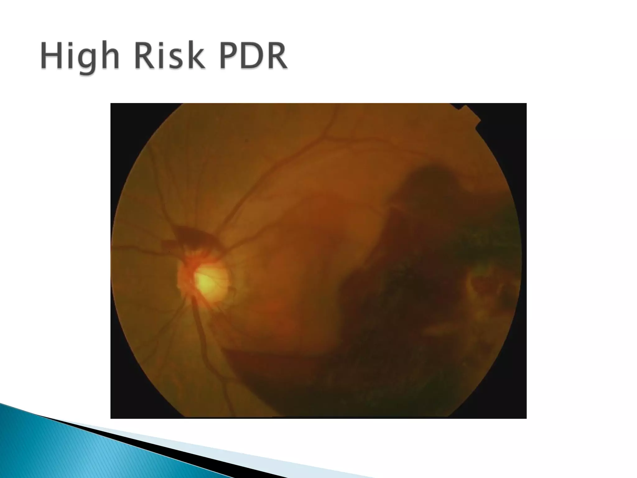
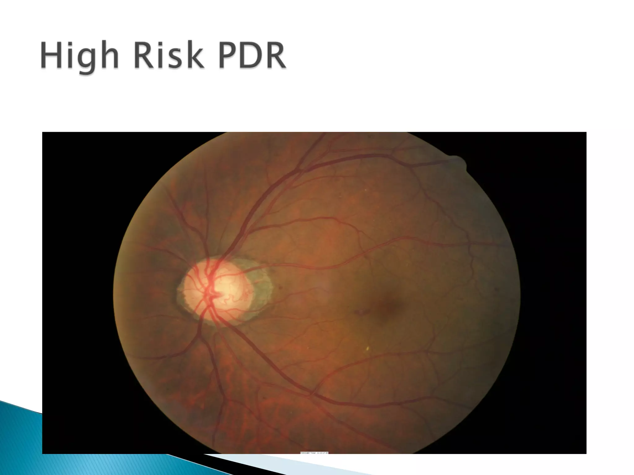
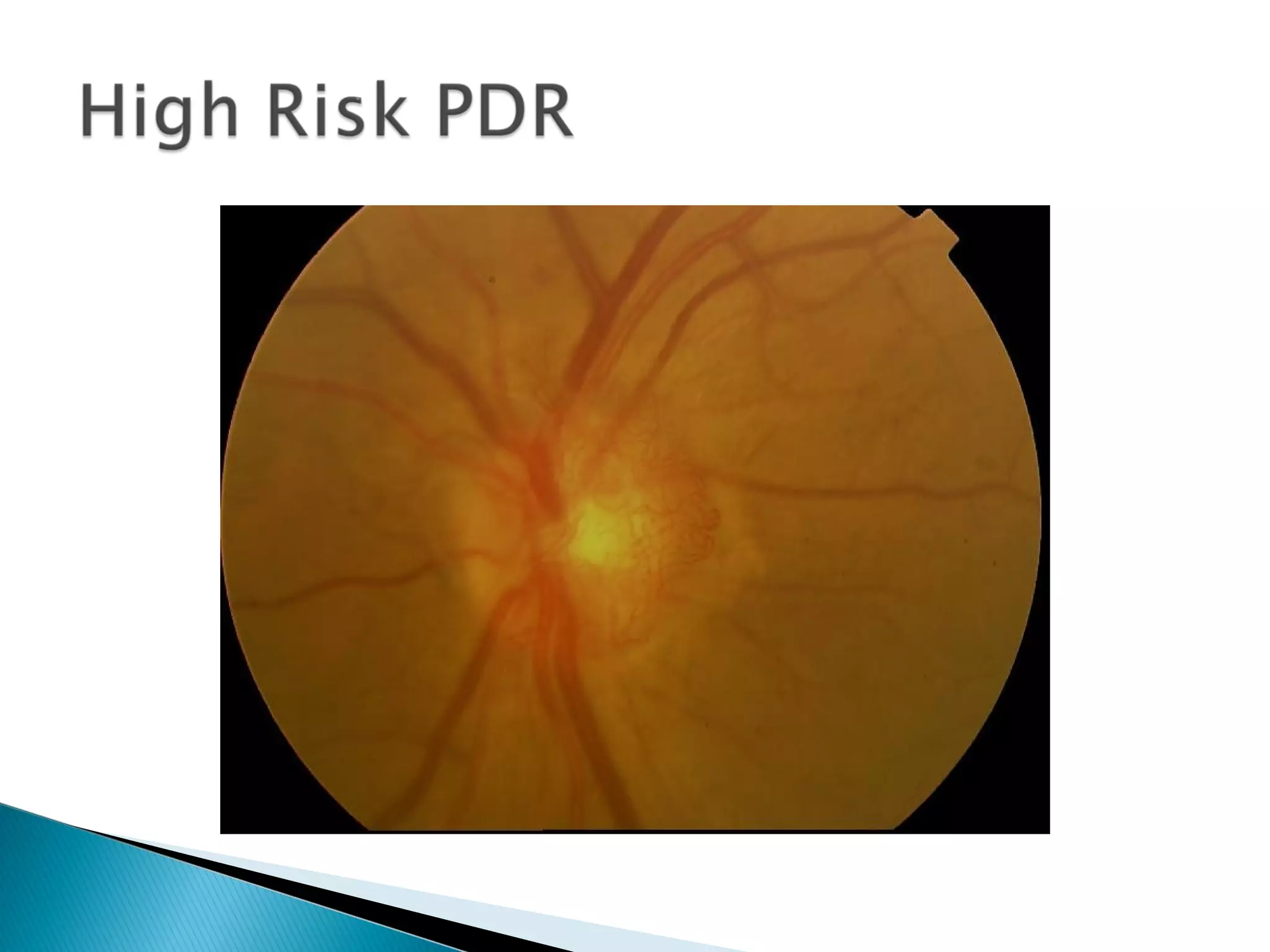
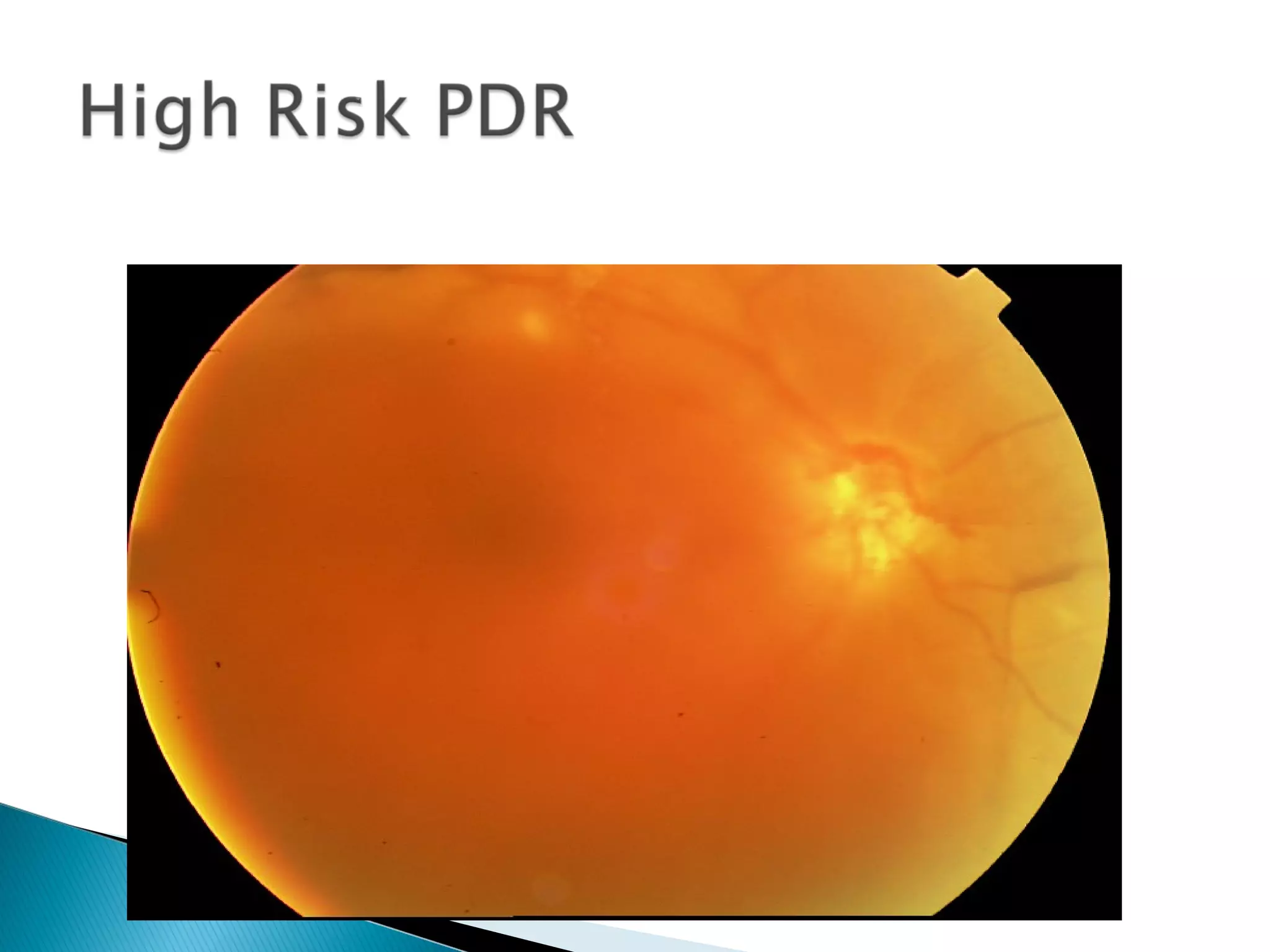
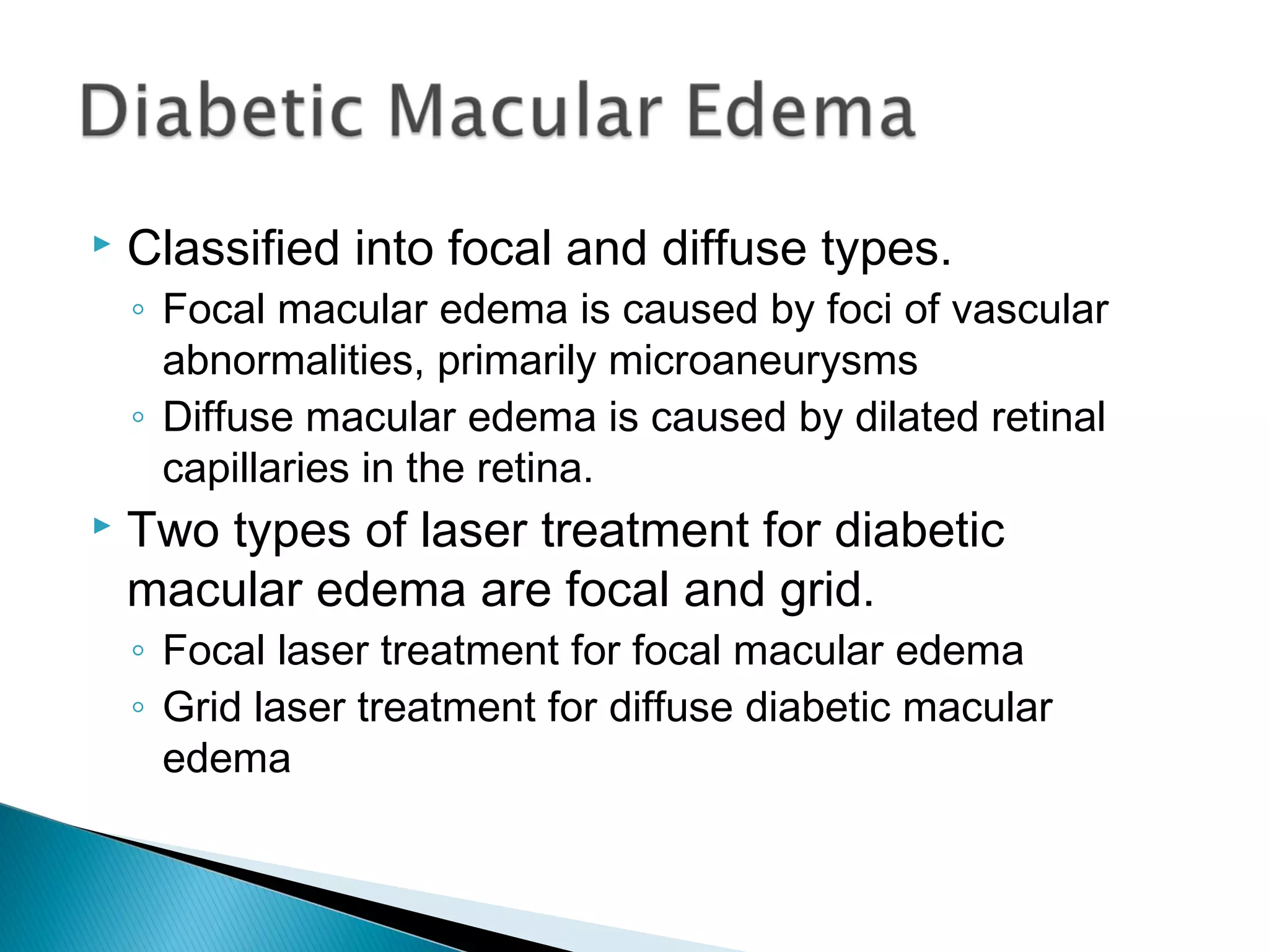
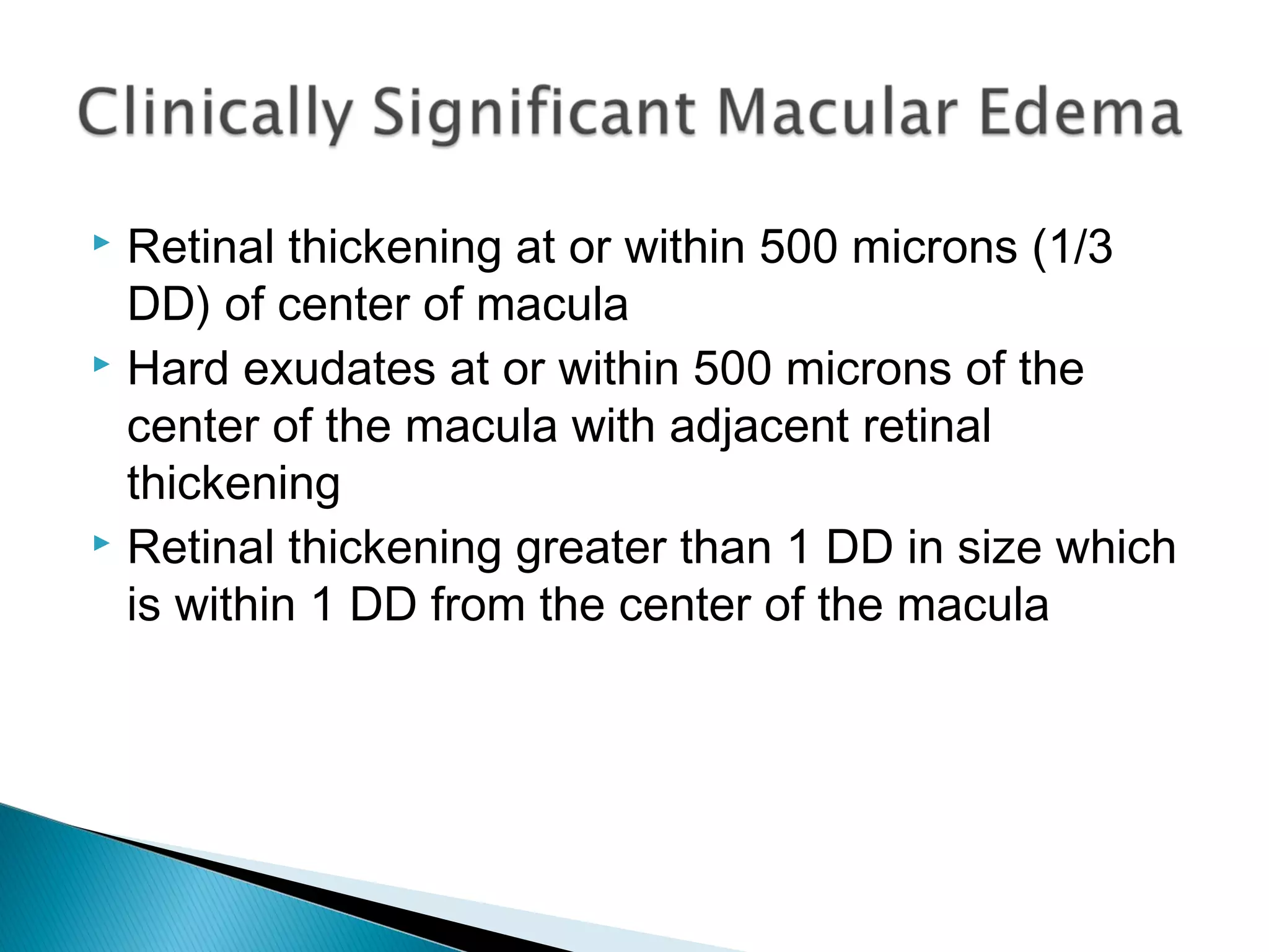
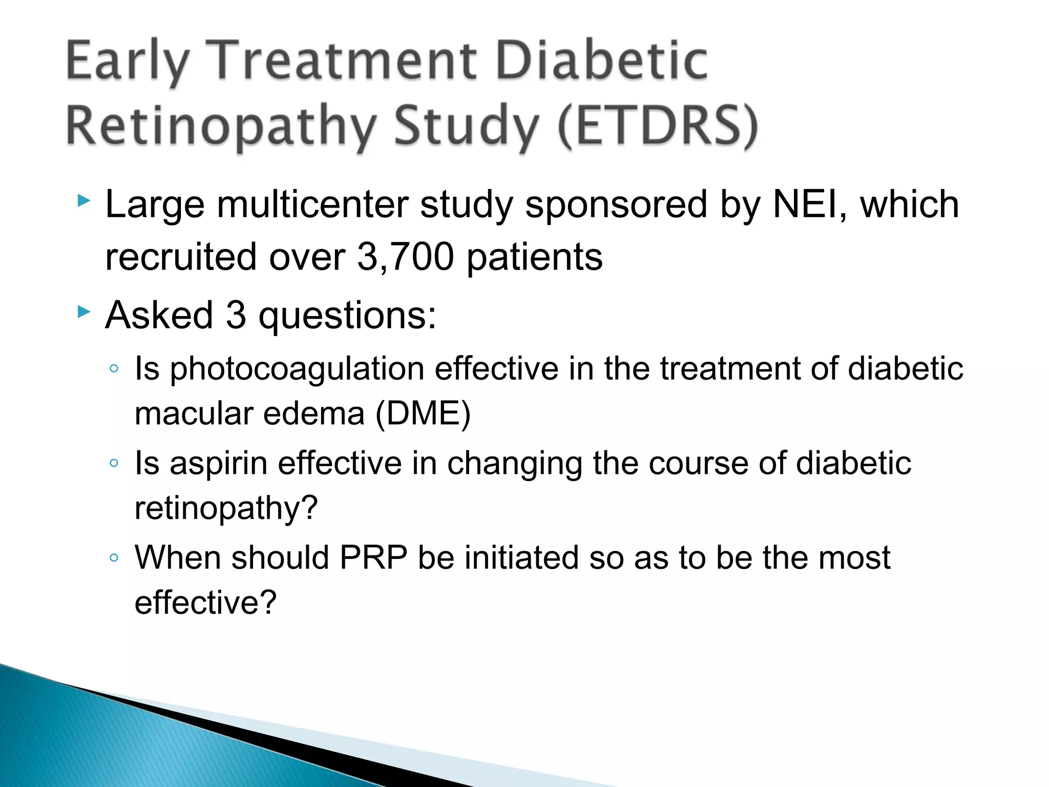
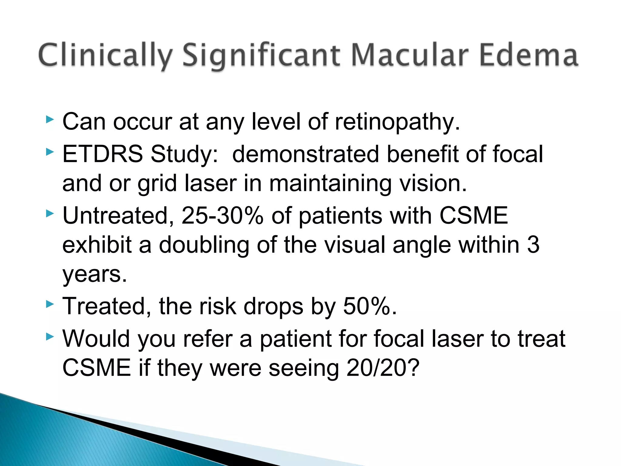
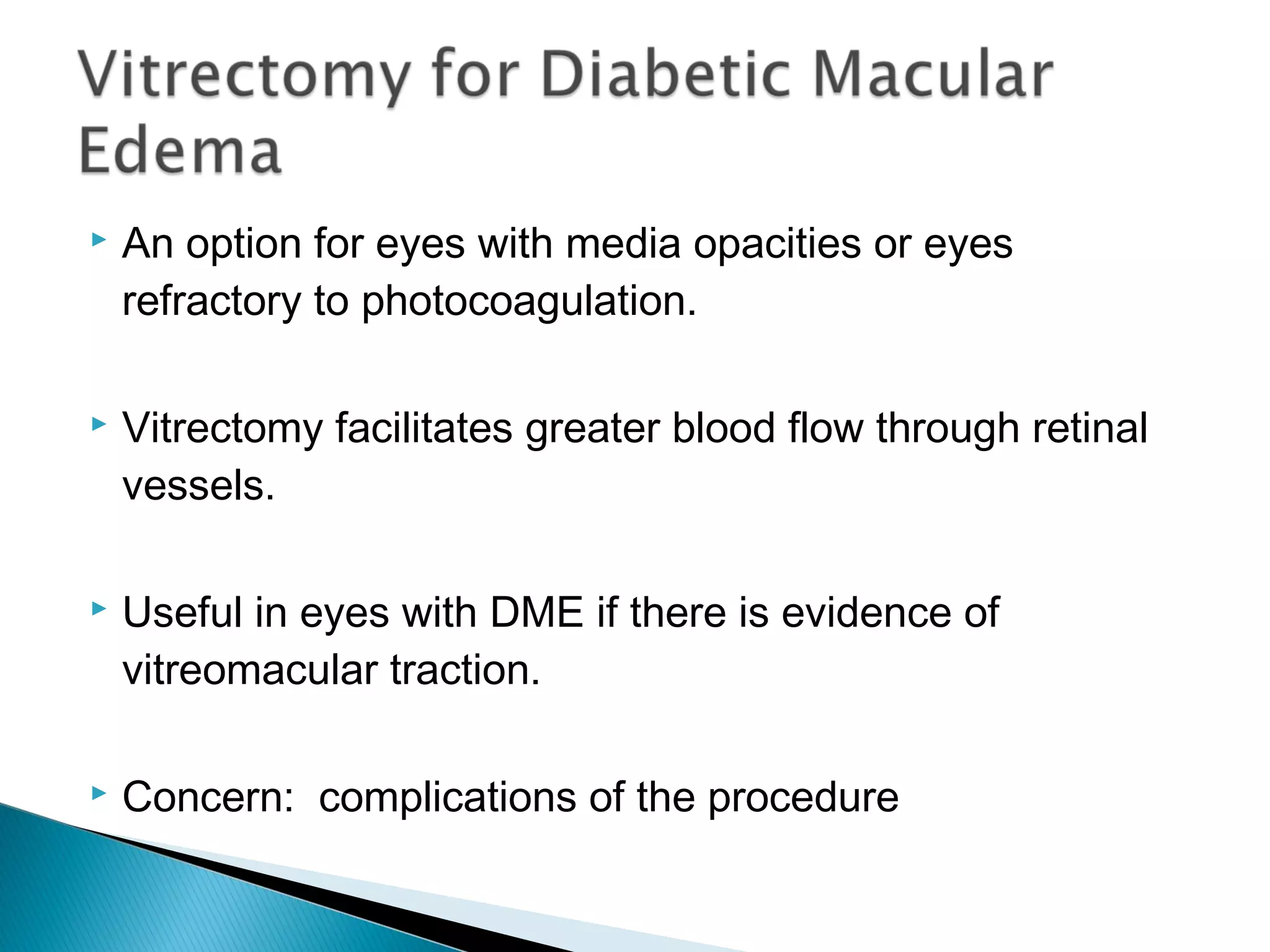
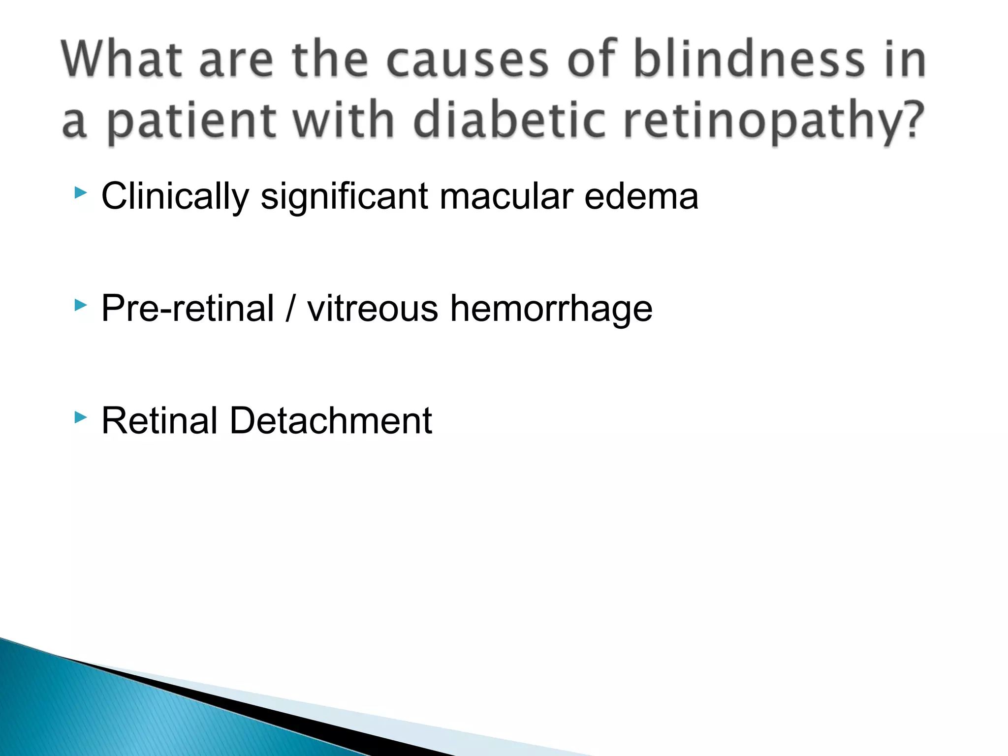
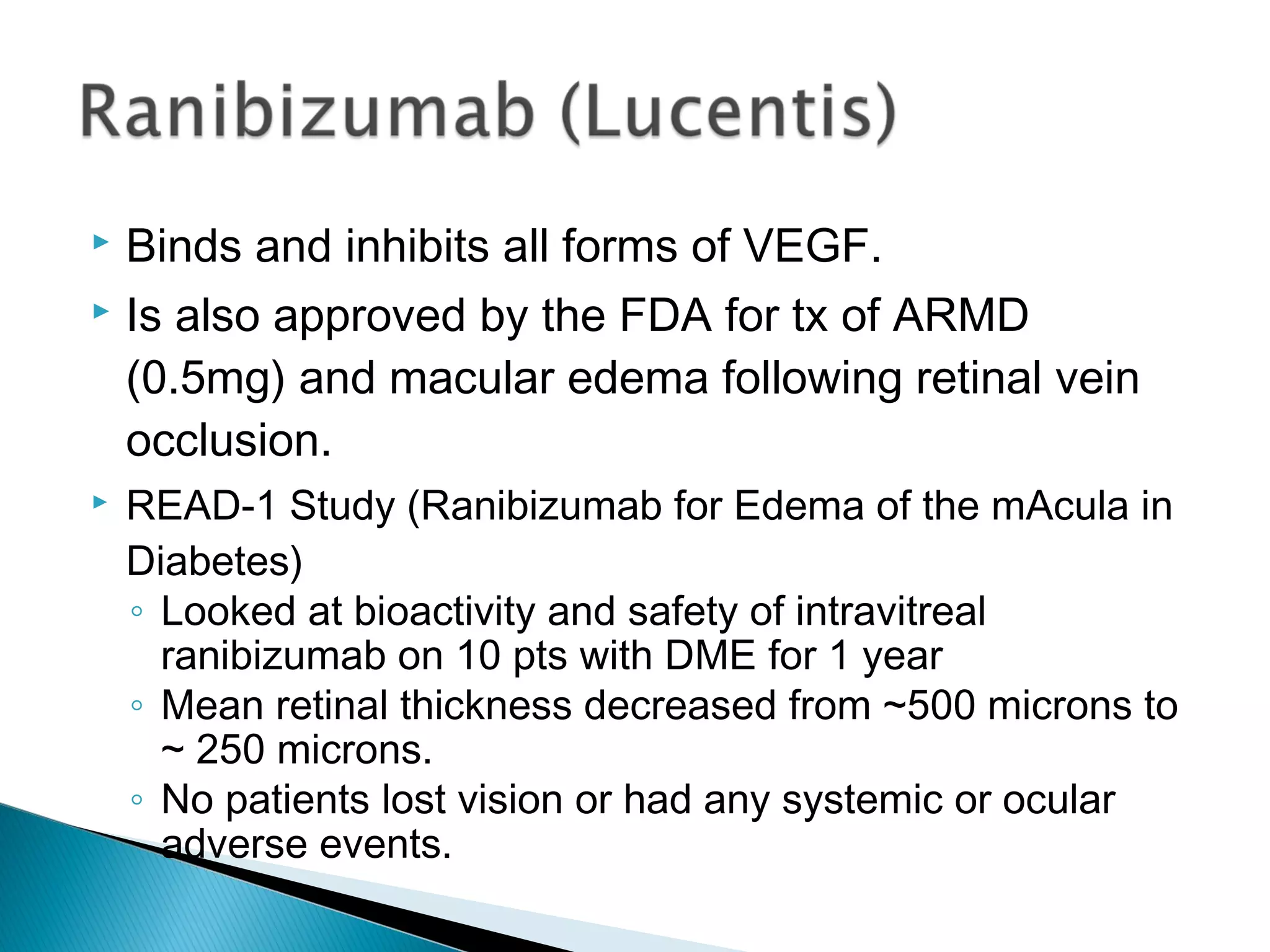
Dr. Raymond R. Maeda
 Students will be able to. . .
◦ Describe the demographics of those individuals who
have diabetes
◦ Identify patients at risk for diabetes
◦ Differentiate the different types of diabetes
◦ Recognize the criteria for diagnosing diabetes
◦ Construct a case history for their patient who has
diabetes
◦ Identify the relevant studies in regards to diabetes and
diabetic retinopathy
◦ Recognize the impact diabetes has on the organs in the
human body
◦ Recognize all of the ocular complications related to
diabetes
◦ Be able to distinguish the different classifications for
diabetic retinopathy and the appropriate treatment
◦ Explain the etiology of the different classifications
diabetic retinopathy
◦ Be familiar with future possible treatments of diabetic
retinopathy
 Diabetes is a group of diseases marked by high
levels of blood glucose resulting from defects in
insulin production, insulin action, or both.
 1.7 million new cases
in 2012
 4% of the US has some
level of diabetic retinopathy
retinopathy
 EVERY 24 HOURS there
are :
◦ 4,100 new cases of diabetes
◦ 810 deaths due to diabetes
◦ 230 amputations
◦ 120 kidney failures
◦ 55 new cases of blindness
29 million people (9.3%)
Total Cost = 245 Billion
Dollars
 Direct Medical Costs
◦ 176 Billion Dollars
◦ (medical expenses)
 Indirect Medical Costs
◦ 69 Billion Dollars
◦ (disability, work loss,
premature death)
 Non-Hispanic Whites 7.6
 Asian Americans 9.0
 Hispanics 12.8
 Non-Hispanic Blacks 13.2
 American Indians/Alaska Natives 15.9
 *2010-2012
 Type II DM appears to be rising parallel with
global trends towards obesity.
 Weight gain of 10-15 pounds can increase the risk
of DM by 50%.
◦ Especially in women
◦ Seeing prevalence of metabolic
syndrome also rise in women.
 65% of Americans are over-
weight or obese
 Type I – bodies immune system destroys pancreatic
beta cells
 Type II – begins as insulin resistance and then gradually
the pancreas loses its’ ability to produce insulin.
 Gestational
◦ Form of glucose intolerance during the 2nd
or 3rd
trimester
◦ 5-10% dx’d with DM
◦ 50% chance of development of DM in 5-10 years
 Associated with obesity, older age, family hx,
impaired glucose metabolism, physical inactivity,
and race/ethnicity
 African Americans, Hispanics, Native Americans,
*Asian Americans, and Pacific Islanders
 Seeing it more frequently dx’d in children
 Recent studies are demonstrating the need for
ethnic-specific guidelines
◦ A1C measures higher in South Asians than White
Europeans (*looked at data from 6040 people aged 40 –
75 between 2005-2009 included 1352 South Asians and
4688 white Europeans) Diabetes Care
◦ A1C level that was associated with an increased
prevalence of retinopathy was significantly lower in blacks
than in whites Annals of Internal Medicine
 National Health and Nutrition Examination Survey
(NHANES) (Dr. May in 2012)
◦ Performed recent analysis on five 2-year cycles looking at
prevalence of prediabetes and diabetes in American teens.
◦ IT HAS MORE THAN DOUBLED BETWEEN 1999
AND 2008 – 9% TO 23%
◦ 26% OF THE OVERWEIGHT AND OBESE
ADOLESCENTS WERE PRE-
HYPERTENSIVE/HYPERTENSIVE AND HAD
BORDERLINE/HIGH LDL-C.
 This is a multicenter study funded by CDC and NIH to
examine diabetes among young children and
adolescents.
◦ From 2008-2009, an estimated 18,436 people younger than
20yo (US) were newly diagnosed with Type I diabetes and 5,089
were newly diagnosed with Type II diabetes.
◦ Non-Hispanic White children and adolescents had the highest
rate of new cases of Type I
◦ The rate of Type II DM was greater among people 10-19 yo than
in younger children, with highest rates in minority populations
 2009-2012: based on fasting glucose or A1C,
37% of U.S. adults 20yo and older had pre-
diabetes (51% of those 65 and older).
 86 million Americans!!
 Impaired fasting glucose (IFG)
◦ Fasting glucose is 100 – 125mg/dL
 Impaired glucose tolerance (IGT)
◦ Glucose level is 140 – 199mg/dl after a 2hr oral glucose tolerance
test
 Approximately 86 million adults have pre-diabetes
◦ 33% will convert to DM if there is no intervention in 10 yr.s
 Progression to DM can be prevented/delayed
 A large prevention study of people at high risk for
DM
◦ Showed that lifestyle intervention to lose weight and
physical activity reduced the development of Type II DM
by 60% (3 yr period)
 Metformin intervention reduced the risk by 30%
and was most effective in younger pt’s (25-44yo)
and in adults with a BMI > 35
 The Diabetes Control and Complications Trial DCCT
◦ Demonstrated benefits of intensive blood glucose control in patients with
Type I DM in regards to development and progression of diabetic
retinopathy.
 The United Kingdom Prospective Diabetes Study (UKPDS)
◦ Showed a 21% reduction in risk for progression of diabetic retinopathy
over a 12 year period for the intensive glycemic control group
 Other studies. .
◦ ACCORD (Action to Control Cardiovascular Risk in Diabetes),
ADVANCE (Action in Diabetes and Vascular Disease, VADT (Veterans
Affairs Diabetes Trial)
 The Wisconsin Epidemiologic Study of Diabetic
Retinopathy (WESDR)
◦ Is an ongoing epidemiologic study on the progression of
diabetic retinopathy.
 Looked at fundus photos, A1C and VA’s
◦ Significant conclusions:
 Severity of diabetic retinopathy is related to the duration of the
disease.
 After 20 years nearly 99% of Type I and 60% of Type II DM
pt’s had some degree of diabetic retinopathy.
*data was limited to primarily to white populations of northern
European descent.
 Early Treatment Diabetic Retinopathy Study (ETDRS)
◦ A prospective, randomized clinical trial evaluating
photocoagulation of pt’s with diabetes.
◦ Provided the definition for clinically significant macular edema**
◦ Demonstrated benefit of focal or grid laser in maintaining vision
◦ Early scatter photocoagulation not indicated in mild/moderate DR
and resulted in a small reduction for the risk of severe vision loss.
◦ Aspirin therapy had no impact on DR progression, risk of vitreous
hemorrhage, VA loss but did reduce risk of CA morbidity and
mortality.
 Periodontal (gum) disease is more common in
people with DM
 There is a clear relationship of the degree of
hyperglycemia and severity of periodontitis.
 Adults with poorly controlled DM (A1C > 9%) were
3x’s more likely to have severe
periodontal disease
 About 60-70% of people with diabetes have mild
to severe forms of nervous system damage.
◦ Impaired sensation or pain in feet or hands
◦ Slowed digestion
◦ Carpal tunnel
◦ Erectile dysfunction
 30% of diabetics over 40 years old
have impaired sensation in their feet.
 Foot ulcers are a common
occurrence (15%)
 50% will develop infection or have other
complications
 25% will have an amputation
 DM is the leading cause of lower-limb amputations
 Diabetes is the leading cause of kidney failure
◦ Accounts for close to 50% of all new cases of kidney
failure
 No sx’s until almost all function is gone
 Tx: ACE inhibitors, low protein
diet, dialysis
 ~200,000 people on dialysis due to
ESRD
 Diabetes is the major cause of heart disease and
stroke
 Adults with DM have heart disease rates 2-4
times higher than adults without DM.
 Risk for stroke is 2-4 times higher among
people with DM
 Diabetes is the 7th
leading cause of death in the
U.S.
◦ Risk for death is twice that of people of similar age
without DM
 Leading cause of kidney failure, non-traumatic
lower limb amputation and new cases of blindness
among adults in U.S.
 Every 1 point drop in A1C can reduce
microvascular complications (eye, kidney nerve
disease) by 40%.
 BP control can reduce the risk of heart disease
and stroke by up to 50%
 Controlling LDL’s can reduce cardiovascular
complications by 20-50%
 Foot care programs (risk assessment) can reduce
amputation rates by 45 – 85%
 Detecting and treating early diabetic kidney
disease by lowering BP can reduce decline in
kidney function by 30 – 70%
Refractive changes

Refractive shift

 Due to increased glucose
levels in the lens
 21% also demonstrate
transient “paralysis” of
accommodation
Dry Eyes
 Reduced corneal sensitivity
◦ Reduce reflex tearing
 Goblet cell density
◦ Produce mucin (stability)
 Affect on lacrimal gland
◦ Correlated to length of DM
 Tear Protein Patterns
◦ Lactoferrin, slgA, albumin,
lipocalin and lysozyme
“Snowflake” cataract
 Common in uncontrolled
Type I diabetic patients
 Sorbitol accumulates in
the lens fibers. Water
enters to correct the
osmotic imbalance
 Lens fibers swell / rupture
Posterior Subcapsular
Cataract
 Earlier onset of age-
related cataracts
 Due to binding of sugars
to lens proteins
 Osmotic imbalance can
also increase cortical
changes
Glaucoma
 LALES found 40% higher
prevalence in Type II
diabetic Latino pt’s
 Neovascular glc
◦ VEGF-induced
neovascularization of the iris
and angle
 Normotensive glc
Sixth Nerve Palsy 50%
 Sudden onset
 Transient
 Absence of other
neurologic involvement
 Resolves in 3-6 months.
CN III Palsy (45%) CN IV Palsy (5%)
Diabetic Papillopathy
 Can be unilateral or
bilateral
 Minimal affect on VA
 Resolved in 2-10 months
 If bilateral, need to r/o
papilledema (imaging or
LP)
 What is a “typical” chief complaint from a diabetic
patient?
 Questions to ask:
◦ Type of DM
◦ Duration of DM
◦ Blood sugars (Fasting / Post-prandial)
◦ A1C
◦ Medications (compliance)
◦ Who do they see / How often
 Does the pt smoke (increase progression)/ drink
(excess = non-compliance)
 Metabolic Syndrome (any 3 of the following):
◦ Central Obesity (waist): > 40” – Men / > 35” Women
◦ Triglycerides > 150mg/dl
◦ HDL Cholesterol: <40mg/dl – Men
<50mg/dl - Women
◦ Blood Pressure: > 130 / 85
◦ FBS > 100mg/dl
◦ Pt’s being treated dyslipidemia,
◦ HTN or DM
 Airlie House Grading System
◦ Developed in 1969
◦ With this grading system, seven-field stereoscopic
photographs are taken and the various lesions of diabetic
retinopathy are graded according to standardized
examples.
◦ Used in 2 landmark studies: The Diabetic Retinopathy
Study (DRS) and the Early Treatment Diabetic
Retinopathy Study (ETDRS).
 ETDRS (1981) – based on the modified Airlie House
classification of diabetic retinopathy.
◦ Gold Standard but not practical in everyday use.
◦ Consists of 30-degree stereoscopic photography of seven
standard fields on color film
 ETDRS (1991) – Annual meeting of the AAO
◦ Divided up retinopathy into NPDR and PDR
◦ Separate category for diabetic macular edema
 Circinate Diffuse
 Ischaemic Mixed
 International Clinical Diabetic Retinopathy (DR)
Disease Severity Scale
◦ In 2001 AAO decided a classification needed to be
evidence based and incorporate data found in excellent
clinical trials (ETDRS, WESDR. . etc)
◦ In 2003 the scale was completed
 Five Scales with increasing severity of retinopathy
 Also developed a grading scale for DME
◦ First determine if there is retinal thickening or not
◦ Second determine level of severity (mild, moderate or
severe).
 Non-Proliferative Diabetic Retinopathy (NPDR)
◦ Mild
◦ Moderate
◦ Severe
◦ Very Severe
 Proliferative
◦ High risk
◦ Low risk
 Also known as background diabetic retinopathy in
the internal medicine literature.
 For non-ophthalmic physicians and patients this
may imply non-progressive condition we all know
this is far from the truth.
 Diabetic retinopathy is a progressive disease.
Microaneurysms
 Pathologic progress
includes the formation of
capillary
microaneurysms,
vascular permeability and
capillary closure.
Diabetic Retinopathy
 4 Levels of Severity
1. Mild 2. Moderate
3. Severe 4. Very Severe
 The extent of intraretinal
microvascular abnormalities
(IRMA), venous
abnormalities, and retinal
heme.s are the determining
factors.
 Microaneurysms
 Intraretinal hemorrhages (dot/blot)
◦ Mild to moderate in less than 4 quadrants
 Hard exudates
◦ Lipoproteins
 Follow-up: 9 months to one year
 Risk of Diabetic Macular Edema is < 5%
 Microaneurysms / hemorrhages
◦ Mild to moderate in 4 quadrants
 Hard exudates
 Cotton wool spots
◦ Indicative of retinal ischemia that causes obstruction of
axoplasmic flow. Subsequent swelling (ends of ruptured
axons) of RNFL give their characteristic appearance.
◦ Histology – cytoid bodies
 Venous beading in less than two quadrants
◦ Occurs when the dilated venular walls have the presence of
saccular microaneurysms.
 Intra-retinal microvascular abnormalities to mild degree
◦ They are dilated capillaries that seem to function as collateral
channels. Occur secondary to hypoxia.
◦ Key ddx is neovascularization.
 Follow-up: 6 months
 4-2-1 Rule: (*at least one of the following)
◦ Microaneurysms / hemorrhaging in all 4 quadrants-
SEVERE (20 / quadrant)
◦ Venous beading in 2 or more quadrants
◦ Moderately severe IRMA in one quadrant
 Any two of the following:
◦ Severe intraretinal hemorrhages in 4 quadrants
◦ Venous beading in 2 quadrants
◦ Moderately severe IRMA in 1 quadrant
 Follow-up: 3 months or retinal consult
◦ Studies show PRP may be beneficial at this stage
(based on ETDRS)
◦ Most retinal surgeons will hold off on PRP until PDR
develops
◦ 50% develop PDR within 15 months.
 Baseline Fundus Photography
◦ Seven Standard Diabetic Photographic Fields
 ONH centered, Macula centered, Temporal to macula,
Superotemporally,
Inferotemporally,
Superonasally and
Inferonasally
(*all exclude ONH
except for #1)
◦ CPT: 92250 (*Bilateral procedure)
 Modifier 52 if performed on only 1 eye
◦ Requires Interpretation and Report
◦ Reimbursement ~ $87.00
◦ Cannot do on the same day as scanning laser, extended
ophthalmoscopy or external photography (slit lamp)
◦ Can be done up to two times per year
 Extended ophthalmoscopy
◦ Presence of pathology / Colored drawings (4”x4”)
◦ CPT: 92225 (*Unilateral code: 92225-RT/92225-LT)
◦ Requires interpretation and report
◦ Reimbursement ~ $28
◦ CPT: 92226 = Subsequent follow-up of the same condition
◦ Requires interpretation and report
◦ Reimbursement ~ $26
 Can be done 6x’s / year for diabetic
 retinopathy / glc (*condition dependent)
 Why did you do it?
 What did you find? (*Clinical Findings)
 What are you going to do? (*Clinical
Management)
 Change in condition
 Amsler Grid
 OCT
 Fluorescein Angiography – assessing vascular
integrity
Diabetic Macular Edema
 CPT: 92133 (ONH) / 92134
(Retina)
 Requires Interpretation and
Report
 Reimbursement ~ $51
 Unilateral or Bilateral
 Cannot do same day as
DFE/Fundus Photos (*if
billing)
 4x’s per year unless acute
condition
 Is a vascular response to retinal hypoxia
 Many theories about the cause of retinal hypoxia. .
◦ Capillary closure
◦ Alterations in capillary b. membrane
◦ Increase blood viscosity
◦ Altered ability of blood to transport oxygen
◦ Abnormal metabolic pathways in the retinal capillaries
◦ Production of VEGF (Vascular Endothelial Growth
◦ Factors)
 VEGF – thought to play a significant role in the
proliferation of neovascularization.
 The neovascularization is initially intraretinal but
breaks through the ILM and lies between it and
the vitreous.
 Fibrous component / ground substance develops
and contracts as the neovascularizaton increases.
 Classification once neovascularization occurs or
if a pre-retinal or vitreous hemorrhage is
present.
 Neovascularization can occur. . .
◦ At the iris (NVI)
◦ Elsewhere (NVE)
◦ At the disc (NVD)
 Gonioscopy – should be performed in presence
of neovascularization of the iris.
 The Diabetic Retinopathy Study (DRS) identified
risk factors for proliferative diabetic retinopathy
(PDR).
 Purpose of the study was to determine:
◦ Does photocoagulation prevent severe vision loss in eyes
with PDR.
◦ Laser photocoagulation was found to reduce the risk of
severe vision loss by more than 50% in eyes with high risk
PDR.
 Low risk
◦ NVD less than 1/4 to 1/3 the disc
◦ NVE (*without presence of traction)
 Obtain a retinal consult within 1 week
 High Risk
◦ NVD greater than 1/4 to 1/3 the disc
◦ NVD less than 1/4 to 1/3 the disc with associated
vitreous/pre-retinal hemorrhage
◦ NVE with associated vitreous/pre-retinal hemorrhage
 Obtain a retinal consult within 24-48 hours
 Pan-retinal photocoagulation (PRP)
◦ DRS proved benefit of immediate PRP
 Argon laser applied throughout the mid-
peripheral and peripheral retina.
 Reduces the retina’s need for oxygen >
decreases hypoxia > decreases vasoproliferative
mediators > regression of new vessel growth
 PRP reduced the rate of severe vision loss by 50% for
eyes with high risks characteristics.
 PRP typically divided into 2 to 3 sessions(w/orbital block)
 1200 – 1600 moderately intense burns placed ½ burn
width apart, using a 500 micron diameter spot size and
exposure time = 0.1 sec.s
 Do not go any closer than 2dd from the center of the
macula (sup/inf/temp) and 1/2dd nasal to the disc.
 The Diabetic Retinopathy Vitrectomy Study
(DRVS) addressed the question of the timing of
vitrectomy surgery for the management of PDR.
(1976-1980: when they typically waited 1 year
before doing a vitrectomy in one with a vitreous
hemorrhage)
◦ Early intervention lead to better outcomes with Type I
DM patients w/vitreous hemorrhage (5mo.s)
 Enhances oxygen to retina by 10 fold
 Use of intra-operative VEGF
 Decreased night vision and dark adaptation
 Decreased visual field / peripheral vision
 Atrophic creep- Becomes problematic when the
laser is applied too close to the macula or nerve
 Choroidal detachment
 Makes RNFL testing / analysis difficult.
 Classified into focal and diffuse types.
◦ Focal macular edema is caused by foci of vascular
abnormalities, primarily microaneurysms
◦ Diffuse macular edema is caused by dilated retinal
capillaries in the retina.
 Two types of laser treatment for diabetic
macular edema are focal and grid.
◦ Focal laser treatment for focal macular edema
◦ Grid laser treatment for diffuse diabetic macular
edema
 Retinal thickening at or within 500 microns (1/3
DD) of center of macula
 Hard exudates at or within 500 microns of the
center of the macula with adjacent retinal
thickening
 Retinal thickening greater than 1 DD in size which
is within 1 DD from the center of the macula
 Large multicenter study sponsored by NEI, which
recruited over 3,700 patients
 Asked 3 questions:
◦ Is photocoagulation effective in the treatment of diabetic
macular edema (DME)
◦ Is aspirin effective in changing the course of diabetic
retinopathy?
◦ When should PRP be initiated so as to be the most
effective?
◦ Is photocoagulation effective in the treatment of diabetic
macular edema (DME)
 YES
◦ Is aspirin effective in changing the course of diabetic
retinopathy?
 NO
◦ When should PRP be initiated so as to be the most
effective?
 Early laser reduced the risk of the need for vitrectomy and
the risk of progression to high-risk retinopathy. For those
with “high risk” characteristics.
 Can occur at any level of retinopathy.
 ETDRS Study: demonstrated benefit of focal
and or grid laser in maintaining vision.
 Untreated, 25-30% of patients with CSME
exhibit a doubling of the visual angle within 3
years.
 Treated, the risk drops by 50%.
 Would you refer a patient for focal laser to treat
CSME if they were seeing 20/20?
 An option for eyes with media opacities or eyes
refractory to photocoagulation.
 Vitrectomy facilitates greater blood flow through retinal
vessels.
 Useful in eyes with DME if there is evidence of
vitreomacular traction.
 Concern: complications of the procedure
 One study demonstrated 61-73% resolution of
edema
 Supplementing with removal of the internal
limiting membrane may improve outcomes.
 Recent analysis demonstrates that this
procedure may only be beneficial for pt’s who
exhibits signs of macular traction.
 Clinically significant macular edema
 Pre-retinal / vitreous hemorrhage
 Retinal Detachment
 Is a potent enhancer of vascular permeability and
a key inducer of angiogenesis (growth of new
blood vessels).
 Angiogenic triggers include hypoxia and retinal
ischemia
 VEGF Isoform 165 is a critical isoform for both
developmental and pathologic retinal
angiogenesis
 VEGF is expressed by retinal endothelial cells
and retinal pigment epithelial cells.
 Anti-VEGF agents are very effective in
managing DME and PDR
◦ Prevents breakdown of blood retinal barrier, decreases
vascular permeability and retinal neovascularization
 Binds to and blocks the effects of VEGF165
 First drug approved by the FDA for tx of
neovascular ARMD (0.3mg dose).
 Studied in a phase II trial for DME – followed for 36
weeks.
 Overall improvement vs the “sham” injection
group. (*reduction of 69microns vs 4 microns)
 Binds and inhibits all forms of VEGF.
 Is also approved by the FDA for tx of ARMD
(0.5mg) and macular edema following retinal vein
occlusion.
 READ-1 Study (Ranibizumab for Edema of the mAcula in
Diabetes)
◦ Looked at bioactivity and safety of intravitreal
ranibizumab on 10 pts with DME for 1 year
◦ Mean retinal thickness decreased from ~500 microns to
~ 250 microns.
◦ No patients lost vision or had any systemic or ocular
adverse events.
 The READ-2 Study was a clinical study evaluating
ranibizumab vs focal laser photo. vs combination
therapy.
◦ The greatest gain in VA was seen in the ranibizumab only group –
8 letter improvement at month 6.
◦ The greatest reduction in retinal thickening (57%) was also seen
in the same group.(*11% - laser / 42% combo)
◦ By year 2, there was no statistically significant difference in mean
VA among the 3 treatment groups.
◦ No associated ocular or systemic adverse events occurred during
the study.
 RIDE & RISE studies
◦ 2 Phase III (36-mo.) multi-center studies to evaluate efficacy
& safety in patients with DME
◦ RIDE study – includes 58 investigators in the U.S. and 7 in
Latin America
◦ RISE study - includes 63 investigators in the US and 2 in
Argentina
◦ Overall, after 24 months 3 line improvement in treatment
group vs placebo group. More likely to achieve 20/40 vision
& less progression.
 In 2012, the FDA has approved Lucentis for the
treatment of diabetic macular edema.
◦ 0.3*-0.5mg dosed monthly for DME
◦ Common adverse effects: subconjunctival hemorrhage,
eye pain, increased IOP and floaters
 Is a mouse-derived monoclonal antibody to all
isoforms of VEGF.
 Is FDA approved for systemic treatment of
metastatic colon cancer but not for ophthalmic
indications.
 “off label” use in conditions such as ARMD,
diabetic retinopathy and DME.
 Short term studies have shown that intravitreal
bevacizumab is associated with rapid regression
of disc, retinal, and iris neo- secondary to PDR,
we well as resolution of macular edema.
 Patients typically need an injection roughly every
12 weeks up to the six month follow up (for DME).
 Back in 2011 - Dept of Veterans Affairs stopped
using Bevacizumab for ophthalmic purposes due
to incidents that have occurred at 3 different
facilities in Florida (12), Tennessee (4) and Los
Angeles (5).
 Diabetic Retinopathy Clinical Research Network
sponsored study – Phase 2 evaluation of Anti-
VEGF Therapy for Diabetic Macular Edema
 2-year study demonstrated that Avastin is more
effective than focal laser in pt’s with diabetic
macular edema.
◦ Injections at baseline, 6 and 12 weeks
◦ Until macular thickness was stable over 3 visits & within
20 microns of the pt’s thinnest recorded measurement.
 This study also found no difference between
1.25mg and 2.5mg of bevacizumab and similar
outcomes were found in other studies
◦ Pan-American Collaborative Retina Study Group
(PACORES)
◦ Study by Lam et al
 Is a fully human VEGF receptor fusion protein
designed to be a potent blocker of all forms of
VEGF-A.
 It binds to VEGF-A with greater affinity than native
receptors and penetrates all layers of the retina.
 A phase 2 randomized clinical trial (DA VINCI
study) was completed with 219 pt.s with 5 different
arms (4 different doses of VEGF Trap and 1 group
receiving focal laser)
 Statistically significant improvement in VA in all
aflibercept treated groups (2.0mg treated group
showed most gains)
◦ Key fact: a considerable number of re-injections were
necessary.
 In 2011aflibercept was recommended by FDA for
tx of wet ARMD. (*completed successful phase III
trials)
 Based on results of a couple of phase 3 trials –
VIEW 1 and VIEW 2
 Has shown promise in preliminary studies for the
treatment of DME.
 Larger studies are ongoing and we should be
getting results shortly.
 Improved delivery will be necessary in order to
avoid repeated intravitreal injections and the
cumulative risk of endophthalmitis.
 Triamcinolone acetonide suppresses
inflammation, reduces extravasation of fluid from
leaking blood vessels, inhibits fibrovascular
proliferation, and down regulates production of
VEGF.
 Desired route is intravitreal.
 Peak action is at 1 week (*3-6 mo.s)
 Complications: elevated IOP’s (50%), cataract
formation, and endophthalmitis (injection-related).
 A phase 3 randomized clinical trial evaluating the
safety and efficacy of preservative-free IVT vs
focal/grid laser
◦ At 4 months the IVT-treated group showed greater gains
in VA but by month 8 the gain narrows and at month 12
the VA gain of the laser treated exceeds the IVT-treated
groups (1mg / 4mg), which persisted to 24 months.
◦ OCT findings regarding retinal thickness were similar.
◦ IOP rise (33%) and cataract development seen in IVT
groups (51%/23% vs 13%)
 Implants are being developed to circumvent the
need for repeated injections.
◦ Ozurdex (Allergan)
◦ Retisert (Bausch & Lomb)
◦ Iluvien (Alimera Sciences)
◦ Ivation (Surmodics)
 Is a biodegradable dexamethasone pellet
(0.7mg)that is delivered to the posterior segment
in-office using an applicator (22 gauge).
 1st
FDA approved drug treatment for macular
edema following RVO (biodegradable)
 20-30% of pt’s experienced 3 line improvement in
BVA that occurred in the first 2-3 months
 Patented drug delivery system – sterile, single use
applicator preloaded with implant
 Designed to last 37 days (dissolves completely)
 ~25% developed increased IOP’s (>10mmhg)
 ~5% developed cataracts
 Consists of a tiny drug reservoir designed to
deliver sustained levels of the fluocinolone
acetonide, for approximately two-and-a-half years
(30 months). FDA approved for chronic
noninfectious uveitis therapy.
 0.59mg implant implanted in posterior segment
 Complications related to the development of
glaucoma and cataracts makes it use limited.
◦ At 3 yr.s 71% developed increase of IOP (>10mmhg)
◦ Nearly all patients developed cataracts
◦ 85% experienced improvement or stabilization in VA
(23% gained 3 lines or more)
 Is a narrow, 3.5mm long / 0.37mm in diameter,
cylindrical fluocinolone acetonide implant (190ug).
 Implanted non-surgically in-office with a 25 gauge
injector (*stays in vitreous base)
 Initial release rate of either 0.23 or 0.45 mcg per
day (lowest available) lasting up to 36 months.
 36 month FAME study completed in October
◦ Fluocinolone Acetonide in Diabetic Macular Edema
◦ ~29% demonstrated improvement in BCVA of 15 letters
from baseline at 3 years.
 Has not been approved by FDA.
◦ Asking for more data
 Has been resubmitted with results of patients being
treated in the UK and Germany.
 Metallic coil coated with triamcinolone
 Inserted with 25 gauge needle
 Can release the drug up to 2 years.
 Phase I study
◦ At 2 yr visit mean reduction in central retinal thickness
was 126 um
◦ VA improved then declined and then improved again
(found to be due to cataract development)
 No sustained elevations in IOP were noted in the
study group (*avg change was < 2mmHg)
 Phase II trials have been suspended
 Intended to evaluate the safety and efficacy
 NEI had released a study suggesting laser tx over
intravitreal injections of TA
 Micropulse Photocoagulation over standard laser
for DME
◦ Subthreshold diode laser
◦ Produces less damage/scarring
 Biodegradable microspheres and nanoparticles
 Looking at encapsulating of either Macugen or
Avastin into these particles for extended delivery.
 These would be biodegradable and intended to be
injected into the vitreous with a small gauge
needle.
 Encapsulated Cell Technology
◦ Involves genetically engineering (plasmid transfection)
certain cells, which encodes the therapeutic factor
which in then incorporated into the cell’s genome.
Cells produce desired therapeutic effect.
◦ The cells within the membrane are implanted and
begin to produce the desired therapeutic factor, which
diffuses throughout the retina.
 Questions??
 rmaeda@westernu.edu

Lecture 3: Diabetic Retinopathy

  • 1.
  • 2.
     Students willbe able to. . . ◦ Describe the demographics of those individuals who have diabetes ◦ Identify patients at risk for diabetes ◦ Differentiate the different types of diabetes ◦ Recognize the criteria for diagnosing diabetes ◦ Construct a case history for their patient who has diabetes ◦ Identify the relevant studies in regards to diabetes and diabetic retinopathy ◦ Recognize the impact diabetes has on the organs in the human body
  • 3.
    ◦ Recognize allof the ocular complications related to diabetes ◦ Be able to distinguish the different classifications for diabetic retinopathy and the appropriate treatment ◦ Explain the etiology of the different classifications diabetic retinopathy ◦ Be familiar with future possible treatments of diabetic retinopathy
  • 4.
     Diabetes isa group of diseases marked by high levels of blood glucose resulting from defects in insulin production, insulin action, or both.  1.7 million new cases in 2012  4% of the US has some level of diabetic retinopathy retinopathy
  • 5.
     EVERY 24HOURS there are : ◦ 4,100 new cases of diabetes ◦ 810 deaths due to diabetes ◦ 230 amputations ◦ 120 kidney failures ◦ 55 new cases of blindness
  • 6.
    29 million people(9.3%) Total Cost = 245 Billion Dollars  Direct Medical Costs ◦ 176 Billion Dollars ◦ (medical expenses)  Indirect Medical Costs ◦ 69 Billion Dollars ◦ (disability, work loss, premature death)
  • 7.
     Non-Hispanic Whites7.6  Asian Americans 9.0  Hispanics 12.8  Non-Hispanic Blacks 13.2  American Indians/Alaska Natives 15.9  *2010-2012
  • 8.
     Type IIDM appears to be rising parallel with global trends towards obesity.  Weight gain of 10-15 pounds can increase the risk of DM by 50%. ◦ Especially in women ◦ Seeing prevalence of metabolic syndrome also rise in women.  65% of Americans are over- weight or obese
  • 9.
     Type I– bodies immune system destroys pancreatic beta cells  Type II – begins as insulin resistance and then gradually the pancreas loses its’ ability to produce insulin.  Gestational ◦ Form of glucose intolerance during the 2nd or 3rd trimester ◦ 5-10% dx’d with DM ◦ 50% chance of development of DM in 5-10 years
  • 11.
     Associated withobesity, older age, family hx, impaired glucose metabolism, physical inactivity, and race/ethnicity  African Americans, Hispanics, Native Americans, *Asian Americans, and Pacific Islanders  Seeing it more frequently dx’d in children
  • 13.
     Recent studiesare demonstrating the need for ethnic-specific guidelines ◦ A1C measures higher in South Asians than White Europeans (*looked at data from 6040 people aged 40 – 75 between 2005-2009 included 1352 South Asians and 4688 white Europeans) Diabetes Care ◦ A1C level that was associated with an increased prevalence of retinopathy was significantly lower in blacks than in whites Annals of Internal Medicine
  • 14.
     National Healthand Nutrition Examination Survey (NHANES) (Dr. May in 2012) ◦ Performed recent analysis on five 2-year cycles looking at prevalence of prediabetes and diabetes in American teens. ◦ IT HAS MORE THAN DOUBLED BETWEEN 1999 AND 2008 – 9% TO 23% ◦ 26% OF THE OVERWEIGHT AND OBESE ADOLESCENTS WERE PRE- HYPERTENSIVE/HYPERTENSIVE AND HAD BORDERLINE/HIGH LDL-C.
  • 15.
     This isa multicenter study funded by CDC and NIH to examine diabetes among young children and adolescents. ◦ From 2008-2009, an estimated 18,436 people younger than 20yo (US) were newly diagnosed with Type I diabetes and 5,089 were newly diagnosed with Type II diabetes. ◦ Non-Hispanic White children and adolescents had the highest rate of new cases of Type I ◦ The rate of Type II DM was greater among people 10-19 yo than in younger children, with highest rates in minority populations
  • 16.
     2009-2012: basedon fasting glucose or A1C, 37% of U.S. adults 20yo and older had pre- diabetes (51% of those 65 and older).  86 million Americans!!
  • 17.
     Impaired fastingglucose (IFG) ◦ Fasting glucose is 100 – 125mg/dL  Impaired glucose tolerance (IGT) ◦ Glucose level is 140 – 199mg/dl after a 2hr oral glucose tolerance test  Approximately 86 million adults have pre-diabetes ◦ 33% will convert to DM if there is no intervention in 10 yr.s  Progression to DM can be prevented/delayed
  • 18.
     A largeprevention study of people at high risk for DM ◦ Showed that lifestyle intervention to lose weight and physical activity reduced the development of Type II DM by 60% (3 yr period)  Metformin intervention reduced the risk by 30% and was most effective in younger pt’s (25-44yo) and in adults with a BMI > 35
  • 19.
     The DiabetesControl and Complications Trial DCCT ◦ Demonstrated benefits of intensive blood glucose control in patients with Type I DM in regards to development and progression of diabetic retinopathy.  The United Kingdom Prospective Diabetes Study (UKPDS) ◦ Showed a 21% reduction in risk for progression of diabetic retinopathy over a 12 year period for the intensive glycemic control group  Other studies. . ◦ ACCORD (Action to Control Cardiovascular Risk in Diabetes), ADVANCE (Action in Diabetes and Vascular Disease, VADT (Veterans Affairs Diabetes Trial)
  • 20.
     The WisconsinEpidemiologic Study of Diabetic Retinopathy (WESDR) ◦ Is an ongoing epidemiologic study on the progression of diabetic retinopathy.  Looked at fundus photos, A1C and VA’s ◦ Significant conclusions:  Severity of diabetic retinopathy is related to the duration of the disease.  After 20 years nearly 99% of Type I and 60% of Type II DM pt’s had some degree of diabetic retinopathy. *data was limited to primarily to white populations of northern European descent.
  • 21.
     Early TreatmentDiabetic Retinopathy Study (ETDRS) ◦ A prospective, randomized clinical trial evaluating photocoagulation of pt’s with diabetes. ◦ Provided the definition for clinically significant macular edema** ◦ Demonstrated benefit of focal or grid laser in maintaining vision ◦ Early scatter photocoagulation not indicated in mild/moderate DR and resulted in a small reduction for the risk of severe vision loss. ◦ Aspirin therapy had no impact on DR progression, risk of vitreous hemorrhage, VA loss but did reduce risk of CA morbidity and mortality.
  • 23.
     Periodontal (gum)disease is more common in people with DM  There is a clear relationship of the degree of hyperglycemia and severity of periodontitis.  Adults with poorly controlled DM (A1C > 9%) were 3x’s more likely to have severe periodontal disease
  • 24.
     About 60-70%of people with diabetes have mild to severe forms of nervous system damage. ◦ Impaired sensation or pain in feet or hands ◦ Slowed digestion ◦ Carpal tunnel ◦ Erectile dysfunction  30% of diabetics over 40 years old have impaired sensation in their feet.
  • 25.
     Foot ulcersare a common occurrence (15%)  50% will develop infection or have other complications  25% will have an amputation  DM is the leading cause of lower-limb amputations
  • 26.
     Diabetes isthe leading cause of kidney failure ◦ Accounts for close to 50% of all new cases of kidney failure  No sx’s until almost all function is gone  Tx: ACE inhibitors, low protein diet, dialysis  ~200,000 people on dialysis due to ESRD
  • 27.
     Diabetes isthe major cause of heart disease and stroke  Adults with DM have heart disease rates 2-4 times higher than adults without DM.  Risk for stroke is 2-4 times higher among people with DM
  • 28.
     Diabetes isthe 7th leading cause of death in the U.S. ◦ Risk for death is twice that of people of similar age without DM  Leading cause of kidney failure, non-traumatic lower limb amputation and new cases of blindness among adults in U.S.
  • 29.
     Every 1point drop in A1C can reduce microvascular complications (eye, kidney nerve disease) by 40%.  BP control can reduce the risk of heart disease and stroke by up to 50%
  • 30.
     Controlling LDL’scan reduce cardiovascular complications by 20-50%  Foot care programs (risk assessment) can reduce amputation rates by 45 – 85%  Detecting and treating early diabetic kidney disease by lowering BP can reduce decline in kidney function by 30 – 70%
  • 31.
    Refractive changes  Refractive shift  Due to increased glucose levels in the lens  21% also demonstrate transient “paralysis” of accommodation
  • 32.
    Dry Eyes  Reducedcorneal sensitivity ◦ Reduce reflex tearing  Goblet cell density ◦ Produce mucin (stability)  Affect on lacrimal gland ◦ Correlated to length of DM  Tear Protein Patterns ◦ Lactoferrin, slgA, albumin, lipocalin and lysozyme
  • 33.
    “Snowflake” cataract  Commonin uncontrolled Type I diabetic patients  Sorbitol accumulates in the lens fibers. Water enters to correct the osmotic imbalance  Lens fibers swell / rupture
  • 34.
    Posterior Subcapsular Cataract  Earlieronset of age- related cataracts  Due to binding of sugars to lens proteins  Osmotic imbalance can also increase cortical changes
  • 35.
    Glaucoma  LALES found40% higher prevalence in Type II diabetic Latino pt’s  Neovascular glc ◦ VEGF-induced neovascularization of the iris and angle  Normotensive glc
  • 36.
    Sixth Nerve Palsy50%  Sudden onset  Transient  Absence of other neurologic involvement  Resolves in 3-6 months.
  • 37.
    CN III Palsy(45%) CN IV Palsy (5%)
  • 38.
    Diabetic Papillopathy  Canbe unilateral or bilateral  Minimal affect on VA  Resolved in 2-10 months  If bilateral, need to r/o papilledema (imaging or LP)
  • 39.
     What isa “typical” chief complaint from a diabetic patient?  Questions to ask: ◦ Type of DM ◦ Duration of DM ◦ Blood sugars (Fasting / Post-prandial) ◦ A1C ◦ Medications (compliance) ◦ Who do they see / How often
  • 40.
     Does thept smoke (increase progression)/ drink (excess = non-compliance)  Metabolic Syndrome (any 3 of the following): ◦ Central Obesity (waist): > 40” – Men / > 35” Women ◦ Triglycerides > 150mg/dl ◦ HDL Cholesterol: <40mg/dl – Men <50mg/dl - Women ◦ Blood Pressure: > 130 / 85 ◦ FBS > 100mg/dl ◦ Pt’s being treated dyslipidemia, ◦ HTN or DM
  • 41.
     Airlie HouseGrading System ◦ Developed in 1969 ◦ With this grading system, seven-field stereoscopic photographs are taken and the various lesions of diabetic retinopathy are graded according to standardized examples. ◦ Used in 2 landmark studies: The Diabetic Retinopathy Study (DRS) and the Early Treatment Diabetic Retinopathy Study (ETDRS).
  • 42.
     ETDRS (1981)– based on the modified Airlie House classification of diabetic retinopathy. ◦ Gold Standard but not practical in everyday use. ◦ Consists of 30-degree stereoscopic photography of seven standard fields on color film  ETDRS (1991) – Annual meeting of the AAO ◦ Divided up retinopathy into NPDR and PDR ◦ Separate category for diabetic macular edema  Circinate Diffuse  Ischaemic Mixed
  • 43.
     International ClinicalDiabetic Retinopathy (DR) Disease Severity Scale ◦ In 2001 AAO decided a classification needed to be evidence based and incorporate data found in excellent clinical trials (ETDRS, WESDR. . etc) ◦ In 2003 the scale was completed  Five Scales with increasing severity of retinopathy  Also developed a grading scale for DME ◦ First determine if there is retinal thickening or not ◦ Second determine level of severity (mild, moderate or severe).
  • 45.
     Non-Proliferative DiabeticRetinopathy (NPDR) ◦ Mild ◦ Moderate ◦ Severe ◦ Very Severe  Proliferative ◦ High risk ◦ Low risk
  • 46.
     Also knownas background diabetic retinopathy in the internal medicine literature.  For non-ophthalmic physicians and patients this may imply non-progressive condition we all know this is far from the truth.  Diabetic retinopathy is a progressive disease.
  • 47.
    Microaneurysms  Pathologic progress includesthe formation of capillary microaneurysms, vascular permeability and capillary closure.
  • 48.
    Diabetic Retinopathy  4Levels of Severity 1. Mild 2. Moderate 3. Severe 4. Very Severe  The extent of intraretinal microvascular abnormalities (IRMA), venous abnormalities, and retinal heme.s are the determining factors.
  • 49.
     Microaneurysms  Intraretinalhemorrhages (dot/blot) ◦ Mild to moderate in less than 4 quadrants  Hard exudates ◦ Lipoproteins  Follow-up: 9 months to one year  Risk of Diabetic Macular Edema is < 5%
  • 52.
     Microaneurysms /hemorrhages ◦ Mild to moderate in 4 quadrants  Hard exudates  Cotton wool spots ◦ Indicative of retinal ischemia that causes obstruction of axoplasmic flow. Subsequent swelling (ends of ruptured axons) of RNFL give their characteristic appearance. ◦ Histology – cytoid bodies
  • 53.
     Venous beadingin less than two quadrants ◦ Occurs when the dilated venular walls have the presence of saccular microaneurysms.  Intra-retinal microvascular abnormalities to mild degree ◦ They are dilated capillaries that seem to function as collateral channels. Occur secondary to hypoxia. ◦ Key ddx is neovascularization.  Follow-up: 6 months
  • 60.
     4-2-1 Rule:(*at least one of the following) ◦ Microaneurysms / hemorrhaging in all 4 quadrants- SEVERE (20 / quadrant) ◦ Venous beading in 2 or more quadrants ◦ Moderately severe IRMA in one quadrant
  • 61.
     Any twoof the following: ◦ Severe intraretinal hemorrhages in 4 quadrants ◦ Venous beading in 2 quadrants ◦ Moderately severe IRMA in 1 quadrant
  • 62.
     Follow-up: 3months or retinal consult ◦ Studies show PRP may be beneficial at this stage (based on ETDRS) ◦ Most retinal surgeons will hold off on PRP until PDR develops ◦ 50% develop PDR within 15 months.
  • 67.
     Baseline FundusPhotography ◦ Seven Standard Diabetic Photographic Fields  ONH centered, Macula centered, Temporal to macula, Superotemporally, Inferotemporally, Superonasally and Inferonasally (*all exclude ONH except for #1)
  • 69.
    ◦ CPT: 92250(*Bilateral procedure)  Modifier 52 if performed on only 1 eye ◦ Requires Interpretation and Report ◦ Reimbursement ~ $87.00 ◦ Cannot do on the same day as scanning laser, extended ophthalmoscopy or external photography (slit lamp) ◦ Can be done up to two times per year
  • 70.
     Extended ophthalmoscopy ◦Presence of pathology / Colored drawings (4”x4”) ◦ CPT: 92225 (*Unilateral code: 92225-RT/92225-LT) ◦ Requires interpretation and report ◦ Reimbursement ~ $28 ◦ CPT: 92226 = Subsequent follow-up of the same condition ◦ Requires interpretation and report ◦ Reimbursement ~ $26  Can be done 6x’s / year for diabetic  retinopathy / glc (*condition dependent)
  • 71.
     Why didyou do it?  What did you find? (*Clinical Findings)  What are you going to do? (*Clinical Management)  Change in condition
  • 72.
     Amsler Grid OCT  Fluorescein Angiography – assessing vascular integrity
  • 73.
    Diabetic Macular Edema CPT: 92133 (ONH) / 92134 (Retina)  Requires Interpretation and Report  Reimbursement ~ $51  Unilateral or Bilateral  Cannot do same day as DFE/Fundus Photos (*if billing)  4x’s per year unless acute condition
  • 76.
     Is avascular response to retinal hypoxia  Many theories about the cause of retinal hypoxia. . ◦ Capillary closure ◦ Alterations in capillary b. membrane ◦ Increase blood viscosity ◦ Altered ability of blood to transport oxygen ◦ Abnormal metabolic pathways in the retinal capillaries ◦ Production of VEGF (Vascular Endothelial Growth ◦ Factors)
  • 77.
     VEGF –thought to play a significant role in the proliferation of neovascularization.  The neovascularization is initially intraretinal but breaks through the ILM and lies between it and the vitreous.  Fibrous component / ground substance develops and contracts as the neovascularizaton increases.
  • 78.
     Classification onceneovascularization occurs or if a pre-retinal or vitreous hemorrhage is present.  Neovascularization can occur. . . ◦ At the iris (NVI) ◦ Elsewhere (NVE) ◦ At the disc (NVD)  Gonioscopy – should be performed in presence of neovascularization of the iris.
  • 85.
     The DiabeticRetinopathy Study (DRS) identified risk factors for proliferative diabetic retinopathy (PDR).  Purpose of the study was to determine: ◦ Does photocoagulation prevent severe vision loss in eyes with PDR. ◦ Laser photocoagulation was found to reduce the risk of severe vision loss by more than 50% in eyes with high risk PDR.
  • 86.
     Low risk ◦NVD less than 1/4 to 1/3 the disc ◦ NVE (*without presence of traction)  Obtain a retinal consult within 1 week
  • 90.
     High Risk ◦NVD greater than 1/4 to 1/3 the disc ◦ NVD less than 1/4 to 1/3 the disc with associated vitreous/pre-retinal hemorrhage ◦ NVE with associated vitreous/pre-retinal hemorrhage  Obtain a retinal consult within 24-48 hours
  • 91.
     Pan-retinal photocoagulation(PRP) ◦ DRS proved benefit of immediate PRP  Argon laser applied throughout the mid- peripheral and peripheral retina.  Reduces the retina’s need for oxygen > decreases hypoxia > decreases vasoproliferative mediators > regression of new vessel growth
  • 92.
     PRP reducedthe rate of severe vision loss by 50% for eyes with high risks characteristics.  PRP typically divided into 2 to 3 sessions(w/orbital block)  1200 – 1600 moderately intense burns placed ½ burn width apart, using a 500 micron diameter spot size and exposure time = 0.1 sec.s  Do not go any closer than 2dd from the center of the macula (sup/inf/temp) and 1/2dd nasal to the disc.
  • 93.
     The DiabeticRetinopathy Vitrectomy Study (DRVS) addressed the question of the timing of vitrectomy surgery for the management of PDR. (1976-1980: when they typically waited 1 year before doing a vitrectomy in one with a vitreous hemorrhage) ◦ Early intervention lead to better outcomes with Type I DM patients w/vitreous hemorrhage (5mo.s)  Enhances oxygen to retina by 10 fold  Use of intra-operative VEGF
  • 101.
     Decreased nightvision and dark adaptation  Decreased visual field / peripheral vision  Atrophic creep- Becomes problematic when the laser is applied too close to the macula or nerve  Choroidal detachment  Makes RNFL testing / analysis difficult.
  • 104.
     Classified intofocal and diffuse types. ◦ Focal macular edema is caused by foci of vascular abnormalities, primarily microaneurysms ◦ Diffuse macular edema is caused by dilated retinal capillaries in the retina.  Two types of laser treatment for diabetic macular edema are focal and grid. ◦ Focal laser treatment for focal macular edema ◦ Grid laser treatment for diffuse diabetic macular edema
  • 105.
     Retinal thickeningat or within 500 microns (1/3 DD) of center of macula  Hard exudates at or within 500 microns of the center of the macula with adjacent retinal thickening  Retinal thickening greater than 1 DD in size which is within 1 DD from the center of the macula
  • 106.
     Large multicenterstudy sponsored by NEI, which recruited over 3,700 patients  Asked 3 questions: ◦ Is photocoagulation effective in the treatment of diabetic macular edema (DME) ◦ Is aspirin effective in changing the course of diabetic retinopathy? ◦ When should PRP be initiated so as to be the most effective?
  • 107.
    ◦ Is photocoagulationeffective in the treatment of diabetic macular edema (DME)  YES ◦ Is aspirin effective in changing the course of diabetic retinopathy?  NO ◦ When should PRP be initiated so as to be the most effective?  Early laser reduced the risk of the need for vitrectomy and the risk of progression to high-risk retinopathy. For those with “high risk” characteristics.
  • 111.
     Can occurat any level of retinopathy.  ETDRS Study: demonstrated benefit of focal and or grid laser in maintaining vision.  Untreated, 25-30% of patients with CSME exhibit a doubling of the visual angle within 3 years.  Treated, the risk drops by 50%.  Would you refer a patient for focal laser to treat CSME if they were seeing 20/20?
  • 112.
     An optionfor eyes with media opacities or eyes refractory to photocoagulation.  Vitrectomy facilitates greater blood flow through retinal vessels.  Useful in eyes with DME if there is evidence of vitreomacular traction.  Concern: complications of the procedure
  • 113.
     One studydemonstrated 61-73% resolution of edema  Supplementing with removal of the internal limiting membrane may improve outcomes.  Recent analysis demonstrates that this procedure may only be beneficial for pt’s who exhibits signs of macular traction.
  • 114.
     Clinically significantmacular edema  Pre-retinal / vitreous hemorrhage  Retinal Detachment
  • 115.
     Is apotent enhancer of vascular permeability and a key inducer of angiogenesis (growth of new blood vessels).  Angiogenic triggers include hypoxia and retinal ischemia  VEGF Isoform 165 is a critical isoform for both developmental and pathologic retinal angiogenesis
  • 116.
     VEGF isexpressed by retinal endothelial cells and retinal pigment epithelial cells.  Anti-VEGF agents are very effective in managing DME and PDR ◦ Prevents breakdown of blood retinal barrier, decreases vascular permeability and retinal neovascularization
  • 117.
     Binds toand blocks the effects of VEGF165  First drug approved by the FDA for tx of neovascular ARMD (0.3mg dose).  Studied in a phase II trial for DME – followed for 36 weeks.  Overall improvement vs the “sham” injection group. (*reduction of 69microns vs 4 microns)
  • 118.
     Binds andinhibits all forms of VEGF.  Is also approved by the FDA for tx of ARMD (0.5mg) and macular edema following retinal vein occlusion.  READ-1 Study (Ranibizumab for Edema of the mAcula in Diabetes) ◦ Looked at bioactivity and safety of intravitreal ranibizumab on 10 pts with DME for 1 year ◦ Mean retinal thickness decreased from ~500 microns to ~ 250 microns. ◦ No patients lost vision or had any systemic or ocular adverse events.
  • 119.
     The READ-2Study was a clinical study evaluating ranibizumab vs focal laser photo. vs combination therapy. ◦ The greatest gain in VA was seen in the ranibizumab only group – 8 letter improvement at month 6. ◦ The greatest reduction in retinal thickening (57%) was also seen in the same group.(*11% - laser / 42% combo) ◦ By year 2, there was no statistically significant difference in mean VA among the 3 treatment groups. ◦ No associated ocular or systemic adverse events occurred during the study.
  • 120.
     RIDE &RISE studies ◦ 2 Phase III (36-mo.) multi-center studies to evaluate efficacy & safety in patients with DME ◦ RIDE study – includes 58 investigators in the U.S. and 7 in Latin America ◦ RISE study - includes 63 investigators in the US and 2 in Argentina ◦ Overall, after 24 months 3 line improvement in treatment group vs placebo group. More likely to achieve 20/40 vision & less progression.
  • 121.
     In 2012,the FDA has approved Lucentis for the treatment of diabetic macular edema. ◦ 0.3*-0.5mg dosed monthly for DME ◦ Common adverse effects: subconjunctival hemorrhage, eye pain, increased IOP and floaters
  • 122.
     Is amouse-derived monoclonal antibody to all isoforms of VEGF.  Is FDA approved for systemic treatment of metastatic colon cancer but not for ophthalmic indications.  “off label” use in conditions such as ARMD, diabetic retinopathy and DME.
  • 123.
     Short termstudies have shown that intravitreal bevacizumab is associated with rapid regression of disc, retinal, and iris neo- secondary to PDR, we well as resolution of macular edema.  Patients typically need an injection roughly every 12 weeks up to the six month follow up (for DME).
  • 124.
     Back in2011 - Dept of Veterans Affairs stopped using Bevacizumab for ophthalmic purposes due to incidents that have occurred at 3 different facilities in Florida (12), Tennessee (4) and Los Angeles (5).
  • 125.
     Diabetic RetinopathyClinical Research Network sponsored study – Phase 2 evaluation of Anti- VEGF Therapy for Diabetic Macular Edema  2-year study demonstrated that Avastin is more effective than focal laser in pt’s with diabetic macular edema. ◦ Injections at baseline, 6 and 12 weeks ◦ Until macular thickness was stable over 3 visits & within 20 microns of the pt’s thinnest recorded measurement.
  • 126.
     This studyalso found no difference between 1.25mg and 2.5mg of bevacizumab and similar outcomes were found in other studies ◦ Pan-American Collaborative Retina Study Group (PACORES) ◦ Study by Lam et al
  • 127.
     Is afully human VEGF receptor fusion protein designed to be a potent blocker of all forms of VEGF-A.  It binds to VEGF-A with greater affinity than native receptors and penetrates all layers of the retina.
  • 128.
     A phase2 randomized clinical trial (DA VINCI study) was completed with 219 pt.s with 5 different arms (4 different doses of VEGF Trap and 1 group receiving focal laser)  Statistically significant improvement in VA in all aflibercept treated groups (2.0mg treated group showed most gains) ◦ Key fact: a considerable number of re-injections were necessary.
  • 129.
     In 2011afliberceptwas recommended by FDA for tx of wet ARMD. (*completed successful phase III trials)  Based on results of a couple of phase 3 trials – VIEW 1 and VIEW 2
  • 130.
     Has shownpromise in preliminary studies for the treatment of DME.  Larger studies are ongoing and we should be getting results shortly.  Improved delivery will be necessary in order to avoid repeated intravitreal injections and the cumulative risk of endophthalmitis.
  • 131.
     Triamcinolone acetonidesuppresses inflammation, reduces extravasation of fluid from leaking blood vessels, inhibits fibrovascular proliferation, and down regulates production of VEGF.  Desired route is intravitreal.  Peak action is at 1 week (*3-6 mo.s)  Complications: elevated IOP’s (50%), cataract formation, and endophthalmitis (injection-related).
  • 132.
     A phase3 randomized clinical trial evaluating the safety and efficacy of preservative-free IVT vs focal/grid laser ◦ At 4 months the IVT-treated group showed greater gains in VA but by month 8 the gain narrows and at month 12 the VA gain of the laser treated exceeds the IVT-treated groups (1mg / 4mg), which persisted to 24 months. ◦ OCT findings regarding retinal thickness were similar. ◦ IOP rise (33%) and cataract development seen in IVT groups (51%/23% vs 13%)
  • 133.
     Implants arebeing developed to circumvent the need for repeated injections. ◦ Ozurdex (Allergan) ◦ Retisert (Bausch & Lomb) ◦ Iluvien (Alimera Sciences) ◦ Ivation (Surmodics)
  • 134.
     Is abiodegradable dexamethasone pellet (0.7mg)that is delivered to the posterior segment in-office using an applicator (22 gauge).  1st FDA approved drug treatment for macular edema following RVO (biodegradable)  20-30% of pt’s experienced 3 line improvement in BVA that occurred in the first 2-3 months
  • 135.
     Patented drugdelivery system – sterile, single use applicator preloaded with implant  Designed to last 37 days (dissolves completely)  ~25% developed increased IOP’s (>10mmhg)  ~5% developed cataracts
  • 137.
     Consists ofa tiny drug reservoir designed to deliver sustained levels of the fluocinolone acetonide, for approximately two-and-a-half years (30 months). FDA approved for chronic noninfectious uveitis therapy.  0.59mg implant implanted in posterior segment
  • 138.
     Complications relatedto the development of glaucoma and cataracts makes it use limited. ◦ At 3 yr.s 71% developed increase of IOP (>10mmhg) ◦ Nearly all patients developed cataracts ◦ 85% experienced improvement or stabilization in VA (23% gained 3 lines or more)
  • 140.
     Is anarrow, 3.5mm long / 0.37mm in diameter, cylindrical fluocinolone acetonide implant (190ug).  Implanted non-surgically in-office with a 25 gauge injector (*stays in vitreous base)  Initial release rate of either 0.23 or 0.45 mcg per day (lowest available) lasting up to 36 months.
  • 141.
     36 monthFAME study completed in October ◦ Fluocinolone Acetonide in Diabetic Macular Edema ◦ ~29% demonstrated improvement in BCVA of 15 letters from baseline at 3 years.  Has not been approved by FDA. ◦ Asking for more data  Has been resubmitted with results of patients being treated in the UK and Germany.
  • 143.
     Metallic coilcoated with triamcinolone  Inserted with 25 gauge needle  Can release the drug up to 2 years.  Phase I study ◦ At 2 yr visit mean reduction in central retinal thickness was 126 um ◦ VA improved then declined and then improved again (found to be due to cataract development)  No sustained elevations in IOP were noted in the study group (*avg change was < 2mmHg)
  • 144.
     Phase IItrials have been suspended  Intended to evaluate the safety and efficacy  NEI had released a study suggesting laser tx over intravitreal injections of TA
  • 147.
     Micropulse Photocoagulationover standard laser for DME ◦ Subthreshold diode laser ◦ Produces less damage/scarring
  • 148.
     Biodegradable microspheresand nanoparticles  Looking at encapsulating of either Macugen or Avastin into these particles for extended delivery.  These would be biodegradable and intended to be injected into the vitreous with a small gauge needle.
  • 149.
     Encapsulated CellTechnology ◦ Involves genetically engineering (plasmid transfection) certain cells, which encodes the therapeutic factor which in then incorporated into the cell’s genome. Cells produce desired therapeutic effect. ◦ The cells within the membrane are implanted and begin to produce the desired therapeutic factor, which diffuses throughout the retina.
  • 150.

Editor's Notes