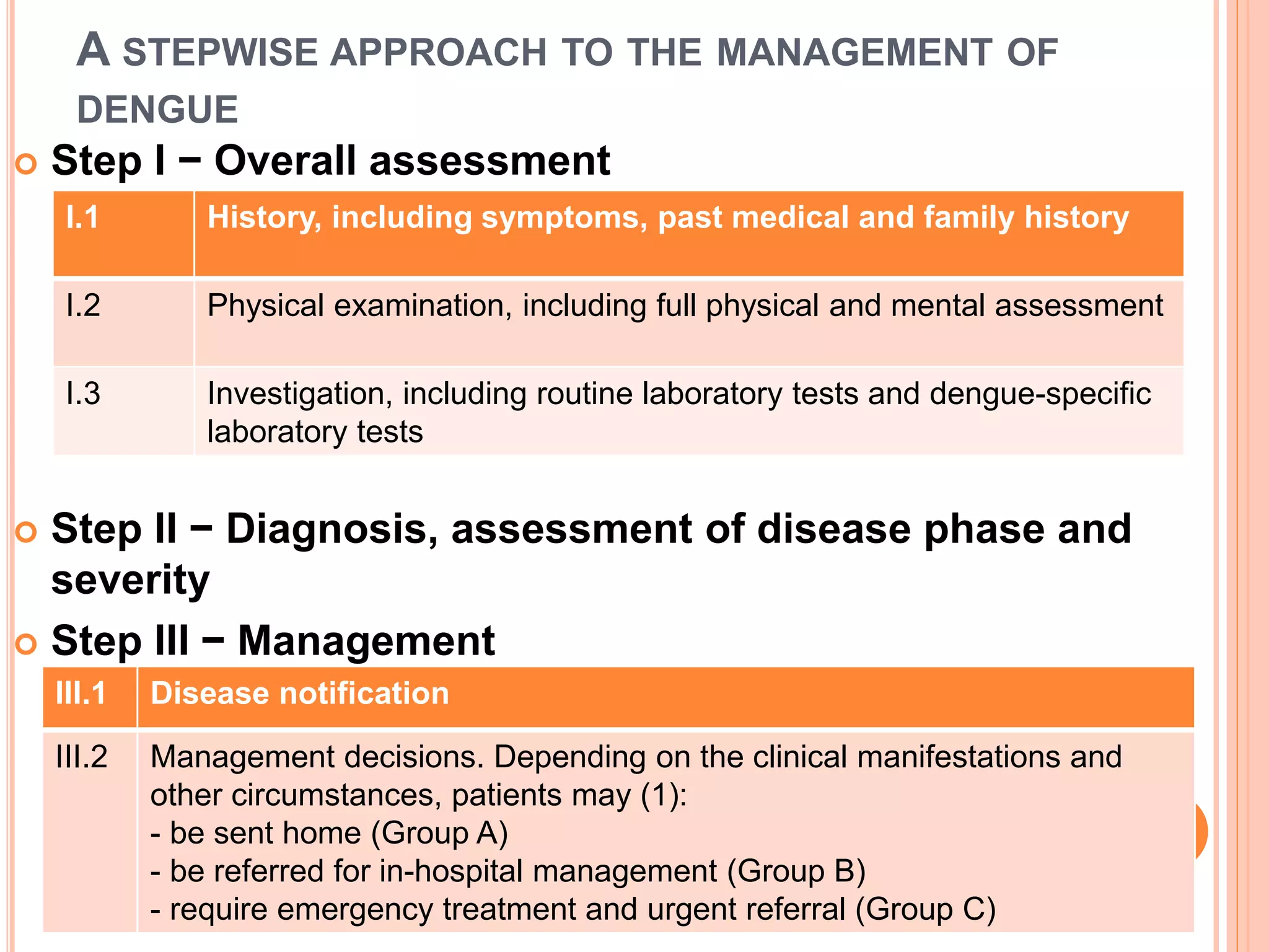
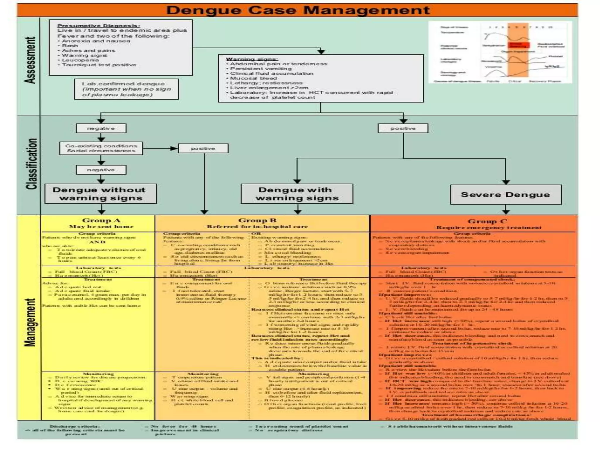
DENGUE MANAGEMENT
BY Dr.Rakesh CH
SR IN PEDIATRICS DEPT,RIMS,ADILABAD
A STEPWISE APPROACH TO THE MANAGEMENT OF
DENGUE
 Step I − Overall assessment
 Step II − Diagnosis, assessment of disease phase and
severity
 Step III − Management
I.1 History, including symptoms, past medical and family history
I.2 Physical examination, including full physical and mental assessment
I.3 Investigation, including routine laboratory tests and dengue-specific
laboratory tests
III.1 Disease notification
III.2 Management decisions. Depending on the clinical manifestations and
other circumstances, patients may (1):
- be sent home (Group A)
- be referred for in-hospital management (Group B)
- require emergency treatment and urgent referral (Group C)
DISEASE NOTIFICATION
 In dengue-endemic countries, cases of suspected,
probable and confirmed dengue should be notified
early so that appropriate public-health measures
can be initiated.
 Laboratory confirmation is not necessary before
notification, but it should be obtained.
 In non-endemic countries, usually only confirmed
cases will be notified.
MANAGEMENT DECISIONS
 Depending on the clinical manifestations and other
circumstances,
 patients may either be sent home (Group A);
 be referred for in-hospital management (Group B);
 or require emergency treatment and urgent referral
(Group C).
GROUP A--- MAY BE SENT HOME
 Group criteria
 Patients who do not have warning signs AND who are
able: tolerate adequate volumes of oral fluids and pass
urine at least once every 6 hours
GROUP A;;;;; TREATMENT
 The key to the success of ambulatory (outpatient)
management is to give clear, definitive advice on the care
that the patient needs to receive at home: i.e. bed rest and
frequent oral fluids
 Patients with ≥ 3 days of illness should be reviewed daily
for disease progression (indicated by decreasing white
blood cell and platelet counts and increasing
haematocrit,defervescence and warning signs) until they
are out of the critical period
 Those with stable haematocrit can be sent home but
should be advised to return to the nearest hospital
immediately if they develop any of the warning signs and
to adhere to the following action plan:
 Adequate oral fluid intake may reduce the number of
hospitalizations
 Small amounts of oral fluids should be given frequently
for those with nausea and anorexia
 The choice of fluids should be based on the local culture:
coconut water in some countries, in others rice water or
barley water
 Oral rehydration solution or soup may be given to
prevent electrolyte imbalance
 Commercial carbonated drinks that exceed the isotonic
level (5% sugar) should be avoided
 Sufficient oral fluid intake should result in a urinary
frequency of at least 4 to 6 times per day
 A record of oral fluid and urine output could be
maintained and reviewed daily
 Give paracetamol for high fever if the patient
 The recommended dose is 10-15 mg/kg/dose, not more
than 3−4 times in 24 hours in children and not more than
3 g/day
 Sponge with tepid water if the patient still has a high fever
 Do not give acetylsalicylic acid (aspirin),or other non-
steroidal antiinflammatory agents (NSAIDs) or
intramuscular injections, as these aggravate gastritis or
bleeding.
 Instruct caregivers that the patient should be brought to
hospital immediately if any of the following occur: no
clinical improvement, deterioration , severe abdominal
pain, persistent vomiting, cold and clammy extremities,
lethargy or irritability/restlessness, bleeding (e.g. black
stools or coffeeground vomiting), shortness of breath, not
passing urine for more than 4−6 hours.
 Admission during the febrile period should be
reserved for those who are unable to manage
adequate oral hydration at home, infants, and those
with warning signs
 Ambulatory patients should be monitored daily for
temperature pattern, volume of fluid intake and
losses, urine output (volume and frequency),
warning signs, signs of plasma leakage and
bleeding and complete blood counts.
GROUP B
 These are patients who should be admitted for in-hospital
management for close observation as they approach the
critical phase.
 These include patients with warning signs,or its
management more complicated (such as, infancy,
obesity,heart failure, renal failure, chronic haemolytic
diseases such as sickle-cell disease and autoimmune
diseases), &
those with certain social circumstances (such as living
alone,or living far from a health facility without reliable
means of transport).
 Rapid fluid replacement in patients with warning signs is
the key to prevent progression to the shock state.
 The action plan should be as follows and applies to infants,
children
 Obtain a reference haematocrit before intravenous fluid
therapy begins. Give only isotonic solutions such as 0.9%
saline, Ringer's lactate .
 Start with 5−7 ml/kg/hour for 1−2 hours, then reduce to 3−5
ml/kg/hour for 2−4 hours, and then reduce to 2−3
ml/kg/hour or less according to the clinical response
 Reassess the clinical status and repeat the haematocrit. If
the haematocrit remainsthe same or rises only minimally,
continue at the same rate (2−3 ml/kg/hour) for another 2−4
hours. If the vital signs are worsening and the haematocrit
is rising rapidly, increase the rate to 5−10 ml/kg/hour for
1−2 hours. Reassess the clinical status, repeat the
haematocrit and review fluid infusion rates accordingly.
 Give the minimum intravenous fluid volume required to
maintain good perfusion and an urine output of about 0.5
ml/kg/hour. Intravenous fluids are usually needed for only
24−48 hours.
 Reduce intravenous fluids gradually when the rate of
plasma leakage decreases towards the end of the critical
phase. This is indicated by urine output and/or oral fluid
intake improving, or the haematocrit decreasing below the
baseline value in a stable patient
 A detailed fluid balance should be maintained.
Parameters that should be monitored include vital signs
and peripheral perfusion (1−4 hourly until the patient is
out of the critical phase), urine output (4−6 hourly),
haematocrit (before and after fluid replacement, then
6−12 hourly), blood glucose and other organ functions
(such as renal profile, liver profile, coagulation profile, as
indicated).
GROUP C
 These are patients with severe dengue who require
emergency treatment and urgentreferral because they
are in the critical phase of the disease and have:
• severe plasma leakage leading to dengue shock and/or
fluid accumulation with respiratory distress;
• severe haemorrhages;
• severe organ impairment (hepatic damage, renal
impairment, cardiomyopathy,encephalopathy or
encephalitis).
 All patients with severe dengue should be admitted to a
hospital with access to blood transfusion facilities
JUDICIOUS INTRAVENOUS FLUID RESUSCITATION IS
THE ESSENTIAL AND USUALLY SOLE INTERVENTION
REQUIRED
 The goals of fluid resuscitation include:
 • improving central and peripheral circulation – i.e.
decreasing tachycardia, improving BP and pulse
volume, warm and pink extremities, a capillary refill
time < 2 seconds;
 • improving end-organ perfusion – i.e. achieving a
stable conscious level (more alert or less restless),
and urine output ≥ 0.5 ml/kg/hour or decreasing
metabolic acidosis
TREATMENT OF SHOCK
WHEN TO STOP INTRAVENOUS FLUID
THERAPY
 • signs of cessation of plasma leakage;
 • stable BP, pulse and peripheral perfusion;
 • haematocrit decreases in the presence of a good
pulse volume;
 • apyrexia (without the use of antipyretics) for more
than 24–48 hours;
 • resolving bowel/abdominal symptoms;
 • improving urine output.
 Continuing intravenous fluid therapy beyond the 48
hours of the critical phase will put the patient at risk
of pulmonary oedema and other complications such
as thrombophlebitis.
TREATMENT OF HAEMORRHAGIC
COMPLICATIONS
 Mucosal bleeding may occur in any patient with
dengue but if the patient remains stable with fluid
resuscitation/replacement, this should be
considered as a minor issue.
 The bleeding usually improves rapidly during the
recovery phase.
 In patients with profound thrombocytopenia, ensure
strict bed rest and protection from trauma. Do not
give intramuscular injections.
 No evidence exists that prophylactic platelet
transfusions are beneficial in haemodynamically
stable patients
PATIENTS AT RISK OF SEVERE BLEEDING ARE
THOSE WHO:
 have profound/prolonged/refractory shock;
 • have hypotensive shock and multi-organ failure or
severe and persistent metabolic acidosis;
 • are given non-steroidal anti-inflammatory agents;
 • have pre-existing peptic ulcer disease;
 • are on anticoagulant therapy;
SEVERE BLEEDING SHOULD BE RECOGNIZED IN THE
FOLLOWING SITUATIONS:
 • persistent and/or severe overt bleeding in the
presence of unstable haemodynamic status,
regardless of the haematocrit level;
 • a decrease in haematocrit after boluses of fluid
resuscitation together with unstable
haemodynamic status;
 • refractory shock that fails to respond to
consecutive fluid resuscitation of 40–60 ml/kg;
 • hypotensive shock with inappropriately low/normal
haematocrit;
 • persistent or worsening metabolic acidosis in
patients with a well-maintained systolicBP,
especially in those with severe abdominal
tenderness and distension.
THE ACTION PLAN FOR THE TREATMENT OF
HAEMORRHAGIC COMPLICATIONS IS AS FOLLOWS:
 • If possible, attempts should be made to stop bleeding if the
source of bleeding is identified e.g. severe epistaxis may be
controlled by nasal adrenaline packing.
 • If blood loss can be quantified, this should be replaced. If
not, give aliquots of
 5−10 ml/kg of fresh -packed red cells or 10−20 ml/kg of fresh
or fairly fresh whole blood (FWB) at an appropriate rate and
observe the clinical response. It is importantthat fresh whole
blood or fresh red cells are given.
 Oxygen delivery at tissue level isoptimal with high levels of 2,3
diphosphoglycerate (2,3 DPG). Stored erythrocytes lose 2,3
DPG, low levels of which impede the oxygen-releasing
capacity of haemoglobin, resulting in functional tissue
hypoxia.
 A good clinical response includes improving haemodynamic
status and acid-base balance.
 • Consider repeating the blood transfusion if there is further
overt blood loss or noappropriate rise in haematocrit after
blood transfusion in an unstable patient
DISCHARGE CRITERIA
Clinical No fever for 48 hours.
Improvement in clinical status (general well-being,
appetite, haemodynamic
status, urine output, no respiratory distress).
Laboratory Increasing trend of platelet count.
Stable haematocrit without intravenous fluids.
Dr.Ch,Rakesh

Dengue management ppt

  • 1.
    DENGUE MANAGEMENT BY Dr.RakeshCH SR IN PEDIATRICS DEPT,RIMS,ADILABAD
  • 2.
    A STEPWISE APPROACHTO THE MANAGEMENT OF DENGUE  Step I − Overall assessment  Step II − Diagnosis, assessment of disease phase and severity  Step III − Management I.1 History, including symptoms, past medical and family history I.2 Physical examination, including full physical and mental assessment I.3 Investigation, including routine laboratory tests and dengue-specific laboratory tests III.1 Disease notification III.2 Management decisions. Depending on the clinical manifestations and other circumstances, patients may (1): - be sent home (Group A) - be referred for in-hospital management (Group B) - require emergency treatment and urgent referral (Group C)
  • 3.
    DISEASE NOTIFICATION  Indengue-endemic countries, cases of suspected, probable and confirmed dengue should be notified early so that appropriate public-health measures can be initiated.  Laboratory confirmation is not necessary before notification, but it should be obtained.  In non-endemic countries, usually only confirmed cases will be notified.
  • 4.
    MANAGEMENT DECISIONS  Dependingon the clinical manifestations and other circumstances,  patients may either be sent home (Group A);  be referred for in-hospital management (Group B);  or require emergency treatment and urgent referral (Group C).
  • 6.
    GROUP A--- MAYBE SENT HOME  Group criteria  Patients who do not have warning signs AND who are able: tolerate adequate volumes of oral fluids and pass urine at least once every 6 hours
  • 7.
    GROUP A;;;;; TREATMENT The key to the success of ambulatory (outpatient) management is to give clear, definitive advice on the care that the patient needs to receive at home: i.e. bed rest and frequent oral fluids  Patients with ≥ 3 days of illness should be reviewed daily for disease progression (indicated by decreasing white blood cell and platelet counts and increasing haematocrit,defervescence and warning signs) until they are out of the critical period  Those with stable haematocrit can be sent home but should be advised to return to the nearest hospital immediately if they develop any of the warning signs and to adhere to the following action plan:
  • 8.
     Adequate oralfluid intake may reduce the number of hospitalizations  Small amounts of oral fluids should be given frequently for those with nausea and anorexia  The choice of fluids should be based on the local culture: coconut water in some countries, in others rice water or barley water  Oral rehydration solution or soup may be given to prevent electrolyte imbalance  Commercial carbonated drinks that exceed the isotonic level (5% sugar) should be avoided  Sufficient oral fluid intake should result in a urinary frequency of at least 4 to 6 times per day  A record of oral fluid and urine output could be maintained and reviewed daily
  • 9.
     Give paracetamolfor high fever if the patient  The recommended dose is 10-15 mg/kg/dose, not more than 3−4 times in 24 hours in children and not more than 3 g/day  Sponge with tepid water if the patient still has a high fever  Do not give acetylsalicylic acid (aspirin),or other non- steroidal antiinflammatory agents (NSAIDs) or intramuscular injections, as these aggravate gastritis or bleeding.  Instruct caregivers that the patient should be brought to hospital immediately if any of the following occur: no clinical improvement, deterioration , severe abdominal pain, persistent vomiting, cold and clammy extremities, lethargy or irritability/restlessness, bleeding (e.g. black stools or coffeeground vomiting), shortness of breath, not passing urine for more than 4−6 hours.
  • 10.
     Admission duringthe febrile period should be reserved for those who are unable to manage adequate oral hydration at home, infants, and those with warning signs  Ambulatory patients should be monitored daily for temperature pattern, volume of fluid intake and losses, urine output (volume and frequency), warning signs, signs of plasma leakage and bleeding and complete blood counts.
  • 11.
    GROUP B  Theseare patients who should be admitted for in-hospital management for close observation as they approach the critical phase.  These include patients with warning signs,or its management more complicated (such as, infancy, obesity,heart failure, renal failure, chronic haemolytic diseases such as sickle-cell disease and autoimmune diseases), & those with certain social circumstances (such as living alone,or living far from a health facility without reliable means of transport).  Rapid fluid replacement in patients with warning signs is the key to prevent progression to the shock state.
  • 12.
     The actionplan should be as follows and applies to infants, children  Obtain a reference haematocrit before intravenous fluid therapy begins. Give only isotonic solutions such as 0.9% saline, Ringer's lactate .  Start with 5−7 ml/kg/hour for 1−2 hours, then reduce to 3−5 ml/kg/hour for 2−4 hours, and then reduce to 2−3 ml/kg/hour or less according to the clinical response  Reassess the clinical status and repeat the haematocrit. If the haematocrit remainsthe same or rises only minimally, continue at the same rate (2−3 ml/kg/hour) for another 2−4 hours. If the vital signs are worsening and the haematocrit is rising rapidly, increase the rate to 5−10 ml/kg/hour for 1−2 hours. Reassess the clinical status, repeat the haematocrit and review fluid infusion rates accordingly.
  • 13.
     Give theminimum intravenous fluid volume required to maintain good perfusion and an urine output of about 0.5 ml/kg/hour. Intravenous fluids are usually needed for only 24−48 hours.  Reduce intravenous fluids gradually when the rate of plasma leakage decreases towards the end of the critical phase. This is indicated by urine output and/or oral fluid intake improving, or the haematocrit decreasing below the baseline value in a stable patient  A detailed fluid balance should be maintained. Parameters that should be monitored include vital signs and peripheral perfusion (1−4 hourly until the patient is out of the critical phase), urine output (4−6 hourly), haematocrit (before and after fluid replacement, then 6−12 hourly), blood glucose and other organ functions (such as renal profile, liver profile, coagulation profile, as indicated).
  • 15.
    GROUP C  Theseare patients with severe dengue who require emergency treatment and urgentreferral because they are in the critical phase of the disease and have: • severe plasma leakage leading to dengue shock and/or fluid accumulation with respiratory distress; • severe haemorrhages; • severe organ impairment (hepatic damage, renal impairment, cardiomyopathy,encephalopathy or encephalitis).  All patients with severe dengue should be admitted to a hospital with access to blood transfusion facilities
  • 16.
    JUDICIOUS INTRAVENOUS FLUIDRESUSCITATION IS THE ESSENTIAL AND USUALLY SOLE INTERVENTION REQUIRED  The goals of fluid resuscitation include:  • improving central and peripheral circulation – i.e. decreasing tachycardia, improving BP and pulse volume, warm and pink extremities, a capillary refill time < 2 seconds;  • improving end-organ perfusion – i.e. achieving a stable conscious level (more alert or less restless), and urine output ≥ 0.5 ml/kg/hour or decreasing metabolic acidosis
  • 17.
  • 20.
    WHEN TO STOPINTRAVENOUS FLUID THERAPY  • signs of cessation of plasma leakage;  • stable BP, pulse and peripheral perfusion;  • haematocrit decreases in the presence of a good pulse volume;  • apyrexia (without the use of antipyretics) for more than 24–48 hours;  • resolving bowel/abdominal symptoms;  • improving urine output.  Continuing intravenous fluid therapy beyond the 48 hours of the critical phase will put the patient at risk of pulmonary oedema and other complications such as thrombophlebitis.
  • 21.
    TREATMENT OF HAEMORRHAGIC COMPLICATIONS Mucosal bleeding may occur in any patient with dengue but if the patient remains stable with fluid resuscitation/replacement, this should be considered as a minor issue.  The bleeding usually improves rapidly during the recovery phase.  In patients with profound thrombocytopenia, ensure strict bed rest and protection from trauma. Do not give intramuscular injections.  No evidence exists that prophylactic platelet transfusions are beneficial in haemodynamically stable patients
  • 22.
    PATIENTS AT RISKOF SEVERE BLEEDING ARE THOSE WHO:  have profound/prolonged/refractory shock;  • have hypotensive shock and multi-organ failure or severe and persistent metabolic acidosis;  • are given non-steroidal anti-inflammatory agents;  • have pre-existing peptic ulcer disease;  • are on anticoagulant therapy;
  • 23.
    SEVERE BLEEDING SHOULDBE RECOGNIZED IN THE FOLLOWING SITUATIONS:  • persistent and/or severe overt bleeding in the presence of unstable haemodynamic status, regardless of the haematocrit level;  • a decrease in haematocrit after boluses of fluid resuscitation together with unstable haemodynamic status;  • refractory shock that fails to respond to consecutive fluid resuscitation of 40–60 ml/kg;  • hypotensive shock with inappropriately low/normal haematocrit;  • persistent or worsening metabolic acidosis in patients with a well-maintained systolicBP, especially in those with severe abdominal tenderness and distension.
  • 24.
    THE ACTION PLANFOR THE TREATMENT OF HAEMORRHAGIC COMPLICATIONS IS AS FOLLOWS:  • If possible, attempts should be made to stop bleeding if the source of bleeding is identified e.g. severe epistaxis may be controlled by nasal adrenaline packing.  • If blood loss can be quantified, this should be replaced. If not, give aliquots of  5−10 ml/kg of fresh -packed red cells or 10−20 ml/kg of fresh or fairly fresh whole blood (FWB) at an appropriate rate and observe the clinical response. It is importantthat fresh whole blood or fresh red cells are given.  Oxygen delivery at tissue level isoptimal with high levels of 2,3 diphosphoglycerate (2,3 DPG). Stored erythrocytes lose 2,3 DPG, low levels of which impede the oxygen-releasing capacity of haemoglobin, resulting in functional tissue hypoxia.  A good clinical response includes improving haemodynamic status and acid-base balance.  • Consider repeating the blood transfusion if there is further overt blood loss or noappropriate rise in haematocrit after blood transfusion in an unstable patient
  • 25.
    DISCHARGE CRITERIA Clinical Nofever for 48 hours. Improvement in clinical status (general well-being, appetite, haemodynamic status, urine output, no respiratory distress). Laboratory Increasing trend of platelet count. Stable haematocrit without intravenous fluids.
  • 26.