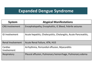
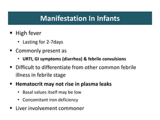
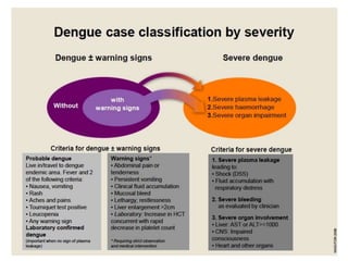
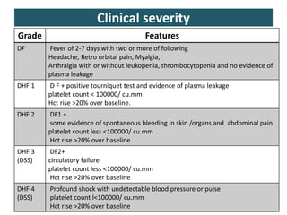
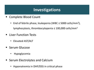
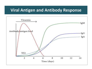
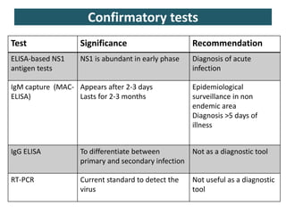
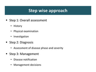
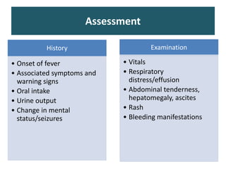
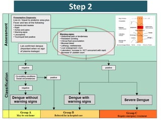
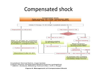
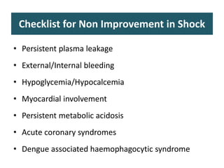
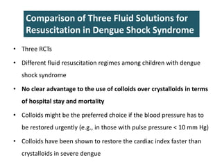
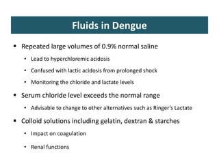
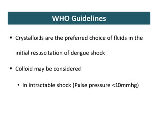
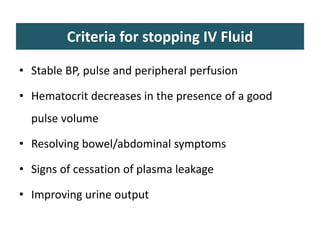
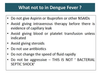
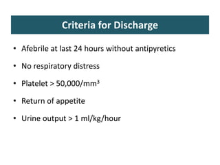
Management of Dengue Fever
Dr. Dinesh
Learning objectives
 Overview of Dengue fever
 Management of Dengue fever
• Practical aspects
• Current guidelines and evidence
Introduction
• Acute systemic viral disease
• Established itself globally in both endemic & epidemic
transmission cycles
Burden of dengue
 40% of the world’s population is at risk
 390 million dengue infections per year
 An estimated 500 000 people with severe dengue
require hospitalization each year and about 2.5% of
those affected die
WHO South-East Asia Journal of Public Health | January-
March 2014
Nature | vol 496 | 25 april 2013
Indian scenario
 36 states/UTs, 35 (all except Lakshadweep) have
reported dengue cases during the last two decades
 Case fatality ratio (CFR – deaths per 100 cases) has
declined from 3.3% in 1996 to 0.3% in 2013
The Virus and the vector
 Single stranded RNA virus
• Flaviridae family
 4 serotypes
• DENV-1, DENV-2, DENV-3, and DENV-4
Genetically & antigenically different
• Infection with one serotype confers life long immunity to that
serotype only.
• Vector: female Aedes aegypti, daytime feeder, eggs and
larvae in artificial water containers.
Pathogenesis
Pathogenesis
Endothelium is the main site
Pathophysiology of bleeding
 Thrombocytopenia
 Coagulation pathway defect
• Decrease fibrinogen level
• Increase level of fibrinogen degradation product (FDP)
• Increase level of D-Dimer
• Consumptive coagulopathy (activation of mononuclear
phagocytes)
• Sequestration of platelets
Symptoms of Dengue fever
 Clinical features
• Fever : mild to moderate (difficult to differentiate from any
other viral fevers in the initial days)
• Headache
• Retro-orbital pain
• Myalgia
• Arthralgia
• Rash
• Hemorrhagic manifestations
Skin bleed, Malena, Hematemesis, Hematuria
Clinical features
 Severe manifestation
• Evidence of plasma leakage
• Shock
• Bleeding
• Organ manifestation
 Life threatening bleeding with out any evidence of
capillary leakage/hemo-concentration
 Multiple organ involvement without bleeding or shock
System Atypical Manifestations
CNS Involvement Encephalopathy, Encephalitis, IC Bleed, Febrile seizures
GI Involvement Acute hepatitis, Cholecystitis, Cholangitis, Acute Pancreatitis,
Renal Involvement Acute Renal Failure, ATN, HUS
Cardiac
Involvement
Arrhythmia, Pericardial effusion, Myocarditis
Respiratory Pleural effusion, Pulmonary hemorrhage, Pulmonary edema
Expanded Dengue Syndrome
Manifestation In Infants
 High fever
• Lasting for 2-7days
 Commonly present as
• URTI, GI symptoms (diarrhea) & febrile convulsions
 Difficult to differentiate from other common febrile
illness in febrile stage
 Hematocrit may not rise in plasma leaks
• Basal values itself may be low
• Concomitant iron deficiency
 Liver involvement commoner
Clinical severity
Grade Features
DF Fever of 2-7 days with two or more of following
Headache, Retro orbital pain, Myalgia,
Arthralgia with or without leukopenia, thrombocytopenia and no evidence of
plasma leakage
DHF 1 D F + positive tourniquet test and evidence of plasma leakage
platelet count < 100000/ cu.mm
Hct rise >20% over baseline.
DHF 2 DF1 +
some evidence of spontaneous bleeding in skin /organs and abdominal pain
platelet count less <100000/ cu.mm
Hct rise >20% over baseline
DHF 3
(DSS)
DF2+
circulatory failure
platelet count less <100000/ cu.mm
Hct rise >20% over baseline
DHF 4
(DSS)
Profound shock with undetectable blood pressure or pulse
platelet count l<100000/ cu.mm
Hct rise >20% over baseline
Natural course of dengue
Critical phase:
• 1-4days after
defervesence
• Plasma leakage and
hemoconcentration
• Pleural effusion and
ascites
• Organ dysfunction
• Metabolic
abnormalities
Dengue classification
Investigations
 Complete Blood Count
• End of febrile phase, leukopenia (WBC ≤ 5000 cells/mm3),
lymphocytosis, thrombocytopenia ≤ 100,000 cells/mm3
 Liver Function Tests
• Elevated AST/ALT
 Serum Glucose
• Hypoglycemia
 Serum Electrolytes and Calcium
• Hyponatremia in DHF/DSS in critical phase
 BUN, Creatinine
• Severity of illness
 Coagulation Profile
• Prolonged partial thromboplastin time (PTT) in 50% of
DHF/DSS
• Prolonged thrombin time (TT) in 10%-20%
 Chest X-ray
• Pleural effusion
Investigations
Viral Antigen and Antibody Response
Test Significance Recommendation
ELISA-based NS1
antigen tests
NS1 is abundant in early phase Diagnosis of acute
infection
IgM capture (MAC-
ELISA)
Appears after 2-3 days
Lasts for 2-3 months
Epidemiological
surveillance in non
endemic area
Diagnosis >5 days of
illness
IgG ELISA To differentiate between
primary and secondary infection
Not as a diagnostic tool
RT-PCR Current standard to detect the
virus
Not useful as a diagnostic
tool
Confirmatory tests
Rapid Diagnostic Tests
• NS1 And IgM/IgG Antibodies
• Quick
• 100-200 Rupees Per Kit
• Not Recommended In National
Vector Borne Disease Control
Program
NVBDCP Recommendations
 First 5 days (ELISA based) NS1 based assays
 >5 days (ELISA based ) IgM based assays
Step wise approach
 Step 1: Overall assessment
• History
• Physical examination
• Investigation
 Step 2: Diagnosis
• Assessment of disease phase and severity
 Step 3: Management
• Disease notification
• Management decisions
History
• Onset of fever
• Associated symptoms and
warning signs
• Oral intake
• Urine output
• Change in mental
status/seizures
Examination
• Vitals
• Respiratory
distress/effusion
• Abdominal tenderness,
hepatomegaly, ascites
• Rash
• Bleeding manifestations
Assessment
Step 2
Group A: Home based care
 Patients who do not have warning signs
 Able to
• Tolerate adequate volumes of oral fluids
• Pass urine at least once every 6 hours
• Don’t belong to high risk category
Group A
 Advice for
• Adequate rest & fluid intake
• Paracetamol, 10-15mg/kg (3 gram max. Per day)
• Patients with >3 days fever should be called daily for review
until they are out of critical phase
• To monitor at home
Temperature pattern
Volume of fluid intake
Urine output volume and frequency
Group A: Monitoring
 Daily review for disease progression
• Decreasing WBC, Rise of hematocrit and fall in platelets
count.
• Defervescence
• Warning signs (until out of critical period)
• Advice for immediate return to hospital if development of
any warning signs
• Written advice of management
Group B: In Hospital Care
 With warning signs/Risk factors
• Obtain a reference HB/TLC/Hematocrit (Hct)
• Isotonic solutions (0.9% saline, RL)
5-7ml/kg for 1-2 hrs
Then 3-5ml/kg for 2-4hrs and then 2-3ml/kg or less as
per response
IV fluids usually needed only for 24-48hrs
• Reassess clinical signs and Hct
 Target Urine output- 0.5ml/kg/hr
Group C: Severe Dengue
• With compensated shock
• With hypotensive shock
Compensated shock
Decompensated shock
Checklist for Non Improvement in Shock
• Persistent plasma leakage
• External/Internal bleeding
• Hypoglycemia/Hypocalcemia
• Myocardial involvement
• Persistent metabolic acidosis
• Acute coronary syndromes
• Dengue associated haemophagocytic syndrome
Which fluid ?
• Crystalloids (NS/RL) or Colloids
• Three RCTs
• Different fluid resuscitation regimes among children with dengue
shock syndrome
• No clear advantage to the use of colloids over crystalloids in terms
of hospital stay and mortality
• Colloids might be the preferred choice if the blood pressure has to
be restored urgently (e.g., in those with pulse pressure < 10 mm Hg)
• Colloids have been shown to restore the cardiac index faster than
crystalloids in severe dengue
Comparison of Three Fluid Solutions for
Resuscitation in Dengue Shock Syndrome
Fluids in Dengue
 Repeated large volumes of 0.9% normal saline
• Lead to hyperchloremic acidosis
• Confused with lactic acidosis from prolonged shock
• Monitoring the chloride and lactate levels
 Serum chloride level exceeds the normal range
• Advisable to change to other alternatives such as Ringer’s Lactate
 Colloid solutions including gelatin, dextran & starches
• Impact on coagulation
• Renal functions
WHO Guidelines
 Crystalloids are the preferred choice of fluids in the
initial resuscitation of dengue shock
 Colloid may be considered
• In intractable shock (Pulse pressure <10mmhg)
Criteria for stopping IV Fluid
• Stable BP, pulse and peripheral perfusion
• Hematocrit decreases in the presence of a good
pulse volume
• Resolving bowel/abdominal symptoms
• Signs of cessation of plasma leakage
• Improving urine output
Who are at risk for bleeding ?
 Profound/prolonged/refractory shock
 MODS
 Severe and persistent metabolic acidosis
 H/O NSAIDs intake
 Pre-existing peptic ulcer disease
 Intramuscular injection
Platelet Transfusion: Threshold
 Significant clinical bleeding
 Prolonged shock with coagulopathy
 Prophylactic:
• <10,000/mm3 in absence of clinical manifestations
What not to in Dengue Fever ?
Criteria for Discharge
• Afebrile at last 24 hours without antipyretics
• No respiratory distress
• Platelet > 50,000/mm3
• Return of appetite
• Urine output > 1 ml/kg/hour
DENGVAXIA (DENGUE VACCINE)
• The first dengue vaccine, DENGVAXIA (CYD-TDV) by
Sanofi Pasteur, was first registered in Mexico in
December, 2015.
• Live attenuated tetravalent dengue vaccine that has
been evaluated as a 3-dose series on a 0/6/12 month
schedule.
• It has been registered for use in individuals 9-45 years
of age living in endemic areas.
THANK YOU
DENGUE ENCEPHALITIS
• The spectrum of neurological manifestations seen
in dengue has been classified into 3 categories.
1. Due to neurotropic effect of the virus –
encephalitis, meningitis, myositis and myelitis.
2.Due to the systemic complications of infection- encephalopathy,
stroke and hypokalemic paralysis.
3.Due to post-infectious complications- encephalomyelitis, optic
neuritis and Guillain Barré syndrome.
DENGUE ENCEPHALITIS
The main symptoms of dengue encephalitis are
• headache,
• Seizures
• Altered consciousness
• Typical symptoms of dengue fever like
myalgias, rash and
bleeding are seen in less than 50% of
encephalitis cases.
Diagnosis
The criteria for dengue encephalitis are:
i) fever;
ii) acute signs of cerebral involvement;
iii) presence of anti-dengue IgM antibodies or
dengue genomic material in the serum and/or
cerebrospinal fluid;
iv) exclusion of other causes of viral encephalitis
and encephalopathy
Neuroimaging
MRI is the modality of choice which shows-
• cerebral edema,
• white matter changes,
• necrosis
• brain atrophy.
• Encephalitis features in brain (Hyperintense
areas) can be seen in global pallidus,temporal
lobes ,thalamus hippocampus,pons and spinal
cord.

Dengue ppt Aiims

  • 1.
    Management of DengueFever Dr. Dinesh
  • 2.
    Learning objectives  Overviewof Dengue fever  Management of Dengue fever • Practical aspects • Current guidelines and evidence
  • 3.
    Introduction • Acute systemicviral disease • Established itself globally in both endemic & epidemic transmission cycles
  • 4.
    Burden of dengue 40% of the world’s population is at risk  390 million dengue infections per year  An estimated 500 000 people with severe dengue require hospitalization each year and about 2.5% of those affected die WHO South-East Asia Journal of Public Health | January- March 2014 Nature | vol 496 | 25 april 2013
  • 5.
    Indian scenario  36states/UTs, 35 (all except Lakshadweep) have reported dengue cases during the last two decades  Case fatality ratio (CFR – deaths per 100 cases) has declined from 3.3% in 1996 to 0.3% in 2013
  • 6.
    The Virus andthe vector  Single stranded RNA virus • Flaviridae family  4 serotypes • DENV-1, DENV-2, DENV-3, and DENV-4 Genetically & antigenically different • Infection with one serotype confers life long immunity to that serotype only. • Vector: female Aedes aegypti, daytime feeder, eggs and larvae in artificial water containers.
  • 7.
  • 8.
  • 9.
    Pathophysiology of bleeding Thrombocytopenia  Coagulation pathway defect • Decrease fibrinogen level • Increase level of fibrinogen degradation product (FDP) • Increase level of D-Dimer • Consumptive coagulopathy (activation of mononuclear phagocytes) • Sequestration of platelets
  • 10.
    Symptoms of Denguefever  Clinical features • Fever : mild to moderate (difficult to differentiate from any other viral fevers in the initial days) • Headache • Retro-orbital pain • Myalgia • Arthralgia • Rash • Hemorrhagic manifestations Skin bleed, Malena, Hematemesis, Hematuria
  • 11.
    Clinical features  Severemanifestation • Evidence of plasma leakage • Shock • Bleeding • Organ manifestation  Life threatening bleeding with out any evidence of capillary leakage/hemo-concentration  Multiple organ involvement without bleeding or shock
  • 12.
    System Atypical Manifestations CNSInvolvement Encephalopathy, Encephalitis, IC Bleed, Febrile seizures GI Involvement Acute hepatitis, Cholecystitis, Cholangitis, Acute Pancreatitis, Renal Involvement Acute Renal Failure, ATN, HUS Cardiac Involvement Arrhythmia, Pericardial effusion, Myocarditis Respiratory Pleural effusion, Pulmonary hemorrhage, Pulmonary edema Expanded Dengue Syndrome
  • 13.
    Manifestation In Infants High fever • Lasting for 2-7days  Commonly present as • URTI, GI symptoms (diarrhea) & febrile convulsions  Difficult to differentiate from other common febrile illness in febrile stage  Hematocrit may not rise in plasma leaks • Basal values itself may be low • Concomitant iron deficiency  Liver involvement commoner
  • 15.
    Clinical severity Grade Features DFFever of 2-7 days with two or more of following Headache, Retro orbital pain, Myalgia, Arthralgia with or without leukopenia, thrombocytopenia and no evidence of plasma leakage DHF 1 D F + positive tourniquet test and evidence of plasma leakage platelet count < 100000/ cu.mm Hct rise >20% over baseline. DHF 2 DF1 + some evidence of spontaneous bleeding in skin /organs and abdominal pain platelet count less <100000/ cu.mm Hct rise >20% over baseline DHF 3 (DSS) DF2+ circulatory failure platelet count less <100000/ cu.mm Hct rise >20% over baseline DHF 4 (DSS) Profound shock with undetectable blood pressure or pulse platelet count l<100000/ cu.mm Hct rise >20% over baseline
  • 16.
    Natural course ofdengue Critical phase: • 1-4days after defervesence • Plasma leakage and hemoconcentration • Pleural effusion and ascites • Organ dysfunction • Metabolic abnormalities
  • 17.
  • 18.
    Investigations  Complete BloodCount • End of febrile phase, leukopenia (WBC ≤ 5000 cells/mm3), lymphocytosis, thrombocytopenia ≤ 100,000 cells/mm3  Liver Function Tests • Elevated AST/ALT  Serum Glucose • Hypoglycemia  Serum Electrolytes and Calcium • Hyponatremia in DHF/DSS in critical phase
  • 19.
     BUN, Creatinine •Severity of illness  Coagulation Profile • Prolonged partial thromboplastin time (PTT) in 50% of DHF/DSS • Prolonged thrombin time (TT) in 10%-20%  Chest X-ray • Pleural effusion Investigations
  • 20.
    Viral Antigen andAntibody Response
  • 21.
    Test Significance Recommendation ELISA-basedNS1 antigen tests NS1 is abundant in early phase Diagnosis of acute infection IgM capture (MAC- ELISA) Appears after 2-3 days Lasts for 2-3 months Epidemiological surveillance in non endemic area Diagnosis >5 days of illness IgG ELISA To differentiate between primary and secondary infection Not as a diagnostic tool RT-PCR Current standard to detect the virus Not useful as a diagnostic tool Confirmatory tests
  • 22.
    Rapid Diagnostic Tests •NS1 And IgM/IgG Antibodies • Quick • 100-200 Rupees Per Kit • Not Recommended In National Vector Borne Disease Control Program
  • 23.
    NVBDCP Recommendations  First5 days (ELISA based) NS1 based assays  >5 days (ELISA based ) IgM based assays
  • 24.
    Step wise approach Step 1: Overall assessment • History • Physical examination • Investigation  Step 2: Diagnosis • Assessment of disease phase and severity  Step 3: Management • Disease notification • Management decisions
  • 25.
    History • Onset offever • Associated symptoms and warning signs • Oral intake • Urine output • Change in mental status/seizures Examination • Vitals • Respiratory distress/effusion • Abdominal tenderness, hepatomegaly, ascites • Rash • Bleeding manifestations Assessment
  • 26.
  • 27.
    Group A: Homebased care  Patients who do not have warning signs  Able to • Tolerate adequate volumes of oral fluids • Pass urine at least once every 6 hours • Don’t belong to high risk category
  • 28.
    Group A  Advicefor • Adequate rest & fluid intake • Paracetamol, 10-15mg/kg (3 gram max. Per day) • Patients with >3 days fever should be called daily for review until they are out of critical phase • To monitor at home Temperature pattern Volume of fluid intake Urine output volume and frequency
  • 29.
    Group A: Monitoring Daily review for disease progression • Decreasing WBC, Rise of hematocrit and fall in platelets count. • Defervescence • Warning signs (until out of critical period) • Advice for immediate return to hospital if development of any warning signs • Written advice of management
  • 30.
    Group B: InHospital Care  With warning signs/Risk factors • Obtain a reference HB/TLC/Hematocrit (Hct) • Isotonic solutions (0.9% saline, RL) 5-7ml/kg for 1-2 hrs Then 3-5ml/kg for 2-4hrs and then 2-3ml/kg or less as per response IV fluids usually needed only for 24-48hrs • Reassess clinical signs and Hct  Target Urine output- 0.5ml/kg/hr
  • 32.
    Group C: SevereDengue • With compensated shock • With hypotensive shock
  • 33.
  • 34.
  • 35.
    Checklist for NonImprovement in Shock • Persistent plasma leakage • External/Internal bleeding • Hypoglycemia/Hypocalcemia • Myocardial involvement • Persistent metabolic acidosis • Acute coronary syndromes • Dengue associated haemophagocytic syndrome
  • 36.
    Which fluid ? •Crystalloids (NS/RL) or Colloids
  • 37.
    • Three RCTs •Different fluid resuscitation regimes among children with dengue shock syndrome • No clear advantage to the use of colloids over crystalloids in terms of hospital stay and mortality • Colloids might be the preferred choice if the blood pressure has to be restored urgently (e.g., in those with pulse pressure < 10 mm Hg) • Colloids have been shown to restore the cardiac index faster than crystalloids in severe dengue Comparison of Three Fluid Solutions for Resuscitation in Dengue Shock Syndrome
  • 38.
    Fluids in Dengue Repeated large volumes of 0.9% normal saline • Lead to hyperchloremic acidosis • Confused with lactic acidosis from prolonged shock • Monitoring the chloride and lactate levels  Serum chloride level exceeds the normal range • Advisable to change to other alternatives such as Ringer’s Lactate  Colloid solutions including gelatin, dextran & starches • Impact on coagulation • Renal functions
  • 39.
    WHO Guidelines  Crystalloidsare the preferred choice of fluids in the initial resuscitation of dengue shock  Colloid may be considered • In intractable shock (Pulse pressure <10mmhg)
  • 40.
    Criteria for stoppingIV Fluid • Stable BP, pulse and peripheral perfusion • Hematocrit decreases in the presence of a good pulse volume • Resolving bowel/abdominal symptoms • Signs of cessation of plasma leakage • Improving urine output
  • 41.
    Who are atrisk for bleeding ?  Profound/prolonged/refractory shock  MODS  Severe and persistent metabolic acidosis  H/O NSAIDs intake  Pre-existing peptic ulcer disease  Intramuscular injection
  • 42.
    Platelet Transfusion: Threshold Significant clinical bleeding  Prolonged shock with coagulopathy  Prophylactic: • <10,000/mm3 in absence of clinical manifestations
  • 43.
    What not toin Dengue Fever ?
  • 44.
    Criteria for Discharge •Afebrile at last 24 hours without antipyretics • No respiratory distress • Platelet > 50,000/mm3 • Return of appetite • Urine output > 1 ml/kg/hour
  • 45.
    DENGVAXIA (DENGUE VACCINE) •The first dengue vaccine, DENGVAXIA (CYD-TDV) by Sanofi Pasteur, was first registered in Mexico in December, 2015. • Live attenuated tetravalent dengue vaccine that has been evaluated as a 3-dose series on a 0/6/12 month schedule. • It has been registered for use in individuals 9-45 years of age living in endemic areas.
  • 46.
  • 47.
    DENGUE ENCEPHALITIS • Thespectrum of neurological manifestations seen in dengue has been classified into 3 categories. 1. Due to neurotropic effect of the virus – encephalitis, meningitis, myositis and myelitis. 2.Due to the systemic complications of infection- encephalopathy, stroke and hypokalemic paralysis. 3.Due to post-infectious complications- encephalomyelitis, optic neuritis and Guillain Barré syndrome.
  • 48.
    DENGUE ENCEPHALITIS The mainsymptoms of dengue encephalitis are • headache, • Seizures • Altered consciousness • Typical symptoms of dengue fever like myalgias, rash and bleeding are seen in less than 50% of encephalitis cases.
  • 49.
    Diagnosis The criteria fordengue encephalitis are: i) fever; ii) acute signs of cerebral involvement; iii) presence of anti-dengue IgM antibodies or dengue genomic material in the serum and/or cerebrospinal fluid; iv) exclusion of other causes of viral encephalitis and encephalopathy
  • 50.
    Neuroimaging MRI is themodality of choice which shows- • cerebral edema, • white matter changes, • necrosis • brain atrophy. • Encephalitis features in brain (Hyperintense areas) can be seen in global pallidus,temporal lobes ,thalamus hippocampus,pons and spinal cord.