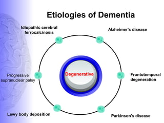
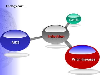
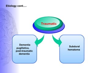
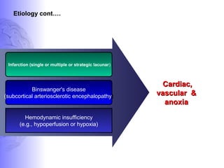
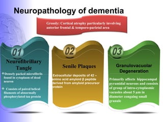
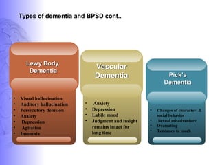
Dr. Santanu Ghosh, MD
Assistant Professor,
Department of Psychiatry
Tripura medical College
What is dementia??
Dementia is defined as a progressive
impairment of cognitive functions
occurring in clear consciousness (that is,
in the absence of delirium)
HISTORY
1 2 3
Dementia = Dementatus
Meaning out of one's mind
Celsus
First used the term
dementia
Oribasius,
first attempts to describe
an etiology beyond old
age
Epidemiology
>65 years
>85 years
(Outpatient Population)
>85 years
(General Population
Chronic Care Facilities
By 2050
5%
15 - 20 %
20 -40 %
50%
• 14 million :Alzheimer's disease
• >18 million people with dementia
Prevalence
Etiologies of Dementia
Degenerative
Alzheimer's disease
Frontotemporal
degeneration
Parkinson's diseaseLewy body deposition
Progressive
supranuclear palsy
Idiopathic cerebral
ferrocalcinosis
Metabolic
Hypercalcemia
Vitamin
deficiency
Endocrinopathies
Uremia
Etiology cont.….
Persistent hemodialysis
Heavy metals
Irradiation
Carbon Monoxide
Anticholinergic Drugs
Drugs
&
toxins
Alcohol
Etiology cont….
Etiology cont….
Cognitive decline
in
schizophrenia
Pseudodementia
of depression
Psychiatric
SyphilisSyphilis
AIDSAIDS
Etiology cont….
InfectionInfection
Prion diseasesPrion diseases
Etiology cont….
Subdural
hematoma
Dementia
pugilistica,
post-traumatic
dementia
Traumatic
Infarction (single or multiple or strategic lacunar)
Binswanger's disease
(subcortical arteriosclerotic encephalopathy)
Hemodynamic insufficiency
(e.g., hypoperfusion or hypoxia)
Cardiac,Cardiac,
vascular &vascular &
anoxiaanoxia
Etiology cont….
Neuropathology of dementia
Densely packed microfibrils
found in cytoplasm of dead
neuron
 Consists of paired helical
filaments of abnormally
phosphorylated tau protein
Primarily affects hippocampal
pyramidal neurons and consists
of group of intra-cytoplasmic
vacuoles about 5 µm in
diameter congaing small
granule
Neurofibrillary
Tangle Senile Plaques Granulovacuolar
Degeneration
Extracellular deposits of 42 –
amino acid amyloid β peptide
derived from amyloid precursor
protein
Grossly: Cortical atrophy particularly involving
anterior frontal & temporo-parietal area
Neuropathology of dementia cont..
1= Congophilic angiopathy consisting of amyloid deposit in the wall of small
arteries
2= Generalized loss of neuronal dendrites
3= Loss of choloinergic neurons nucleus basalis of Meynert
4= Loss of noradrenergic neurons in locus coeruleus, loss of serotonergic neurons
of the dorsal raphe nucleus
Neuropathology
Neurotransmitters in Dementia
Serotonin Dopamine
Glutamine
Acetyl
Choline
Types of dementia and BPSD
Aspontinity, aggression,
wandering
Incontinence, Paranoid
symptoms
Alzheimer Disease
Types of dementia and BPSD cont..
• Visual hallucination
• Auditory hallucination
• Persecutory delusion
• Anxiety
• Depression
• Agitation
• Insomnia
Lewy BodyLewy Body
DementiaDementia
• Changes of character &
social behavior
• Sexual misadventure
• Overeating
• Tendency to touch
Pick’sPick’s
DementiaDementia
• Anxiety
• Depression
• Labile mood
• Judgment and insight
remains intact for
long time
VascularVascular
DementiaDementia
Assessment
Other medical
conditions
Behavioral problems,
psychotic symptoms, or
depression
Mini Mental status Examination
• Severe : 0-10
• Moderate : 11-17
• Mild : 18-23
• Normal : 24-30
Daily function
•Feeding
•Bathing
•dressing,
• Mobility
• Manage finances and
medications
1. ALZHEIMERS DISEASE
Alzheimr’s Disease (AD)
• Is the most common cause of dementia.
• > 90% sporadic , age usually >65yrs.
• <10% familial, age usually <60 yrs.
• Clinically: Initially normal- develops ‘Mild Cognitive
Impairment’ – goes on to develop AD.
• Pathology: AD is characterized by senile plaques,
neurofibrillary tangles, decreased synaptic density, neuron
loss and cerebral atrophy.
sMRI- Alzehimer’s Disease
Assessment of cerebral atrophy of hemisphere
particularly posterior temporal and parietal lobes & specific
anatomic areas like hippocampus and medial temporal
lobe
1. Visual ranking system: Mild / moderate / severe.
2. Quantitative measurement : Linear / area / volumetric*.
Measurements must be adjusted for age, gender and head
size and then referenced to an appropriate control
population.
Among these measurements of hippocampus was most sensitive
marker of pathology of AD early in disease.
Normal ALZHEIMER’S DISEASE
ALZHEIMER’S DISEASENORMAL
MR Perfusion study in Alzheimer’s disease
A characteristic bilateral temporoparietal decrease in blood flow
is noted.
STRUCTURAL ANALYSIS
Amygdala
Hippocampus
Rest of cortex
PET: Reduced uptake of
glucose in temporal lobe.
SPECT/MR perfusion
Reduced perfusion
MR SPECTROSCOPY:
-Decrease in NAA.
-Increase in myoinositol.
Alzheimer’s disease
2. DEMENTIA WITH LEWY BODIES
• It is characterized by
presence of lewy bodies in the
cortical neurons on histology.
• It is 2nd
/ 3rd
most common
cause of dementia in elderly.
• To date, no MRI features
have been identified that to
characterize DLB.
• “THE ABSENCE OF
SIGNIFICANT MEDIAL
TEMPORAL LOBE
ATROPHY” in a elderly
demented patient suggests
DLB etiology rather than AD
etiology.
3. FRONTOTEMPORAL DEMENTIA
• Age of onset: 50-65 yrs.
• Genetically linked to chromosome 3 and 17
• FTD is a term used to describe a family of
neurodegenerative disorders characterized by
degeneration of frontal and temporal lobes.
• The three most common HISTOLOGICALLY
classified(Not radiological) FTD syndromes are
 Pick's disease.
 Frontal-lobe degeneration &
 FTD with amyotrophic lateral sclerosis.
MRI features of FTD
• MR features:
 Severe sharply localized atrophy- bilaterally symmetric-
“knife-blade atrophy.”
 Hyperintense signal in the cortex and underlying white
matter of the affected areas.
• Areas involved: frontal lobe, anterior temporal lobes,
extra pyramidal nuclei especially the caudate nucleus,
insular cortex & anterior corpus callosum.
• Areas spared: Posterior two thirds of the superior
temporal gyrus, occipital lobes, parietal lobes &
perirolandic region
• These MR findings in an appropriate clinical setting
may support the diagnosis of FTD.
Frontotemporal dementia
4. DEMENTIA OF VASCULAR ETIOLOGY
• Dementia due to chronic cerebrovascular disease is
2nd
/ 3rd
most common cause of dementia in elderly(AD
and dementia with Lewy bodies).
• Three main forms are recognized:
 Multi infarct dementia.
 Sub cortical vascular dementia/ Binswanger’s
disease
 Cerebral amyloid angiopathy.
SPECT-Vascular Dementia
• 99mTc-HMPAO
SPECT of the
brain in vascular
dementia shows
multiple patchy
perfusion defects.
5. NORMAL PRESSURE HYDROCEPHALUS
• TRIAD: Dementia + gait disturbance + Urinary incontinence.
• AGE: usually after 60 yrs.
• Theories:
Impaired extraventricluar CSF absorption due to prior
subarachnoid hemorrhage / meningitis.
Decrease white matter tensile strength due to deep white
matter infarction / ischemic changes.
• Three primary MR findings have been described in NPH:
Enlargement of the ventricular system out of proportion to the
subarachnoid space
A prominent periventricular halo and
A prominent CSF flow void in the cerebral aqueduct.
MRS- Dementia
• NAA loss is consistently seen in Alzheimer's disease.
• NAA loss is also seen in Parkinson’s disease &
Huntington’s disease.
• Significantly elevated myoinositol in grey matter of
Alzheimer's disease.
Pharmacological Intervention
Cognitive enhancer
Rivastigmine
Donepezil
Galantamine
Mamentine
Piracetam
4. Neurotonic
Dose: 400-800
mg/day
5. NMDA -receptor
antagonist
Dose: 5 mg OD - 10
mg BD
1. Acetyl
Cholinesterase inhibitor
Dose: 1.5 mg BD to 6 mg BD
2. Acetyl
Cholinesterase
inhibitor
Dose: 5mg - 10 mg
daily
3. Acetyl Cholinesterase
inhibitor
Dose4 mg BD - 12 mg BD
Guidelines for prescribing cholinesterase inhibitors
A. Prescription only for patients-
a) Fulfilling criteria for probable AD
b) Duration of illness being more than 6 months
c) MMSE score more than 10 (i.E., Mild or moderately severe dementia)
B. Three phase evaluation of response-
a) Early (2 weeks) for assessing tolerance and side effects,
b) Later (3 months) for cognitive state,
c) Continued (6 months) for disease state
C. Stop treatment-
a) If early evaluation shows poor tolerance or compliance
b) If deterioration continues at pre-treatment rate after 3-6 months of
treatment
c) If even after reaching maintenance dose accelerating deterioration
continues
Anti-amyloid therapy
Vaccines to prevent amyloid plaque formation :
Under experimental stage
Secretase
Inhibitor
Fibrilogenesis
inhibitor
e.g. cliniquinol
Cholesterine
Newer
drugs
Other drugs
Quetiapine is
safest
Valproate is
preferable
Sertraline is
preferable
Non
benzodiazepines
are preferable
Sedatives
Antidepressants
Mood
stabilisers
Antipsychotics
Non-pharmacological Intervention
I. Environmental modifications
1 2 3
Enhanced Environment
Simulated home environment
with appropriate visual auditory
and olfactory stimuli which
may decrease the chance of
trespassing, exit seeking and
other agitation behaviors
Reduced Stimulation
Environment
Camouflaged doors, neutral
colors and pictures on walls,
no televisions, radios, stereo
players or ringing telephones,
added with a consistent daily
routine, and slow and soft
speech for communication
Natural Environment
Mimic natural surroundings
consisting of recorded
songs of birds, babbling
brooks or small animals,
together with large bright
picture
Environment
Calm, non-taxing environments,
good lighting, prominent placement
of frequently required objects
Soft wall colours, non-skid
flooring contrast between the
wall and floor, handrails are
useful
Clear and repetitive instructions,
visual direction to different rooms
through colour lines and pictures
decrease confusion
without frequent change of furniture
minimum change of caregivers or
staff.
Environmental modifications contd..
II. Social interactions
““simulated presence therapy,”simulated presence therapy,”““simulated presence therapy,”simulated presence therapy,”
Audio or videos containing a relative’s portion of interaction is
played, and pauses are given that allow the patient to respond to
the relative’s questions. Displaying photos and names of
family and friends in the patient’s living area is helpful.
LanguagePet therapyInteraction
Non-pharmacological Intervention cont.
One to one interaction for 30 min
per day for 10 days to be
effective in decreasing verbally
disruptive behaviour.
Interaction in the mother
tongue and regular
intensive interaction help in
reality orientation.
Spending time with pets or
having a pet at home (pet
therapy) decreases agitation and
verbal aggression
III. Minimize the impact of sensory deficits
Provide favourite foods
Devices
Patience
Rapport
Food
Slow and repetitive explanations
reduce confusion and agitation.
Massage and touch, aromatherapy
Minimize the impact of
sensory deficits Corrective
eyeglasses and hearing aids
decrease risk of
disorientation
Non-pharmacological Intervention cont.
IV. Medical and nursing interventions
Music
Therapy
Proper
food
Sleep
Hygiene
Light
Therapy
Frequent
outing
Pain
Management
Personal
Hygiene
Non-pharmacological Intervention cont.
Title in here
Reinforcements
&
Extinction
Stimulus control
Spiritual
&
religious activities
VI. Cognitive activities
Mind gameMind game
Crossword
Puzzles
Chess Su-doku
CARE NACSP ADL
Calming Aggressive
Reactions In Elderly
Nursing Assistant
Communication Skill
Program
Abilities-focused program of
morning care, and training
on activities of daily living
(ADL)
Nonpharmacological Intervention cont.
Dementia-  recent updates

Dementia- recent updates

  • 1.
    Dr. Santanu Ghosh,MD Assistant Professor, Department of Psychiatry Tripura medical College
  • 2.
    What is dementia?? Dementiais defined as a progressive impairment of cognitive functions occurring in clear consciousness (that is, in the absence of delirium)
  • 3.
    HISTORY 1 2 3 Dementia= Dementatus Meaning out of one's mind Celsus First used the term dementia Oribasius, first attempts to describe an etiology beyond old age
  • 4.
    Epidemiology >65 years >85 years (OutpatientPopulation) >85 years (General Population Chronic Care Facilities By 2050 5% 15 - 20 % 20 -40 % 50% • 14 million :Alzheimer's disease • >18 million people with dementia Prevalence
  • 5.
    Etiologies of Dementia Degenerative Alzheimer'sdisease Frontotemporal degeneration Parkinson's diseaseLewy body deposition Progressive supranuclear palsy Idiopathic cerebral ferrocalcinosis
  • 6.
  • 7.
    Persistent hemodialysis Heavy metals Irradiation CarbonMonoxide Anticholinergic Drugs Drugs & toxins Alcohol Etiology cont….
  • 8.
  • 9.
  • 10.
  • 11.
    Infarction (single ormultiple or strategic lacunar) Binswanger's disease (subcortical arteriosclerotic encephalopathy) Hemodynamic insufficiency (e.g., hypoperfusion or hypoxia) Cardiac,Cardiac, vascular &vascular & anoxiaanoxia Etiology cont….
  • 12.
    Neuropathology of dementia Denselypacked microfibrils found in cytoplasm of dead neuron  Consists of paired helical filaments of abnormally phosphorylated tau protein Primarily affects hippocampal pyramidal neurons and consists of group of intra-cytoplasmic vacuoles about 5 µm in diameter congaing small granule Neurofibrillary Tangle Senile Plaques Granulovacuolar Degeneration Extracellular deposits of 42 – amino acid amyloid β peptide derived from amyloid precursor protein Grossly: Cortical atrophy particularly involving anterior frontal & temporo-parietal area
  • 13.
    Neuropathology of dementiacont.. 1= Congophilic angiopathy consisting of amyloid deposit in the wall of small arteries 2= Generalized loss of neuronal dendrites 3= Loss of choloinergic neurons nucleus basalis of Meynert 4= Loss of noradrenergic neurons in locus coeruleus, loss of serotonergic neurons of the dorsal raphe nucleus
  • 14.
  • 15.
    Neurotransmitters in Dementia SerotoninDopamine Glutamine Acetyl Choline
  • 17.
    Types of dementiaand BPSD Aspontinity, aggression, wandering Incontinence, Paranoid symptoms Alzheimer Disease
  • 18.
    Types of dementiaand BPSD cont.. • Visual hallucination • Auditory hallucination • Persecutory delusion • Anxiety • Depression • Agitation • Insomnia Lewy BodyLewy Body DementiaDementia • Changes of character & social behavior • Sexual misadventure • Overeating • Tendency to touch Pick’sPick’s DementiaDementia • Anxiety • Depression • Labile mood • Judgment and insight remains intact for long time VascularVascular DementiaDementia
  • 20.
    Assessment Other medical conditions Behavioral problems, psychoticsymptoms, or depression Mini Mental status Examination • Severe : 0-10 • Moderate : 11-17 • Mild : 18-23 • Normal : 24-30 Daily function •Feeding •Bathing •dressing, • Mobility • Manage finances and medications
  • 22.
  • 23.
    Alzheimr’s Disease (AD) •Is the most common cause of dementia. • > 90% sporadic , age usually >65yrs. • <10% familial, age usually <60 yrs. • Clinically: Initially normal- develops ‘Mild Cognitive Impairment’ – goes on to develop AD. • Pathology: AD is characterized by senile plaques, neurofibrillary tangles, decreased synaptic density, neuron loss and cerebral atrophy.
  • 24.
    sMRI- Alzehimer’s Disease Assessmentof cerebral atrophy of hemisphere particularly posterior temporal and parietal lobes & specific anatomic areas like hippocampus and medial temporal lobe 1. Visual ranking system: Mild / moderate / severe. 2. Quantitative measurement : Linear / area / volumetric*. Measurements must be adjusted for age, gender and head size and then referenced to an appropriate control population.
  • 25.
    Among these measurementsof hippocampus was most sensitive marker of pathology of AD early in disease. Normal ALZHEIMER’S DISEASE
  • 26.
  • 27.
    MR Perfusion studyin Alzheimer’s disease A characteristic bilateral temporoparietal decrease in blood flow is noted.
  • 28.
    STRUCTURAL ANALYSIS Amygdala Hippocampus Rest ofcortex PET: Reduced uptake of glucose in temporal lobe. SPECT/MR perfusion Reduced perfusion MR SPECTROSCOPY: -Decrease in NAA. -Increase in myoinositol. Alzheimer’s disease
  • 29.
    2. DEMENTIA WITHLEWY BODIES
  • 30.
    • It ischaracterized by presence of lewy bodies in the cortical neurons on histology. • It is 2nd / 3rd most common cause of dementia in elderly. • To date, no MRI features have been identified that to characterize DLB. • “THE ABSENCE OF SIGNIFICANT MEDIAL TEMPORAL LOBE ATROPHY” in a elderly demented patient suggests DLB etiology rather than AD etiology.
  • 31.
  • 32.
    • Age ofonset: 50-65 yrs. • Genetically linked to chromosome 3 and 17 • FTD is a term used to describe a family of neurodegenerative disorders characterized by degeneration of frontal and temporal lobes. • The three most common HISTOLOGICALLY classified(Not radiological) FTD syndromes are  Pick's disease.  Frontal-lobe degeneration &  FTD with amyotrophic lateral sclerosis.
  • 33.
    MRI features ofFTD • MR features:  Severe sharply localized atrophy- bilaterally symmetric- “knife-blade atrophy.”  Hyperintense signal in the cortex and underlying white matter of the affected areas. • Areas involved: frontal lobe, anterior temporal lobes, extra pyramidal nuclei especially the caudate nucleus, insular cortex & anterior corpus callosum. • Areas spared: Posterior two thirds of the superior temporal gyrus, occipital lobes, parietal lobes & perirolandic region • These MR findings in an appropriate clinical setting may support the diagnosis of FTD.
  • 35.
  • 36.
    4. DEMENTIA OFVASCULAR ETIOLOGY
  • 37.
    • Dementia dueto chronic cerebrovascular disease is 2nd / 3rd most common cause of dementia in elderly(AD and dementia with Lewy bodies). • Three main forms are recognized:  Multi infarct dementia.  Sub cortical vascular dementia/ Binswanger’s disease  Cerebral amyloid angiopathy.
  • 39.
    SPECT-Vascular Dementia • 99mTc-HMPAO SPECTof the brain in vascular dementia shows multiple patchy perfusion defects.
  • 40.
    5. NORMAL PRESSUREHYDROCEPHALUS
  • 41.
    • TRIAD: Dementia+ gait disturbance + Urinary incontinence. • AGE: usually after 60 yrs. • Theories: Impaired extraventricluar CSF absorption due to prior subarachnoid hemorrhage / meningitis. Decrease white matter tensile strength due to deep white matter infarction / ischemic changes. • Three primary MR findings have been described in NPH: Enlargement of the ventricular system out of proportion to the subarachnoid space A prominent periventricular halo and A prominent CSF flow void in the cerebral aqueduct.
  • 43.
    MRS- Dementia • NAAloss is consistently seen in Alzheimer's disease. • NAA loss is also seen in Parkinson’s disease & Huntington’s disease. • Significantly elevated myoinositol in grey matter of Alzheimer's disease.
  • 45.
  • 46.
    Cognitive enhancer Rivastigmine Donepezil Galantamine Mamentine Piracetam 4. Neurotonic Dose:400-800 mg/day 5. NMDA -receptor antagonist Dose: 5 mg OD - 10 mg BD 1. Acetyl Cholinesterase inhibitor Dose: 1.5 mg BD to 6 mg BD 2. Acetyl Cholinesterase inhibitor Dose: 5mg - 10 mg daily 3. Acetyl Cholinesterase inhibitor Dose4 mg BD - 12 mg BD
  • 47.
    Guidelines for prescribingcholinesterase inhibitors A. Prescription only for patients- a) Fulfilling criteria for probable AD b) Duration of illness being more than 6 months c) MMSE score more than 10 (i.E., Mild or moderately severe dementia) B. Three phase evaluation of response- a) Early (2 weeks) for assessing tolerance and side effects, b) Later (3 months) for cognitive state, c) Continued (6 months) for disease state C. Stop treatment- a) If early evaluation shows poor tolerance or compliance b) If deterioration continues at pre-treatment rate after 3-6 months of treatment c) If even after reaching maintenance dose accelerating deterioration continues
  • 48.
    Anti-amyloid therapy Vaccines toprevent amyloid plaque formation : Under experimental stage Secretase Inhibitor Fibrilogenesis inhibitor e.g. cliniquinol Cholesterine Newer drugs
  • 49.
    Other drugs Quetiapine is safest Valproateis preferable Sertraline is preferable Non benzodiazepines are preferable Sedatives Antidepressants Mood stabilisers Antipsychotics
  • 50.
    Non-pharmacological Intervention I. Environmentalmodifications 1 2 3 Enhanced Environment Simulated home environment with appropriate visual auditory and olfactory stimuli which may decrease the chance of trespassing, exit seeking and other agitation behaviors Reduced Stimulation Environment Camouflaged doors, neutral colors and pictures on walls, no televisions, radios, stereo players or ringing telephones, added with a consistent daily routine, and slow and soft speech for communication Natural Environment Mimic natural surroundings consisting of recorded songs of birds, babbling brooks or small animals, together with large bright picture
  • 51.
    Environment Calm, non-taxing environments, goodlighting, prominent placement of frequently required objects Soft wall colours, non-skid flooring contrast between the wall and floor, handrails are useful Clear and repetitive instructions, visual direction to different rooms through colour lines and pictures decrease confusion without frequent change of furniture minimum change of caregivers or staff. Environmental modifications contd..
  • 52.
    II. Social interactions ““simulatedpresence therapy,”simulated presence therapy,”““simulated presence therapy,”simulated presence therapy,” Audio or videos containing a relative’s portion of interaction is played, and pauses are given that allow the patient to respond to the relative’s questions. Displaying photos and names of family and friends in the patient’s living area is helpful. LanguagePet therapyInteraction Non-pharmacological Intervention cont. One to one interaction for 30 min per day for 10 days to be effective in decreasing verbally disruptive behaviour. Interaction in the mother tongue and regular intensive interaction help in reality orientation. Spending time with pets or having a pet at home (pet therapy) decreases agitation and verbal aggression
  • 53.
    III. Minimize theimpact of sensory deficits Provide favourite foods Devices Patience Rapport Food Slow and repetitive explanations reduce confusion and agitation. Massage and touch, aromatherapy Minimize the impact of sensory deficits Corrective eyeglasses and hearing aids decrease risk of disorientation Non-pharmacological Intervention cont.
  • 54.
    IV. Medical andnursing interventions Music Therapy Proper food Sleep Hygiene Light Therapy Frequent outing Pain Management Personal Hygiene Non-pharmacological Intervention cont.
  • 55.
    Title in here Reinforcements & Extinction Stimuluscontrol Spiritual & religious activities
  • 56.
    VI. Cognitive activities MindgameMind game Crossword Puzzles Chess Su-doku
  • 57.
    CARE NACSP ADL CalmingAggressive Reactions In Elderly Nursing Assistant Communication Skill Program Abilities-focused program of morning care, and training on activities of daily living (ADL) Nonpharmacological Intervention cont.