
Recommended
More Related Content
Similar to Gastrointestinal assessment in a patients
Similar to Gastrointestinal assessment in a patients (20)
More from SachinDwivedi57
More from SachinDwivedi57 (20)
Recently uploaded
Recently uploaded (20)
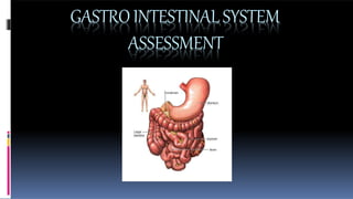
Gastrointestinal assessment in a patients
- 2. HISTORYCOLLECTION Past health history Medications Surgery or other treatments Health management pattern Nutritional pattern Elimination pattern Sleep pattern
- 3. MOUTH INSPECTION For colour, Symmetry, cracking, cyanosis, ulcers, Buccal mucosa and lesions.
- 4. The pharynx is inspected by head tilting, and Depressing the Tongue. • To Inspect uvula, Tonsils, soft palate ask the patient to say ‘ah’. The Uvula and soft palate should rise and remain in the midline. .
- 5. PALPATION • For nodules, ulcers and areas of tenderness,should be noted. • Dentures to be removed during oral examination.
- 6. ABDOMEN INSPECTION AUSCULTATION PERCUSSION PALPATION
- 7. ABDOMINAL QUADRANTS ABDOMINAL REGIONS Four quadrants perpendicular line by a from the sternum to the pubic line and a horizontal line across the abdomen at the umblicus It has umblicus, hypogastric epigastic regions nine regions but and are commonly addressed.
- 8. 9 REGIONS OF ABDOMEN
- 9. INSPECTION Assess the abdomen for skin changes (colour, texture, scars, striae, dilated veins, rashes and lesions). Inspect umbilicus for symmetry, contour, masses. Look across the abdomen for peristalsis, is not visible normally in adult but may visible in thin person.
- 10. AUSCULTATION It is done immediately after the inspection to prevent alteration in bowel sounds. It includes listening for increased or decreased bowel sounds and vascular sounds. Gently warm up the stethoscope in the hands to prevent abdominal muscle contraction.
- 11. The diaphragm of the stethoscope Is used to auscultate high pitched sounds and Bell to detect low pitched sounds. Normally 5 to 35 times the bowel sound is heard per minute like high pitched clicks or gurgles. Listen in the epigastrium and in all four quadrants for bowel sounds for 2 to 5 minutes
- 12. Normally the sounds will be high pitched and gurgling and loud gurgles indicate hyperperistalsis. If the intestines are under tension, such as intestinal obstruction the bowel sounds will be rushes and tinkling Listen for decreased or absence of bowel sounds. Present, absent, increased, decreased, high pitched, tinkling, gurgling and rushing are the terms used to describe the bowel sounds.
- 13. PERCUSSION It is done to determine the presence of fluid, distention and masses. The presence of air produces a higher pitched, hollow sound it is termed as tympany. (Gas in GI) The presence of fluid or masses a short, high pitched sound with little resonance it is termed as dullness. (Organ, fluid or Feces) Hyperresonant- Air (Lungs)
- 14. Liver Percussion should start below the umbilicus in the right mid clavicular line and percuss upward until dullness is heard, thus determine the lower border of the liver. From the nipple line in the mid clavicular line and percuss downward between the ribs to the area of dullness indicating the upper boarder of the liver.
- 15. The height or vertical space between the two areas should be measured to determine the size of the liver. The normal height in the midclavicular line is 2.4 to 5 inches (6 to 12cm). 8cm
- 16. PALPATION The pads of the fingertips is used for palpation depressing the abdominal wall up to 0.4inch (1cm). Light palpation is used to detect tenderness, muscular resistance, masses and swelling. Smooth movements should be used and all quadrants need to be Palpated.
- 17. PALPATION • Deep palpation is used to delineate abdominal organs and masses, the pads of finger tips used to press more deeply in all the quadrants. • During palpation note the location size, shape and presence of tenderness. • The patients facial expression should be noted to know the non-verbal cues of pain or discomfort.
- 18. To palpate liver the left hand is placed behind patient to support the right eleventh and twelfth ribs, then press the left hand forward and place the right hand on the patient’s right abdomen lateral to rectus muscle, gently press in and up.
- 19. To palpate spleen, move to left side of the patient, place right hand under the patient and supports and press the patient’s lower rib cage forward. Then the left hand in the left coastal margin and press it in toward the spleen and ask the patient to breathe deeply, if the spleen is enlarged will be felt by the finger tips. Normally spleen palpated in Mid axillary line but abnormally anterior axillary line.
- 20. RECTUM ANDANUS Inspect the perianal and anal areas for texture, and rashes, colour, lumps scars, erythema, fissures and external hemorrhoids. If there is any lump or unusual areas should be palpated with a gloved hand.
- 21. Palpation of Rectum & Anus For the digital examination of rectum, the gloved lubricated index finger is placed against the anus while the patients strains. Then as the sphincters relaxes, the finger is inserted as much as possible and is pointed towards umbilicus and surfaces are palpated. A sample for stool can be taken and check for occulted blood.
- 22. THANK YOU