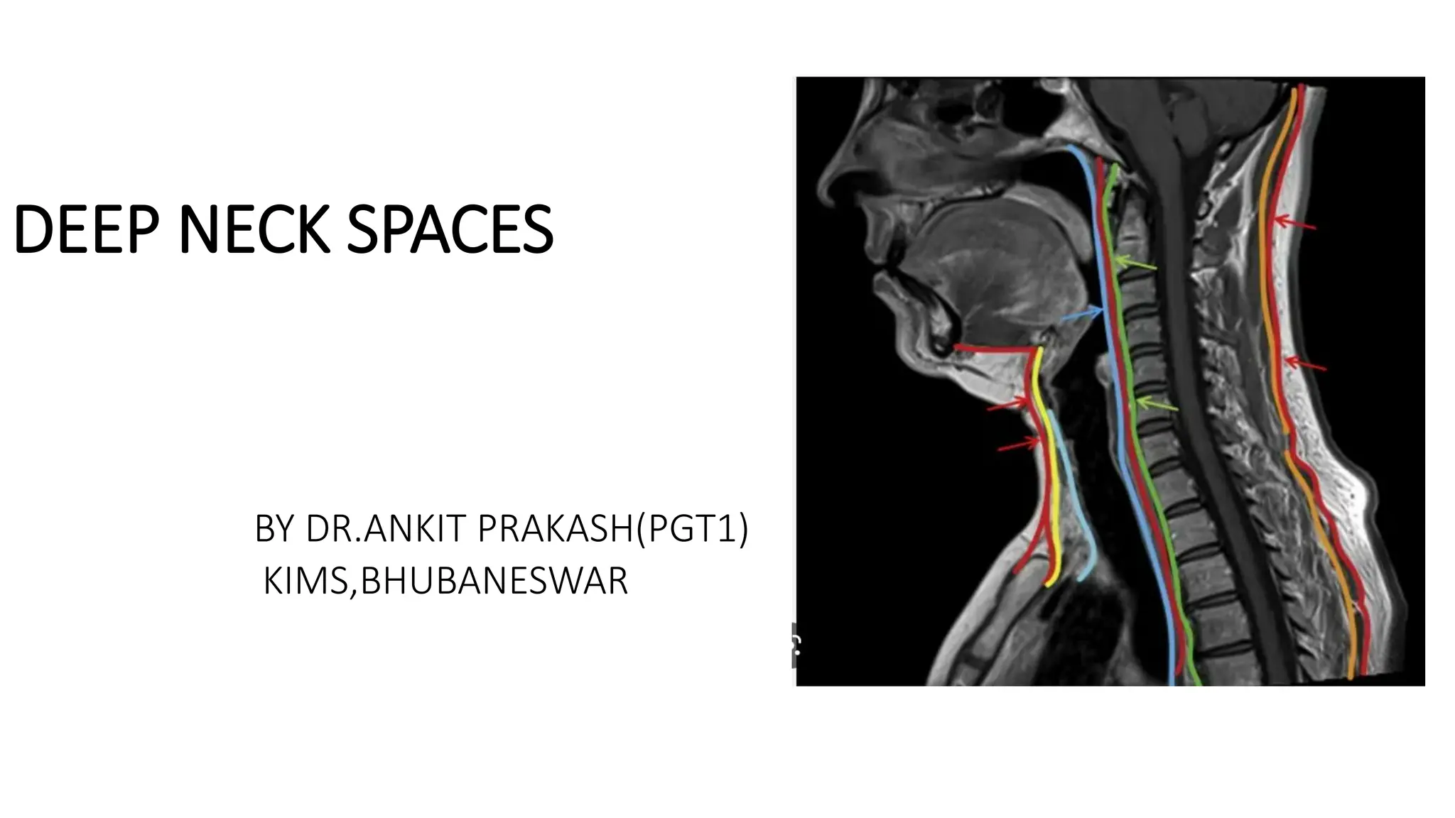
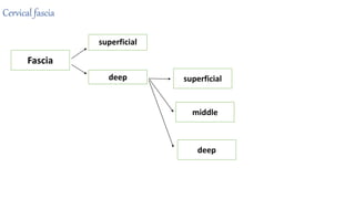
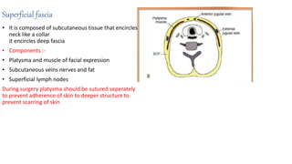
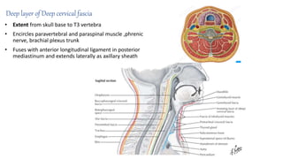
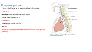
The document summarizes the anatomy of the deep neck spaces. It describes the layers of cervical fascia that divide the neck into compartments containing specific structures. Key points include:
- There are 3 layers of deep cervical fascia that invest the neck muscles and organs.
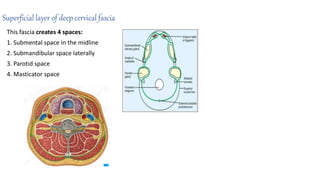
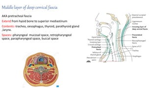
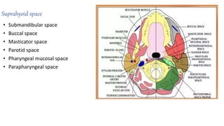
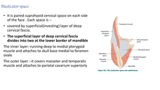
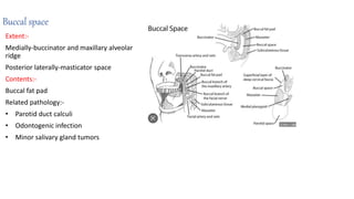
- The spaces include the masticator space containing the muscles of mastication, parotid space containing the parotid gland, and parapharyngeal space containing nerves and vessels.
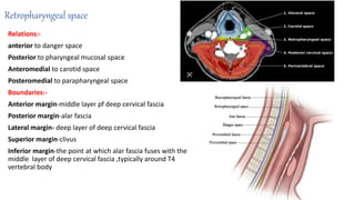
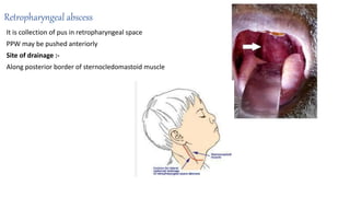
- Infections can spread between spaces through connections in the fascial planes, potentially reaching the mediastinum through the retropharyngeal and danger spaces.
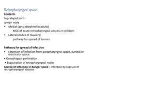
- Specific infections like peritonsillar and parapharyngeal abs
INFRATEMPORAL FOSSA AND PTERYGOPALATINE FOSSA NEW.pptxSudin Kayastha
INFRA TEMPORAL FOSSA
Irregularly shaped space deep & inferior to zygomatic arch, deep to ramus of mandible & posterior to maxilla
Communicates with temporal fossa through interval between (deep to) zygomatic arch & (superficial to) cranial bones
Temporal fossa is superior to zygomatic arch In
the fascial planes of the neck is very important in the spread and containment of infections, as well as being surgical dissection plane during neck surgery.
infections are rare but need to be identified early and treated appropriately to reduce the mortality and morbidity
this is a slightly well illustrated ppt of the previously uploaded one in february 2015
These simplified slides by Dr. Sidra Arshad present an overview of the non-respiratory functions of the respiratory tract.
Learning objectives:
1. Enlist the non-respiratory functions of the respiratory tract
2. Briefly explain how these functions are carried out
3. Discuss the significance of dead space
4. Differentiate between minute ventilation and alveolar ventilation
5. Describe the cough and sneeze reflexes
Study Resources:
1. Chapter 39, Guyton and Hall Textbook of Medical Physiology, 14th edition
2. Chapter 34, Ganong’s Review of Medical Physiology, 26th edition
3. Chapter 17, Human Physiology by Lauralee Sherwood, 9th edition
4. Non-respiratory functions of the lungs https://academic.oup.com/bjaed/article/13/3/98/278874
Explore natural remedies for syphilis treatment in Singapore. Discover alternative therapies, herbal remedies, and lifestyle changes that may complement conventional treatments. Learn about holistic approaches to managing syphilis symptoms and supporting overall health.
263778731218 Abortion Clinic /Pills In Harare ,sisternakatoto
263778731218 Abortion Clinic /Pills In Harare ,ABORTION WOMEN’S CLINIC +27730423979 IN women clinic we believe that every woman should be able to make choices in her pregnancy. Our job is to provide compassionate care, safety,affordable and confidential services. That’s why we have won the trust from all generations of women all over the world. we use non surgical method(Abortion pills) to terminate…Dr.LISA +27730423979women Clinic is committed to providing the highest quality of obstetrical and gynecological care to women of all ages. Our dedicated staff aim to treat each patient and her health concerns with compassion and respect.Our dedicated group ABORTION WOMEN’S CLINIC +27730423979 IN women clinic we believe that every woman should be able to make choices in her pregnancy. Our job is to provide compassionate care, safety,affordable and confidential services. That’s why we have won the trust from all generations of women all over the world. we use non surgical method(Abortion pills) to terminate…Dr.LISA +27730423979women Clinic is committed to providing the highest quality of obstetrical and gynecological care to women of all ages. Our dedicated staff aim to treat each patient and her health concerns with compassion and respect.Our dedicated group of receptionists, nurses, and physicians have worked together as a teamof receptionists, nurses, and physicians have worked together as a team wwww.lisywomensclinic.co.za/
Ozempic: Preoperative Management of Patients on GLP-1 Receptor Agonists Saeid Safari
Preoperative Management of Patients on GLP-1 Receptor Agonists like Ozempic and Semiglutide
ASA GUIDELINE
NYSORA Guideline
2 Case Reports of Gastric Ultrasound
Knee anatomy and clinical tests 2024.pdfvimalpl1234
This includes all relevant anatomy and clinical tests compiled from standard textbooks, Campbell,netter etc..It is comprehensive and best suited for orthopaedicians and orthopaedic residents.
The Gram stain is a fundamental technique in microbiology used to classify bacteria based on their cell wall structure. It provides a quick and simple method to distinguish between Gram-positive and Gram-negative bacteria, which have different susceptibilities to antibiotics
NVBDCP.pptx Nation vector borne disease control programSapna Thakur
NVBDCP was launched in 2003-2004 . Vector-Borne Disease: Disease that results from an infection transmitted to humans and other animals by blood-feeding arthropods, such as mosquitoes, ticks, and fleas. Examples of vector-borne diseases include Dengue fever, West Nile Virus, Lyme disease, and malaria.
Flu Vaccine Alert in Bangalore Karnatakaaddon Scans
As flu season approaches, health officials in Bangalore, Karnataka, are urging residents to get their flu vaccinations. The seasonal flu, while common, can lead to severe health complications, particularly for vulnerable populations such as young children, the elderly, and those with underlying health conditions.
Dr. Vidisha Kumari, a leading epidemiologist in Bangalore, emphasizes the importance of getting vaccinated. "The flu vaccine is our best defense against the influenza virus. It not only protects individuals but also helps prevent the spread of the virus in our communities," he says.
This year, the flu season is expected to coincide with a potential increase in other respiratory illnesses. The Karnataka Health Department has launched an awareness campaign highlighting the significance of flu vaccinations. They have set up multiple vaccination centers across Bangalore, making it convenient for residents to receive their shots.
To encourage widespread vaccination, the government is also collaborating with local schools, workplaces, and community centers to facilitate vaccination drives. Special attention is being given to ensuring that the vaccine is accessible to all, including marginalized communities who may have limited access to healthcare.
Residents are reminded that the flu vaccine is safe and effective. Common side effects are mild and may include soreness at the injection site, mild fever, or muscle aches. These side effects are generally short-lived and far less severe than the flu itself.
Healthcare providers are also stressing the importance of continuing COVID-19 precautions. Wearing masks, practicing good hand hygiene, and maintaining social distancing are still crucial, especially in crowded places.
Protect yourself and your loved ones by getting vaccinated. Together, we can help keep Bangalore healthy and safe this flu season. For more information on vaccination centers and schedules, residents can visit the Karnataka Health Department’s official website or follow their social media pages.
Stay informed, stay safe, and get your flu shot today!
CDSCO and Phamacovigilance {Regulatory body in India}NEHA GUPTA
The Central Drugs Standard Control Organization (CDSCO) is India's national regulatory body for pharmaceuticals and medical devices. Operating under the Directorate General of Health Services, Ministry of Health & Family Welfare, Government of India, the CDSCO is responsible for approving new drugs, conducting clinical trials, setting standards for drugs, controlling the quality of imported drugs, and coordinating the activities of State Drug Control Organizations by providing expert advice.
Pharmacovigilance, on the other hand, is the science and activities related to the detection, assessment, understanding, and prevention of adverse effects or any other drug-related problems. The primary aim of pharmacovigilance is to ensure the safety and efficacy of medicines, thereby protecting public health.
In India, pharmacovigilance activities are monitored by the Pharmacovigilance Programme of India (PvPI), which works closely with CDSCO to collect, analyze, and act upon data regarding adverse drug reactions (ADRs). Together, they play a critical role in ensuring that the benefits of drugs outweigh their risks, maintaining high standards of patient safety, and promoting the rational use of medicines.
3. Superficial fascia
• It is composed of subcutaneous tissue that encircles
neck like a collar
it encircles deep fascia
• Components :-
• Platysma and muscle of facial expression
• Subcutaneous veins nerves and fat
• Superficial lymph nodes
During surgery platysma should be sutured seperately
to prevent adherence of skin to deeper structure to
prevent scarring of skin
4. Deep cervical fascia
Superficial layer/investing layer
Middle layer of deep cervical fascia (pre-tracheal fascia)
Deep layer of deep cervical fascia (prevertebral fascia)
It forms complete collar around neck
Attachments :-
• Skull base, Mandible, hyoid bone, clavicle, sternum, scapula
• Posteriorly:-ligamentum nuchae and cervical spinous process
• It splits to cover SCM and trapezius muscle
Capsule:-
• Parotid
• Submandibular
Spaces:-
• Masticator space
• Parotid space
• Submandibular and sublingual space
5. Superficial layer of deep cervical fascia
Superiorly : attached to the nuchal ridge,
mandible, zygoma, mastoid, and hyoid
bones.
Inferiorly: bounded by clavicles, sternum,
scapula and acromion.
Anteriorly: symphysis menti and hyoid
bone
Posteriorly: ligamentum nuchae and spine
of C7
It invests SCM, trapezius and masseter
and envelops parotid and submaxillary
glands.
6. Superficial layer of deep cervical fascia
This fascia creates 4 spaces:
1. Submental space in the midline
2. Submandibular space laterally
3. Parotid space
4. Masticator space
7. Middle layer of deep cervical fascia
AKA pretracheal fascia
Extend from hyoid bone to superior mediastinum
Contents:-trachea, oesophagus, thyroid, parathyroid gland
,larynx.
Spaces:-pharyngeal mucosal space, retropharyngeal
space, parapharyngeal space, buccal space
8. Deep layer of Deep cervical fascia
• Extent from skull base to T3 vertebra
• Encircles paravertebral and paraspinal muscle ,phrenic
nerve, brachial plexus trunk
• Fuses with anterior longitudinal ligament in posterior
mediastinum and extends laterally as axillary sheath
9. Deep neck spaces
• Our neck is divided into spaces and compartments by
arrangement of layers of superficial and deep layers of
cervical fascia
• They contain loose areolar tissue
• May provide conduit for spread of infection
• it is described in relation to hyoid bone.
Supra hyoid space
Infra hyoid space
Space extending to full length of neck
10. Suprahyoid space
• Submandibular space
• Buccal space
• Masticator space
• Parotid space
• Pharyngeal mucosal space
• Parapharyngeal space
12. Spaces common to both
• Carotid space
• Retropharyngeal space
• Prevertebral space
• Danger space
13. Masticator space
• It is paired suprahyoid cervical space on each side
of the face . Each space is :-
• covered by superficial(investing) layer of deep
cervical fascia.
• The superficial layer of deep cervical fascia
divides into two at the lower border of mandible
The inner layer:-running deep to medial pterygoid
muscle and attaches to skull base medial to foramen
ovale
The outer layer :-it covers masseter and temporalis
muscle and attaches to parietal cavarium superiorly
14. Contents of masticator space
Contents:-
• Muscles of mastication
• Ramus and body of mandible
• Mandibular division of trigeminal nerve
it enters masticator space via foramen ovale
15. Boundaries and relations of masticator space
• Anteriorly:-buccal space
• Posterolaterally:- parotid space
• Medially:-parapharyngeal space
• Inferomedially :-submandibular space
Clinical
Masticator space malignancy can spread to middle
cranial fossa by perineural involvement (mandibular
division of trigeminal nerve )
Related pathologies:-
Masticator space abscess infection of 2nd and 3rd molar
Osteomyelitis
Direct spread of scc
Lymphoma
Minor salivary gland tumors
16. Parotid space
Boundaries:-
Investing fascia (ascending layer of deep cervical
fascia )splitting at angle of mandible-it ecloses parotid
space
Extends
Superiorly to external acoustic meatus
Inferiorly up to mandible
Contents
Parotid gland, proximal part of parotid duct
intra parotid lymphnode
External carotid artery
Retromandibular vein
Parotid gland is divided by facial nerve into
superficial and deep
17. Parotid space
Relations:-
• Lateral to parapharyngeal space
• Medial to superficial space and subcutaneous tissue
• Anterior to carotid space
• Posterior to masticator space
Boundaries :-
This space is circumscribed by superficial layer of deep cervical fascia
Superior margin:-external auditory canal, apex of mastoid process
Inferior margin:-inferior margin of mandible (but can extend further
inferiorly to angle of mandible )
Anterior margin:-masticator space
18. Parotid space
Related pathology :-
• Source of infection is from infection of oral cavity via stenson’s
duct
Any mass originating from parotid space will be centered within
parotid gland
A large mass arising within deep lobe of parotid will medially
displace fat in parapharyngeal space and cause posteromedial
dislocation of posterior belly of digastric muscle and carotid space
Congenital :-
• First branchial cleft cyst
• Cystic hygroma/lymphangioma
• Salivary gland tumor
• Parotid cyst
• Abscess/cellulitis
• Sjogren’s syndrome
19. Parapharyngeal space
It is pyramid shaped space
Base-petrous portion of temporal bone
Apex-at level of hyoid bone
Anterior-pterygo-mandibular raphe
Posterior-pre vertebral fascia
Medial-buccopharyngeal fascia
Lateral-parotid gland,ramus of mandible, medial
pterygoid muscle, fascia covering posterior belly of
digastric muscle
Crossed by styloid process which divides into pre
styloid and post styloid compartment.
20. Parapharyngeal space
Pre styloid compartment:-
• it is anterior to styloid process.
• it contains- fat, deep lobe of parotid gland,connective
tissue ,maxillary artery, inferior alveolar nerve, lingual
nerve and auriculo temporal nerve
Post styloid compartment :-
It is posterior to styloid process
It contains-glossopharyngeal and hypoglossal nerve
,sympathetic chain ,and lymph nodes
Source of infection:-
• Peritonsillar abscess
• Parotid abscess
• Submandibular gland infection
21.
22. parapharyngeal abscess
It is collection of pus in parapharyngeal space .
Causes:-
tonsillitis (acute,chronic)
Peritonsillar abscess, trauma
immuno compromised patients
Causative organism :-
Strep.viridens
Klebsiella
Pseudomonas(ass.with HIV patients )
Present as fluctuant tender swelling in neck
It pushes tonsil medially(bulging is present in lower pole of tonsil)
23. PRESENTING SYMPTOMS OF PARAPHARYNGEAL SPACE LESION
Mostly asymptomatic
Medial displacement of lateral pharyngeal wall
and tonsil is hallmark
Neck & oropharyngeal swelling
Pain
Cranial nerve palsy
Dysphagia , Dyspnea
Trismus, Drooling
ETD
Hoarsness of voice
Hypertension , flushing
Fever
25. Buccal space
Extent:-
Medially-buccinator and maxillary alveolar
ridge
Posterior laterally-masticator space
Contents:-
Buccal fat pad
Related pathology:-
• Parotid duct calculi
• Odontogenic infection
• Minor salivary gland tumors
26. Retropharyngeal space
Extent:-skull base to t4 level/tracheal bifurcation
2 parts :-
Anterior-true retropharyngeal space
Posterior-danger space
Contents:-
SHN-lymph node and fat
IHN-fat
Infection from neck can reach mediastinum through this
pathway
27. Retropharyngeal space
Relations:-
anterior to danger space
Posterior to pharyngeal mucosal space
Anteromedial to carotid space
Posteromedial to parapharyngeal space
Boundaries:-
Anterior margin-middle layer pf deep cervical fascia
Posterior margin-alar fascia
Lateral margin- deep layer of deep cervical fascia
Superior margin-clivus
Inferior margin-the point at which alar fascia fuses with the
middle layer of deep cervical fascia ,typically around T4
vertebral body
28. Retropharyngeal space
Contents
Suprahyoid part:-
Lymph node
• Medial (gets atrophied in adults)
MCC of acute retropharyngeal abscess in children
• Lateral (nodes of rouviere)
pathway for spread of tumors
Pathway for spread of infection
• Extension of infection from parapharyngeal space, parotid or
masticator space
• Oesophageal perforation
• Suppuration of retropharyngeal nodes
Source of infection in danger space :-Infection by rupture of
retropharyngeal abscess
29. Retropharyngeal abscess
It is collection of pus in retropharyngeal space
PPW may be pushed anteriorly
Site of drainage :-
Along posterior border of sternocledomastoid muscle
30. Danger space
Posterior to retropharyngeal space
Bounded by alar fascia anteriorly and prevertebral portion
proper posteriorly
Extends from skull base to diaphragm
Contents-loose areolar tissue which provides easy pathway
for spread of infection
Danger space
At inferior border it is continuous with posterior mediastinum
containing venacava ,arch of aorta, theoracic duct, trachea
and esophagus
erosion of major blood vessel ,lower airway and upper
digestive tract
may cause death of patient
31. Carotid space
It is roughly cylindrical space that extends from skull base to aortic
arch .it is circumscribed by all 3 layers of deep cervical fascia
forming carotid sheath
Lateral to retropharyngeal space
The bifurcation of common carotid usually occurs at boundaries of
suprahyoid and infrahyoid spaces
Relations:-
Suprahyoid carotid space
anteriorly-masticator space ,parapharyngeal space
Laterally-parotid space
Posteriorly-prevertebral space
The suprahyoid portion of carotid space is often synonymous with
post styloid compartment of parapharyngeal space
32. Carotid space
Boundaries :-
Superior margin:-lower border of jugular foramen
Inferior margin:- aortic arch
Anterolateral margin:- sternocledomastoid muscle
Contents :-
• Common carotid artery inferiorly and internal carotid artery superiorly
• Internal juglar vein
• Glossopharyngeal nerve (CN IX) it is anterior to vessel
• Vagus nerve (CN X) it is posterior to posterior notch
• Spinal accessory nerve (CN XI)
• Hypoglossal nerve (CN XII)
• Sympathetic nerves:-medial to vessels and lateral to retropharyngeal
space(behind the sheath )
• Deep cervical lymph node chain
33.
34. Submandibular space
Extent
It extends from myelohyoid superiorly and hyoid bone
inferiorly
Communicates freely with sublingual space
Contents :-
Superficial portion of submandibular gland
Submantle lymph node
Submandibular lymph nodes
Facial artery and veins
35. Submandibular space
Ludwig’s angina :-
It is acute infection of bilateral submandibular and sublingual
spaces and submantle spaces.
Consequence of anterior dental infection
Oedema of floor of mouth and of anterior tongue leads to gross
swelling
Significant risk of life threatening airway obstruction.
Site of drainage
Surgical drainage is done via horizontal incision 2 finger breadth
below the mandibular margin from one angle of the mandible to
the other over the area of induration.
36. Sublingual space
Extent:-
Above myelohyoid muscle
Contents:-
Sublingual gland and their ducts ,lymphnodes, lingual artery and veins
,deeper portion of submandibular gland
RANULA:-
cystic translucent lesion seen in the floor of mouth on one side of the
frenulum and pushing the tongue up.
Arises from sublingual gland due to obstruction of its ducts.
37. Peritonsillar space
Lies between superior constrictor muscle and the tonsillar
capsule.
Clinical:
Infection involving looser areolar tissue causing collection of pus
within the space is called quinsy or peritonsillar abscess.
38. Peritonsillar abscess
It is collection of pus in peritonsillar space
Mostly in young age group (20-30 years)
Causes:-
Acute tonsillitis
Weber’s gland(they are minor salivary gland present in supra tonsillar area)
It may present with
Dysphagia
Referred otalgia(via CN IX)
Neck pain
Site of drainage :-
Should do tonsillectomy 4-6 weeks after drainage