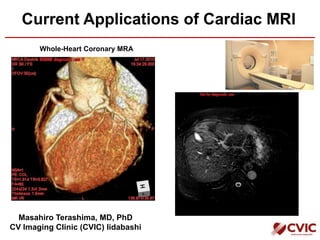
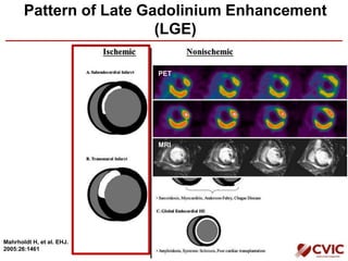
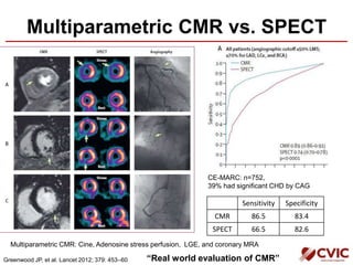
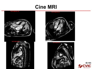
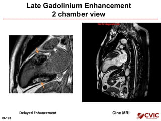
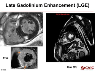
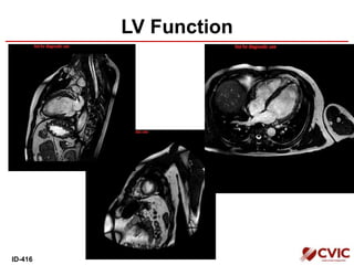
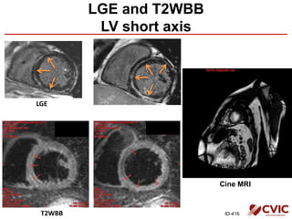
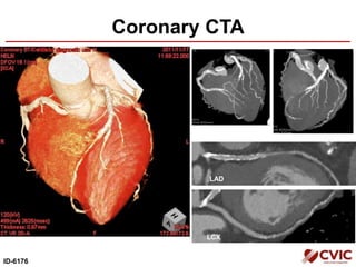
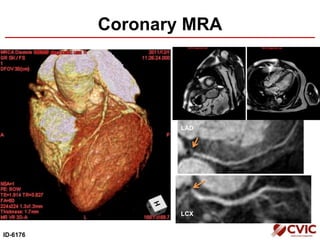
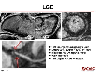
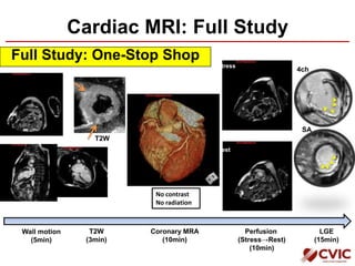
Current Applications of Cardiac MRI
Masahiro Terashima, MD, PhD
CV Imaging Clinic (CVIC) Iidabashi
Whole-Heart Coronary MRA
Today’s Agenda
• Cardiac MRI in Japan
– Recent trends of cardiac MRI in Japan
• Cardiac MRI@CVIC
– Ischemic
– Non-ischemic
• Combination of cardiac CT and MRI
– Calcified lesions
– Unrecognized MI
Today’s Agenda
• Cardiac MRI in Japan
– Recent trends of cardiac MRI in Japan
• Cardiac MRI@CVIC
– Ischemic
– Non-ischemic
• Combination of cardiac CT and MRI
– Calcified lesions
– Unrecognized MI
Trends of Coronary CTA and
Cardiac MRI in Japan
27,131
86,256
150,309
218,053
273,370
346,412
360,275
25,593 17,975 20,900 21,670 20,452 24,511 27,128
0
50,000
100,000
150,000
200,000
250,000
300,000
350,000
400,000
2004 2006 2007 2008 2009 2010 2011
Coronary CTA
Cardiac MRI
日本循環器学会:循環器疾患診療実態調査
Cardiac MRI in Japan
CVIC 冠動脈CT 月間250件 3,000/year
CVIC 心臓MRI (Share 10%) 月間200件 2,400/year
日本循環器学会:循環器疾患診療実態調査
Today’s Agenda
• Cardiac MRI in Japan
– Recent trends of cardiac MRI in Japan
• Cardiac MRI@CVIC
– Ischemic
– Non-ischemic
• Combination of cardiac CT and MRI
– Calcified lesions
– Unrecognized MI
Cardiac MRI: Full Study
Full Study: One-Stop Shop
Wall motion
(5min)
Perfusion
(Stress→Rest)
(10min)
Stress
Rest
LGE
(15min)
Coronary MRA
(10min)
4ch
SA
No contrast
No radiation
T2W
(3min)
T2W
Whole-Heart Coronary MRA
RCA
LAD
LCX
ID-193
Kato, et al. J Am Coll Cardiol 2010;56:983–91
127/138 (92%)
(9.5+/-3.5 min)
Today’s Agenda
• Cardiac MRI in Japan
– Recent trends of cardiac MRI in Japan
• Cardiac MRI@CVIC
– Ischemic
– Non-ischemic
• Combination of cardiac CT and MRI
– Calcified lesions
– Unrecognized MI
Pattern of Late Gadolinium Enhancement
(LGE)
Mahrholdt H, et al. EHJ.
2005:26:1461
PET
MRI
Assessment of Myocardial Viability by MRI
Kim RJ, et al. NEJM. 2000;343:1445
• 12 segments model
• % of HE area = 100 x area A ÷ (area A + area B)
N=50
Transmural extent of hyperenhancement
Mechanism of Delayed Hyperenhancement
Gd diffuses rapidly from
the intravascular to the
extracellular space.
Expanded volume of
distribution of Gd in scar
tissue.
Ruptured cell membrane
increases the potential
interstitial volume.
Higgins, CB. Cardiovascular MRI and MRA. 2002
Myocardial Thinning and Scarring in CAD
JAMA. 2013;309(9):909-918
end-diastolic wall thickness<5.5 mm
N=201/1055 (wall thinning)
Limited scar burden (<50%): 37/201 (18%)
Repeated CMR (n=42)
Improved LV Function in Patients with Thinning
and Limited Scar Burden
JAMA. 2013;309(9):909-918
Case Presentation
• Asymptomatic
• ECG: V1~V3 poor R
(Normal in 2008)
• Coronary Risk Factor: HLP, HT, Smoking
67M
ID-67 6/25/2008 7/6/2009
LV function (Short Axis)
HypokinesisHypokinesis
ID-67
LV function (2ch)
HypokinesisHypokinesis
ID-67
LGE (Late Gadolinium Enhancement)
2ch 4ch
ID-67
LAD
LCx
Coronary MRA
First-Pass Perfusion
Stress Rest
ID-67
Multiparametric CMR vs. SPECT
CE-MARC: n=752,
39% had significant CHD by CAG
Greenwood JP, et al. Lancet 2012; 379: 453–60
Sensitivity Specificity
CMR 86.5 83.4
SPECT 66.5 82.6
Multiparametric CMR: Cine, Adenosine stress perfusion, LGE, and coronary MRA
“Real world evaluation of CMR”
Today’s Agenda
• Cardiac MRI in Japan
– Recent trends of cardiac MRI in Japan
• Cardiac MRI@CVIC
– Ischemic
– Non-ischemic
• Combination of cardiac CT and MRI
– Calcified lesions
– Unrecognized MI
Pattern of LGE
Mahrholdt H, et al. EHJ.
2005:26:1461
54M
Coronary Risk Factor: HT, DM (IGT?), Smoking
Case Presentation
ID-193
Cine MRI
ID-193
Late Gadolinium Enhancement
2 chamber view
Delayed Enhancement Cine MRI
ID-193
Cine MRI
Late Gadolinium Enhancement (LGE)
LGE
ID-193
T2W
T2W MRI of HCM: Myocardial Edema
Our case
LGE T2W
(Abdel-Aty H, et al. JMRI. 2008;28:242)
LGE
T2W
Poor blood
suppression
Stress Perfusion MRI in HCM
ID-193
Diffuse perfusion defect (Microvascular dysfunction?)
LGE of HCM
• LGE (+) in 50% of HCM patients. (Harris SR, et al.
Magn Reson Imaging Clin N Am. 2008;16:165)
• Most frequent in RV insertion points (73%)
(Rubinshtein R. Circ Heart Fail. 2010;3:51) (Adabag SA. JACC. 2008;51:1369)
Our case
Bruder O, et al. JACC.2010;56:875
Teraoka K, et al. MRI. 2004;22: 155
Case Presentation
57M
CHF due to AR
DCM (First diagnosed in 1995)
ID-5
LV Function
ID-5
LGE of DCM
• LV Mid-wall enhancement in DCM
• RV Mid-wall
ID-5
Midwall Fibrosis in CMR and Histology
SCD
Tx
Small amounts of perivascular fibrosis
Extensive replacement fibrosis
JAMA. 2013;309(9):896-908
Prognostic Significance of
Midwall Enhancement
N=472 DCM patients
N=142 (30%) midwall LGE+, 38 deaths (26.8%)
N=330 (70%) LGE-, 35 deaths (10.6%)
Hazard ratio=2.96
JAMA. 2013;309(9):896-908
Case Presentation
40M
CHF, s/o DCM, History of cardiac arrest
ID-416
LV Function
ID-416
Cine MRI
LGE
ID-416
LGE and T2WBB
LV short axis
T2WBB
CMR of Myocarditis
Friedrich MG, et al. JACC2009;53:1475Cocker M, et al. Curr Cardiol Rep (2010) 12:82–89
23M
LGE
T2W
LGE pattern of myocarditis
Today’s Agenda
• Cardiac MRI in Japan
– Recent trends of cardiac MRI in Japan
• Cardiac MRI@CVIC
– Ischemic
– Non-ischemic
• Combination of cardiac CT and MRI
– Calcified lesions
– Unrecognized MI
Case Presentation
71M
• Jaw pain early in the morning (rest and effort)
• Coronary Risk Factor: HT, HLP
Coronary CTA
ID-6176
LAD
LCX
Coronary MRA
ID-6176
LAD
LCX
LGE
ID-6176
 12/1 Emergent CAG@Tokyo Univ.
 LMT#5-99%, LAD#6-100%, #11-99%
 Moderate AS (AV flow=2.7m/s)
 IABP insertion
 12/3 Urgent CABG with AVR
CAG
ID-6176
LCA (Missing LAD) RCA (Collateral to LAD)
Today’s Agenda
• Cardiac MRI in Japan
– Recent trends of cardiac MRI in Japan
• Cardiac MRI@CVIC
– Ischemic
– Non-ischemic
• Combination of cardiac CT and MRI
– Calcified lesions
– Unrecognized MI
Unrecognized MI by Contrast-Enhanced MRI
• 44/195 (23%) pts had positive on
DEMRI.
• Even a very small scar (<2% mean
LV mass) was associated with a >7-
fold increase in MACE hazards.
Kwong RY, et al. Circulation. 2006;113:2733
73M, DM, 3VD, SCD at 8 mo later
68M, UAP at 12 mo later (LAD and RCA)
Unrecognized MI more common, dangerous in
older adults than previously suspected.
Schelbert EB, et al. JAMA. 2012;308(9):890-897
陳旧性心筋梗塞
無症候性心筋梗塞
無症候性心筋梗塞
死亡率に差は無し
73%
17%
10%
No MI
Unrecognized MI
Recognized MI
Older adults (n=936, age: 67-93y.o.)
• ECGではUMIの5%しか検出できず。
• UMIはDM+で21%、DM-で14%
UMI: Aspirin 50%, Beta-blockers 45%, Statin 36%
Cardiac MRI: Full Study
Full Study: One-Stop Shop
Wall motion
(5min)
Perfusion
(Stress→Rest)
(10min)
Stress
Rest
LGE
(15min)
Coronary MRA
(10min)
4ch
SA
No contrast
No radiation
T2W
(3min)
T2W

Cvic tmdu 071913_share

  • 1.
    Current Applications ofCardiac MRI Masahiro Terashima, MD, PhD CV Imaging Clinic (CVIC) Iidabashi Whole-Heart Coronary MRA
  • 2.
    Today’s Agenda • CardiacMRI in Japan – Recent trends of cardiac MRI in Japan • Cardiac MRI@CVIC – Ischemic – Non-ischemic • Combination of cardiac CT and MRI – Calcified lesions – Unrecognized MI
  • 3.
    Today’s Agenda • CardiacMRI in Japan – Recent trends of cardiac MRI in Japan • Cardiac MRI@CVIC – Ischemic – Non-ischemic • Combination of cardiac CT and MRI – Calcified lesions – Unrecognized MI
  • 4.
    Trends of CoronaryCTA and Cardiac MRI in Japan 27,131 86,256 150,309 218,053 273,370 346,412 360,275 25,593 17,975 20,900 21,670 20,452 24,511 27,128 0 50,000 100,000 150,000 200,000 250,000 300,000 350,000 400,000 2004 2006 2007 2008 2009 2010 2011 Coronary CTA Cardiac MRI 日本循環器学会:循環器疾患診療実態調査
  • 5.
    Cardiac MRI inJapan CVIC 冠動脈CT 月間250件 3,000/year CVIC 心臓MRI (Share 10%) 月間200件 2,400/year 日本循環器学会:循環器疾患診療実態調査
  • 6.
    Today’s Agenda • CardiacMRI in Japan – Recent trends of cardiac MRI in Japan • Cardiac MRI@CVIC – Ischemic – Non-ischemic • Combination of cardiac CT and MRI – Calcified lesions – Unrecognized MI
  • 7.
    Cardiac MRI: FullStudy Full Study: One-Stop Shop Wall motion (5min) Perfusion (Stress→Rest) (10min) Stress Rest LGE (15min) Coronary MRA (10min) 4ch SA No contrast No radiation T2W (3min) T2W
  • 8.
    Whole-Heart Coronary MRA RCA LAD LCX ID-193 Kato,et al. J Am Coll Cardiol 2010;56:983–91 127/138 (92%) (9.5+/-3.5 min)
  • 9.
    Today’s Agenda • CardiacMRI in Japan – Recent trends of cardiac MRI in Japan • Cardiac MRI@CVIC – Ischemic – Non-ischemic • Combination of cardiac CT and MRI – Calcified lesions – Unrecognized MI
  • 10.
    Pattern of LateGadolinium Enhancement (LGE) Mahrholdt H, et al. EHJ. 2005:26:1461 PET MRI
  • 11.
    Assessment of MyocardialViability by MRI Kim RJ, et al. NEJM. 2000;343:1445 • 12 segments model • % of HE area = 100 x area A ÷ (area A + area B) N=50 Transmural extent of hyperenhancement
  • 12.
    Mechanism of DelayedHyperenhancement Gd diffuses rapidly from the intravascular to the extracellular space. Expanded volume of distribution of Gd in scar tissue. Ruptured cell membrane increases the potential interstitial volume. Higgins, CB. Cardiovascular MRI and MRA. 2002
  • 13.
    Myocardial Thinning andScarring in CAD JAMA. 2013;309(9):909-918 end-diastolic wall thickness<5.5 mm N=201/1055 (wall thinning) Limited scar burden (<50%): 37/201 (18%) Repeated CMR (n=42)
  • 14.
    Improved LV Functionin Patients with Thinning and Limited Scar Burden JAMA. 2013;309(9):909-918
  • 15.
    Case Presentation • Asymptomatic •ECG: V1~V3 poor R (Normal in 2008) • Coronary Risk Factor: HLP, HT, Smoking 67M ID-67 6/25/2008 7/6/2009
  • 16.
    LV function (ShortAxis) HypokinesisHypokinesis ID-67
  • 17.
  • 18.
    LGE (Late GadoliniumEnhancement) 2ch 4ch ID-67
  • 19.
  • 20.
  • 21.
    Multiparametric CMR vs.SPECT CE-MARC: n=752, 39% had significant CHD by CAG Greenwood JP, et al. Lancet 2012; 379: 453–60 Sensitivity Specificity CMR 86.5 83.4 SPECT 66.5 82.6 Multiparametric CMR: Cine, Adenosine stress perfusion, LGE, and coronary MRA “Real world evaluation of CMR”
  • 22.
    Today’s Agenda • CardiacMRI in Japan – Recent trends of cardiac MRI in Japan • Cardiac MRI@CVIC – Ischemic – Non-ischemic • Combination of cardiac CT and MRI – Calcified lesions – Unrecognized MI
  • 23.
    Pattern of LGE MahrholdtH, et al. EHJ. 2005:26:1461
  • 24.
    54M Coronary Risk Factor:HT, DM (IGT?), Smoking Case Presentation ID-193
  • 25.
  • 26.
    Late Gadolinium Enhancement 2chamber view Delayed Enhancement Cine MRI ID-193
  • 27.
    Cine MRI Late GadoliniumEnhancement (LGE) LGE ID-193 T2W
  • 28.
    T2W MRI ofHCM: Myocardial Edema Our case LGE T2W (Abdel-Aty H, et al. JMRI. 2008;28:242) LGE T2W Poor blood suppression
  • 29.
    Stress Perfusion MRIin HCM ID-193 Diffuse perfusion defect (Microvascular dysfunction?)
  • 30.
    LGE of HCM •LGE (+) in 50% of HCM patients. (Harris SR, et al. Magn Reson Imaging Clin N Am. 2008;16:165) • Most frequent in RV insertion points (73%) (Rubinshtein R. Circ Heart Fail. 2010;3:51) (Adabag SA. JACC. 2008;51:1369) Our case Bruder O, et al. JACC.2010;56:875 Teraoka K, et al. MRI. 2004;22: 155
  • 31.
    Case Presentation 57M CHF dueto AR DCM (First diagnosed in 1995) ID-5
  • 32.
  • 33.
    LGE of DCM •LV Mid-wall enhancement in DCM • RV Mid-wall ID-5
  • 34.
    Midwall Fibrosis inCMR and Histology SCD Tx Small amounts of perivascular fibrosis Extensive replacement fibrosis JAMA. 2013;309(9):896-908
  • 35.
    Prognostic Significance of MidwallEnhancement N=472 DCM patients N=142 (30%) midwall LGE+, 38 deaths (26.8%) N=330 (70%) LGE-, 35 deaths (10.6%) Hazard ratio=2.96 JAMA. 2013;309(9):896-908
  • 36.
    Case Presentation 40M CHF, s/oDCM, History of cardiac arrest ID-416
  • 37.
  • 38.
    Cine MRI LGE ID-416 LGE andT2WBB LV short axis T2WBB
  • 39.
    CMR of Myocarditis FriedrichMG, et al. JACC2009;53:1475Cocker M, et al. Curr Cardiol Rep (2010) 12:82–89 23M LGE T2W LGE pattern of myocarditis
  • 40.
    Today’s Agenda • CardiacMRI in Japan – Recent trends of cardiac MRI in Japan • Cardiac MRI@CVIC – Ischemic – Non-ischemic • Combination of cardiac CT and MRI – Calcified lesions – Unrecognized MI
  • 41.
    Case Presentation 71M • Jawpain early in the morning (rest and effort) • Coronary Risk Factor: HT, HLP
  • 42.
  • 43.
  • 44.
    LGE ID-6176  12/1 EmergentCAG@Tokyo Univ.  LMT#5-99%, LAD#6-100%, #11-99%  Moderate AS (AV flow=2.7m/s)  IABP insertion  12/3 Urgent CABG with AVR
  • 45.
    CAG ID-6176 LCA (Missing LAD)RCA (Collateral to LAD)
  • 46.
    Today’s Agenda • CardiacMRI in Japan – Recent trends of cardiac MRI in Japan • Cardiac MRI@CVIC – Ischemic – Non-ischemic • Combination of cardiac CT and MRI – Calcified lesions – Unrecognized MI
  • 47.
    Unrecognized MI byContrast-Enhanced MRI • 44/195 (23%) pts had positive on DEMRI. • Even a very small scar (<2% mean LV mass) was associated with a >7- fold increase in MACE hazards. Kwong RY, et al. Circulation. 2006;113:2733 73M, DM, 3VD, SCD at 8 mo later 68M, UAP at 12 mo later (LAD and RCA)
  • 48.
    Unrecognized MI morecommon, dangerous in older adults than previously suspected. Schelbert EB, et al. JAMA. 2012;308(9):890-897 陳旧性心筋梗塞 無症候性心筋梗塞 無症候性心筋梗塞 死亡率に差は無し 73% 17% 10% No MI Unrecognized MI Recognized MI Older adults (n=936, age: 67-93y.o.) • ECGではUMIの5%しか検出できず。 • UMIはDM+で21%、DM-で14% UMI: Aspirin 50%, Beta-blockers 45%, Statin 36%
  • 49.
    Cardiac MRI: FullStudy Full Study: One-Stop Shop Wall motion (5min) Perfusion (Stress→Rest) (10min) Stress Rest LGE (15min) Coronary MRA (10min) 4ch SA No contrast No radiation T2W (3min) T2W

Editor's Notes

  • #5 日本循環器学会の循環器疾患診療実態調査の結果より年次ごとの冠動脈CTA、心臓MRI検査件数の推移を示す。