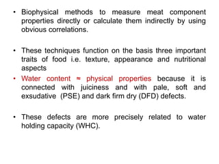
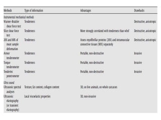
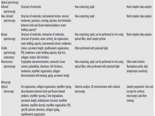
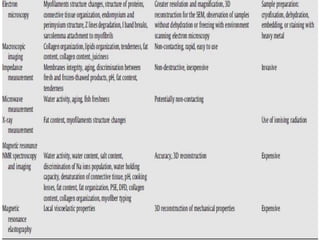
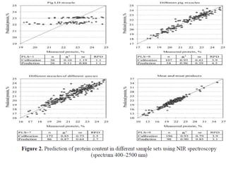
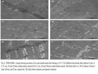
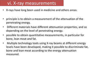
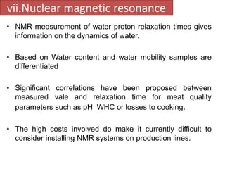
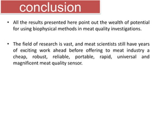
Credit seminar -2 
Meat quality assessment using 
biophysical methods 
Major advisor 
Dr. B.B. Nayak, 
Principal Scientist 
Post Harvest Technology, CIFE, 
Mumbai . 
Presented by 
Naresh Kumar Mehta 
(PHT-PA01-04) 
Ph.D. (Batch: 2011-14), 
Post Harvest Technology 
CIFE, Mumbai .
Introduction 
• Factor influencing the meat properties are related to 
breed, age and sex. 
• Meat toughness depends mainly on MFP and 
conjunctive tissue. 
• MFP structure influenced by zoo-technical conditions. 
‘Greenwood et al., 2007 and Gondret et al., 2005 studied 
on myofibril types the lamb in and out door rearing.’
• Biophysical methods to measure meat component 
properties directly or calculate them indirectly by using 
obvious correlations. 
• These techniques function on the basis three important 
traits of food i.e. texture, appearance and nutritional 
aspects 
• Water content ≈ physical properties because it is 
connected with juiciness and with pale, soft and 
exsudative (PSE) and dark firm dry (DFD) defects. 
• These defects are more precisely related to water 
holding capacity (WHC).
Why does need arise? 
• Chemical indices- time consuming 
• Sensory indices - perceptional 
• To obtain reliable information on meat quality throughout 
the production process 
• To reduce cumbersome data handling 
• To be fast, accurate and non invasive techniques for 
predicting technological and sensory qualities. 
• Majority existing techniques are invasive
Biophysical methods 
Mechanical methods, 
Optical methods, 
X-ray measurements 
Nuclear magnetic resonance (NMR) 
measurements
1. Mechanical methods 
• Instrumental methods – 
• TA measurements(Compression, piercing) 
• Rheometry (shearing) 
• Ultrasound methods- analyzing the acoustic parameters of 
wave propagating makes it possible to assess the 
characteristic of the propagating medium and to characterize 
it. 
Monin (1998) reported that ultrasonic measurements give a 
good prediction of meat texture on live animals and whole 
carcass, while at the same time being inexpensive and non-invasive.
2.Optical methods 
a. Spectroscopic methods 
I. Infrared spectroscopy 
II. Raman spectroscopy 
III. Visible spectroscopy and colorimetry 
IV. Fluorescence spectroscopy 
b. Imaging 
I. Microscopic imaging 
II. Optical microscopy 
III. Histology 
IV. Confocal laser scanning microscopy 
V. Electron microscopy 
VI. Scanning electron microscopy 
VII. Transmission electron microscopy 
VIII. Macroscopic imaging
I. Infrared spectroscopy 
• Principle-Infrared spectroscopy (800-2500nm) is based on the 
principle that the chemical bonds in organic molecules absorb or 
emit infrared light when their vibrational state changes. 
• Uddin et al.,2005 used for fresh and thawed fish to control of 
fraudulent freezing-thawing cycle.
Tehnologija mesa 51 (2010) 2, 133–142
ii. Raman spectroscopy 
• Raman spectroscopy is also a vibrational spectroscopic technique 
used in condensed matter physics, biomedical applications and 
chemistry to study vibrational, rotational, and other low-frequency 
modes in a system. 
• It relies on inelastic scattering of monochromatic light, usually from 
a laser in the visible, IR, or near-UV spectra. It gives similar but 
complementary information to IR spectroscopy.
iii.Visible spectroscopy and colorimetry 
• It covers the visible spectra and the CIE 
L*a*b*colour space as objective and non-destructive 
tools for tissue characterization. 
• Fish muscle absorbs different components 
of light differently, depending on the 
composition and state of the muscles. 
• spectra change depending on the degree of 
spoilage during chilled or frozen storage. 
• Early detection of pale, soft and exsudative 
(PSE) meat is a major potential application 
of visible spectroscopy and colorimetry for 
both pork and poultry meat
Figure 10A is showing a colour picture of the fillet composed of three colours - red (641 
nm), green (552 nm) and blue (458 nm). This is a colour composition that is close to 
what we actually can see with our eyes. 
Figure 10B, is focusing on oxy- and deoxyhaemoglobin (572 nm) and blood is visualised 
as dark spots. 
Figure 10C, is focusing on methaemoglobin (630 nm) and blood is visualised as luminous 
spots.
iv. Fluorescence spectroscopy 
• is a type of electromagnetic spectroscopy which analyzes 
fluorescence from a sample. 
• For a well ordered biological tissue, fluorescence is 
anisotropic and this anisotropy tends to disappear with 
structural degradation. 
• De-structuring processes like ageing, grounding or heating 
have been successfully investigated. 
• Tryptophan is an important intrinsic fluorescent probe that can 
be used to assess the nature of the tryptophan 
microenvironment. 
• Proteins that lack tryptophan can be attached to an extrinsic 
fluorophore probe.
2.i Imaging 
Microscopic imaging 
Optical microscopy- 
• Optical microscopy offers the simplest way to obtain 
magnified images of biological tissues. 
• This field covers a large range of techniques that have been 
used for years to characterize meat and meat product 
structures. 
• Techniques can be classed simply depending on whether 
samples must be prepared in thin cuts or not. 
• Detection of A and I band.
ii.Histology 
• Histology is a widely used as a tool for controlling meat 
texture in food science. 
• The technique may or may not require tissue staining with 
specific dyes. 
• Histology always needs very thin sample cuts. 
• Fiber disorganization, fiber misalignment and increase in 
fibers spacing have been analyzed.
iii.Confocal laser scanning microscopy 
• is a fluorescence technique for obtaining high-longitudinal 
resolution optical images. 
• Evolution of the more traditional fluorescence microscopy. 
• Key feature being the ability to produce point-by-point in-focus 
images of thick specimens. 
• Allowing 3D reconstructions of complex tissues. 
• Because this technique depends on fluorescence, samples 
usually need to be treated with fluorescent dyes to make 
objects. 
• visible, but contrary to the histological techniques, there is 
no need for thin cuts
iv. Scanning electron microscopy (SEM) 
• SEM gives images with great depth-of-field yielding a 
characteristic 3D display that provides greater insight into the 
surface structure of a biological sample 
• Cryofixation, dehydration, embedding and staining
Fig. 6. SEM (200) images showing transverse section of raw and cooked muscle after heating at 
121.1 1C for different time periods. The horizontal dotted bars indicate 150 mm. A, C, E: raw, 
20 and 120 min cooked salmon muscle; B, D, F: raw, 20 and 120 min cooked chicken muscle
v. Transmission electron microscopy (TEM) 
• In TEM electrons are passed through the sample. 
• Resolution is higher than in SEM and the sample can be stained 
with heavy metals to improve image quality
vi. X-ray measurements 
• X-rays have long been used in medicine and others areas. 
• principle is to obtain a measurement of the attenuation of the 
penetrating energy. 
• Different materials have different attenuation properties, and so 
depending on the level of penetrating energy. 
• possible to obtain quantitative measurements, in particular for 
bone, lean meat and fat. 
• Multiple technology tools using X-ray beams at different energy 
levels have been developed, making it possible to discriminate fat, 
bone and lean meat according to the energy attenuation 
measured.
vii.Nuclear magnetic resonance 
• NMR measurement of water proton relaxation times gives 
information on the dynamics of water. 
• Based on Water content and water mobility samples are 
differentiated 
• Significant correlations have been proposed between 
measured vale and relaxation time for meat quality 
parameters such as pH WHC or losses to cooking. 
• The high costs involved do make it currently difficult to 
consider installing NMR systems on production lines.
conclusion 
• All the results presented here point out the wealth of potential 
for using biophysical methods in meat quality investigations. 
• The field of research is vast, and meat scientists still have years 
of exciting work ahead before offering to meat industry a 
cheap, robust, reliable, portable, rapid, universal and 
magnificent meat quality sensor.
Thank 
you

Credit semanr no.2

  • 1.
    Credit seminar -2 Meat quality assessment using biophysical methods Major advisor Dr. B.B. Nayak, Principal Scientist Post Harvest Technology, CIFE, Mumbai . Presented by Naresh Kumar Mehta (PHT-PA01-04) Ph.D. (Batch: 2011-14), Post Harvest Technology CIFE, Mumbai .
  • 2.
    Introduction • Factorinfluencing the meat properties are related to breed, age and sex. • Meat toughness depends mainly on MFP and conjunctive tissue. • MFP structure influenced by zoo-technical conditions. ‘Greenwood et al., 2007 and Gondret et al., 2005 studied on myofibril types the lamb in and out door rearing.’
  • 3.
    • Biophysical methodsto measure meat component properties directly or calculate them indirectly by using obvious correlations. • These techniques function on the basis three important traits of food i.e. texture, appearance and nutritional aspects • Water content ≈ physical properties because it is connected with juiciness and with pale, soft and exsudative (PSE) and dark firm dry (DFD) defects. • These defects are more precisely related to water holding capacity (WHC).
  • 4.
    Why does needarise? • Chemical indices- time consuming • Sensory indices - perceptional • To obtain reliable information on meat quality throughout the production process • To reduce cumbersome data handling • To be fast, accurate and non invasive techniques for predicting technological and sensory qualities. • Majority existing techniques are invasive
  • 5.
    Biophysical methods Mechanicalmethods, Optical methods, X-ray measurements Nuclear magnetic resonance (NMR) measurements
  • 9.
    1. Mechanical methods • Instrumental methods – • TA measurements(Compression, piercing) • Rheometry (shearing) • Ultrasound methods- analyzing the acoustic parameters of wave propagating makes it possible to assess the characteristic of the propagating medium and to characterize it. Monin (1998) reported that ultrasonic measurements give a good prediction of meat texture on live animals and whole carcass, while at the same time being inexpensive and non-invasive.
  • 10.
    2.Optical methods a.Spectroscopic methods I. Infrared spectroscopy II. Raman spectroscopy III. Visible spectroscopy and colorimetry IV. Fluorescence spectroscopy b. Imaging I. Microscopic imaging II. Optical microscopy III. Histology IV. Confocal laser scanning microscopy V. Electron microscopy VI. Scanning electron microscopy VII. Transmission electron microscopy VIII. Macroscopic imaging
  • 11.
    I. Infrared spectroscopy • Principle-Infrared spectroscopy (800-2500nm) is based on the principle that the chemical bonds in organic molecules absorb or emit infrared light when their vibrational state changes. • Uddin et al.,2005 used for fresh and thawed fish to control of fraudulent freezing-thawing cycle.
  • 12.
    Tehnologija mesa 51(2010) 2, 133–142
  • 14.
    ii. Raman spectroscopy • Raman spectroscopy is also a vibrational spectroscopic technique used in condensed matter physics, biomedical applications and chemistry to study vibrational, rotational, and other low-frequency modes in a system. • It relies on inelastic scattering of monochromatic light, usually from a laser in the visible, IR, or near-UV spectra. It gives similar but complementary information to IR spectroscopy.
  • 15.
    iii.Visible spectroscopy andcolorimetry • It covers the visible spectra and the CIE L*a*b*colour space as objective and non-destructive tools for tissue characterization. • Fish muscle absorbs different components of light differently, depending on the composition and state of the muscles. • spectra change depending on the degree of spoilage during chilled or frozen storage. • Early detection of pale, soft and exsudative (PSE) meat is a major potential application of visible spectroscopy and colorimetry for both pork and poultry meat
  • 16.
    Figure 10A isshowing a colour picture of the fillet composed of three colours - red (641 nm), green (552 nm) and blue (458 nm). This is a colour composition that is close to what we actually can see with our eyes. Figure 10B, is focusing on oxy- and deoxyhaemoglobin (572 nm) and blood is visualised as dark spots. Figure 10C, is focusing on methaemoglobin (630 nm) and blood is visualised as luminous spots.
  • 17.
    iv. Fluorescence spectroscopy • is a type of electromagnetic spectroscopy which analyzes fluorescence from a sample. • For a well ordered biological tissue, fluorescence is anisotropic and this anisotropy tends to disappear with structural degradation. • De-structuring processes like ageing, grounding or heating have been successfully investigated. • Tryptophan is an important intrinsic fluorescent probe that can be used to assess the nature of the tryptophan microenvironment. • Proteins that lack tryptophan can be attached to an extrinsic fluorophore probe.
  • 18.
    2.i Imaging Microscopicimaging Optical microscopy- • Optical microscopy offers the simplest way to obtain magnified images of biological tissues. • This field covers a large range of techniques that have been used for years to characterize meat and meat product structures. • Techniques can be classed simply depending on whether samples must be prepared in thin cuts or not. • Detection of A and I band.
  • 19.
    ii.Histology • Histologyis a widely used as a tool for controlling meat texture in food science. • The technique may or may not require tissue staining with specific dyes. • Histology always needs very thin sample cuts. • Fiber disorganization, fiber misalignment and increase in fibers spacing have been analyzed.
  • 21.
    iii.Confocal laser scanningmicroscopy • is a fluorescence technique for obtaining high-longitudinal resolution optical images. • Evolution of the more traditional fluorescence microscopy. • Key feature being the ability to produce point-by-point in-focus images of thick specimens. • Allowing 3D reconstructions of complex tissues. • Because this technique depends on fluorescence, samples usually need to be treated with fluorescent dyes to make objects. • visible, but contrary to the histological techniques, there is no need for thin cuts
  • 22.
    iv. Scanning electronmicroscopy (SEM) • SEM gives images with great depth-of-field yielding a characteristic 3D display that provides greater insight into the surface structure of a biological sample • Cryofixation, dehydration, embedding and staining
  • 23.
    Fig. 6. SEM(200) images showing transverse section of raw and cooked muscle after heating at 121.1 1C for different time periods. The horizontal dotted bars indicate 150 mm. A, C, E: raw, 20 and 120 min cooked salmon muscle; B, D, F: raw, 20 and 120 min cooked chicken muscle
  • 24.
    v. Transmission electronmicroscopy (TEM) • In TEM electrons are passed through the sample. • Resolution is higher than in SEM and the sample can be stained with heavy metals to improve image quality
  • 26.
    vi. X-ray measurements • X-rays have long been used in medicine and others areas. • principle is to obtain a measurement of the attenuation of the penetrating energy. • Different materials have different attenuation properties, and so depending on the level of penetrating energy. • possible to obtain quantitative measurements, in particular for bone, lean meat and fat. • Multiple technology tools using X-ray beams at different energy levels have been developed, making it possible to discriminate fat, bone and lean meat according to the energy attenuation measured.
  • 27.
    vii.Nuclear magnetic resonance • NMR measurement of water proton relaxation times gives information on the dynamics of water. • Based on Water content and water mobility samples are differentiated • Significant correlations have been proposed between measured vale and relaxation time for meat quality parameters such as pH WHC or losses to cooking. • The high costs involved do make it currently difficult to consider installing NMR systems on production lines.
  • 28.
    conclusion • Allthe results presented here point out the wealth of potential for using biophysical methods in meat quality investigations. • The field of research is vast, and meat scientists still have years of exciting work ahead before offering to meat industry a cheap, robust, reliable, portable, rapid, universal and magnificent meat quality sensor.
  • 29.

Editor's Notes

  • #4 Biophysical methods of assessment can either measure meat component properties directly or calculate them indirectly by using obvious correlations between one or several biophysical measurements and meat component properties.
  • #5 The purpose of this method is to measure physical changes over time related to any indicator (e.g., health, nutrition, agriculture, credit) using any accepted measurement unit and procedure
  • #10 Ultrasonic wave propagation in meat depends not only on the composition (e.g., water and lipid content) but also the structure (e.g., orientation of muscle fibers, organization of connective tissue)
  • #13 Fourier transform infrared (FT-IR) spectroscopy result may have practical implications for sorting meat into a high quality class, which could be branded and sold at a higher price (Andres et al., 2007)
  • #18 It involves using a beam of light, usually UV light, that excites the electrons in molecules of certain compounds and causes them to emit a lower-energy light
  • #23 For SEM, samples require preparation, such as cryofixation, dehydration, embedding (in resin. . .), or staining (with heavy metal).
  • #24  gaps between muscle fibers were visible in cooked samples due to solubilization and gelation of collagen (perimysium and endomysium). Fibre diameter increased 9 % and 76 % in chicken and salmon respectively. swelling of fish muscle fiber can be clearly seen when comparing chicken.
  • #25 Unfortunately, samples have to be prepared in very thin slices and put on a grid for the observation, making the technique difficult to implement.
  • #26 Talk abt sarcomere shortening more in fish 32% than in chicken 24% during heating meat toughness + tively correlated with sarcomere shortening May be becoz of low content of connective tissue in fish
  • #27 Over the last 30 years, the meat industry has been using low-energy X-ray systems like the Anyl- Ray system