Congestive heart failure
•Download as DOC, PDF•
45 likes•4,091 views

Congestive heart failure

Recommended
More Related Content
What's hot
What's hot (20)
Similar to Congestive heart failure
Similar to Congestive heart failure (20)
More from Jack Frost
More from Jack Frost (20)
Recently uploaded
Recently uploaded (20)
Congestive heart failure
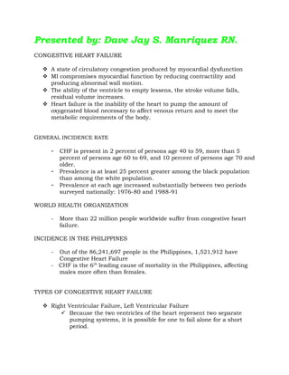
- 1. Presented by: Dave Jay S. Manriquez RN. CONGESTIVE HEART FAILURE A state of circulatory congestion produced by myocardial dysfunction MI compromises myocardial function by reducing contractility and producing abnormal wall motion. The ability of the ventricle to empty lessens, the stroke volume falls, residual volume increases. Heart failure is the inability of the heart to pump the amount of oxygenated blood necessary to affect venous return and to meet the metabolic requirements of the body. GENERAL INCIDENCE RATE - CHF is present in 2 percent of persons age 40 to 59, more than 5 percent of persons age 60 to 69, and 10 percent of persons age 70 and older. - Prevalence is at least 25 percent greater among the black population than among the white population. - Prevalence at each age increased substantially between two periods surveyed nationally: 1976-80 and 1988-91 WORLD HEALTH ORGANIZATION - More than 22 million people worldwide suffer from congestive heart failure. INCIDENCE IN THE PHILIPPINES - Out of the 86,241,697 people in the Philippines, 1,521,912 have Congestive Heart Failure - CHF is the 6th leading cause of mortality in the Philippines, affecting males more often than females. TYPES OF CONGESTIVE HEART FAILURE Right Ventricular Failure, Left Ventricular Failure Because the two ventricles of the heart represent two separate pumping systems, it is possible for one to fail alone for a short period.
- 2. Most heart failure begins with left ventricular failure and progresses to failure of both ventricles Acute pulmonary edema, a medical emergency, results from left ventricular failure. If pulmonary edema is not treated, death will occur from suffocation because the client literally drowns in his or her own fluids Forward Failure, Backward Failure In forward failure, an inadequate output of the affected ventricle causes decreased perfusion to vital signs. In backward failure, blood backs up behind the affected ventricle, causing increased pressure in the atrium behind the affected ventricle. Low Output, High Output In low-output failure, not enough cardiac output is available to meet the demands of the body. High-output failure occurs when a condition causes the heart to work harder to meet the demands of the body. Systolic Failure, Diastolic Failure Systolic failure leads to problems with contraction and ejection of blood. Diastolic failure leads to problems with the heart relaxing and filling with blood. CAUSES OF CONGESTIVE HEART FAILURE Intrinsic Myocardial Infarction Cardiomyopathy/myocarditis Congenital heart disease Valvular heart defects Percarditis/cardiac tamponade Extrinsic Systemic hypertension Chronic obstructive pulmonary disease Pulmonary embolism Anemia Thyrotoxicosis
- 3. Metabolic/respiratory acidosis Blood volume excess/polycythemia Drug toxicity Cardiac dysrhythmias Metabolic diseases PATHOPHYSIOLOGY (see separate page for pathophysiology) Congestive Heart Failure Left-sided CHF Right-sided CHF SIGNS AND SYMPTOMS OF CONGESTIVE HEART FAILURE Comparison of Left and Right CHF Left-sided Congestive Heart Failure Right-sided Congestive Heart Failure Signs of pulmonary congestion Dyspnea Tachypnea Crackles in the lungs Dry, hacking cough Paroxysmal nocturnal dyspnea Increased BP (from fluid volume excess) Dependent edema (legs and sacrum) Jugular vein distention Abdominal distention Hepatomegaly Splenomegaly Anorexia and nausea Nocturnal diuresis Swelling of the fingers and hands Increased BP (from fluid volume excess) *** Assessment Findings of Acute Pulmonary Edema • Severe dyspnea and orthopnea • Pallor • Tachycardia • Expectoration of large amounts of blood-tinged, frothy sputum • Wheezing and crackles on auscultation • Bubbling respirations • Acute anxiety, apprehension, restlessness • Profuse sweating • Cold, clammy skin • Cyanosis • Nasal flaring • Use of accessory breathing muscles
- 4. • Tachypnea • Hypocapnia, evidenced by muscle cramps, weakness, dizziness and paresthesias COLLABORATIVE MANAGEMENT Medications Digitalis Therapy Major therapy for CHF Has positive inotropic (strengthens force of cardiac contractility) and negative chronotropic effects (decreases heart rate) DOC: Lanoxin (Digoxin) Antidote for Toxicity: Digibind Nursing Responsibilities • Assess heart rate before administration; if below 60 bpm or above 120 bpm, withhold the drug. • Monitor serum potassium • Assess for signs of Digitalis toxicity - Bradycardia - GI manifestations (anorexia, nausea, vomiting and diarrhea) - Dysrhythmias - Altered visual perceptions - In males: gynecomastia, decreased libido and impotence Diuretic Therapy To decrease cardiac workload by reducing circulating volume and thereby reduce preload Commonly used diuretics: • Thiazides: Chlorthiazide (Diuril) • Loop diuretics: Furosemide (Lasix) • Potassium-Sparing: Spironolactone (Aldactone) Nursing Responsibilities • Assess for signs of hypokalemia when administering loop and thiazide diuretics. • Give potassium supplement and potassium-rich foods. • Administer early in the morning or early in the afternoon to prevent sleep pattern disturbance related to nocturia.
- 5. Vasodilators To decrease afterload by decreasing resistance to ventricular emptying Commonly used vasodilators: • Nitroprusside (Nipride) • Hydralazine (Apresoline) • Nifedipine • Captopril (Capoten) Other Drugs Sympathomimetics • Dopamine • Dobutamine TREATMENT Diet: sodium-restricted diet to prevent fluid excess Activity: balanced program of activity and rest Oxygen Therapy: to increase oxygen supply NURSING MANAGEMENT Providing Oxygenation Administer oxygen therapy per nasal cannula at 2-6 LPM as ordered Evaluate ABG analysis results Semi-Fowler’s or High-Fowler’s position to promote greater lung expansion Promoting Rest and Activity Bed rest or limited activity may be necessary during the acute phase Provide an overbed table close to the patient to allow resting the head and arms Use pillows for added support when in High-Fowler’s position Administer Diazepam (Valium) 2-10 mg 3-4x a day as ordered to allay apprehension Gradual ambulation is encouraged to prevent risk of venous thrombosis and embolism due to prolonged immobility Activities should progress through dangling, sitting up on a chair and then walking in increased distances under close supervision Assess for signs of activity intolerance (dyspnea, fatigue and increased pulse rate that does not stabilize readily)
- 6. Decreasing Anxiety Allow verbalization of feelings Identify strengths that can be used for coping Learn what can be done to decrease anxiety *** Anxiety causes increased breathlessness which may be perceived by the client as an increase in the severity of the heart failure and this in turn increases anxiety. Facilitating Fluid Balance Control of sodium intake Administer diuretics and digitalis as prescribed Monitor I and O, weight and V/S Dry phlebotomy (rotating tourniquets) Providing Skin Care Edematous skin is poorly nourished and susceptible to pressure sores Change position at frequent intervals Assess the sacral area regularly Use protective devices to prevent pressure sores Promoting Nutrition Provide bland, low-calorie, low-residue with vitamin supplement during acute phase Frequent small feedings minimize exertion and reduce gastroistestinal blood requirements There may be no need to severely restrict sodium intake of the client who receives diuretics. “No added salt” diet is prescribed. No processed foods in the diet. Promoting Elimination Advise to avoid straining at defecation which involves Valsalva manoeuvre. Administer laxative as ordered Encourage use of bedside commode Facilitating Learning Teach the client and his family about the disorder and self-care Monitor signs and symptoms of recurring CHF (weight gain, loss of appetite, dyspnea, orthopnea, edema of the legs, persistent cough and report these to the physician)
- 7. Avoid fatigue, balance rest with activity Observe prescribed sodium restrictions SFF rather than 3 large meals a day Take prescribed medications at regular basis Observe regular follow-up care as directed *** If acute pulmonary edema occurs in the client with CHF, the following are the appropriate management: High-fowler’s position Morphine Sulfate 10-15mg/IV as ordered to allay anxiety, reduce preload and afterlaod Oxygen therapy at 40-70% by nasal cannula or face mask Aminophylline IV to relieve bronchospasm, increase urinary output and increase cardiac output Rapid digitalization Diuretic therapy Dopamine and Dobutamine Monitor serum potassium. Diuresis may result to hypokalemia. PROGNOSIS - The prognosis depends on the patient's age, the severity of the heart failure, the severity of the underlying heart disease and other factors. - When congestive heart failure develops suddenly and has a treatable underlying cause, patients can sometimes return to normal heart function after treatment. - With appropriate treatment, even individuals who develop congestive heart failure as a result of long- standing heart disease can often enjoy many years of productive life.
- 8. PATHOPHYSIOLOGY OF CONGESTIVE HEART FAILURE CAUSES • Heart Damage • Ventricular Overload • Decreased Ventricular Contraction Tachycardia Ventricular Dilatation Myocardial Hypertrophy Decreased Cardiac Output Decreased Renal Perfusion Increased Sodium Restriction Increased Osmotic Pressure Increased ADH Increased Water Reabsorption Fluid Overload Edema
- 9. PATHOPHYSIOLOGY OF LEFT-SIDED CONGESTIVE HEART FAILURE CAUSES: • MI • HPN • Aortic Stenosis/ Insufficiency • Mitral Stenosis/ Insufficiency Reduced Myocardial Contractility Increased Cardiac Workload Decreased Diastolic Filling Obstruction of Left Atrial Emptying Increased Left Atrial Pressure Left-Sided Congestive Heart Failure Blood damns back into the pulmonary capillary bed Pressure of blood into the pulmonary capillary bed increasesFluid shifts into the intra- and interalveolar spacesPulmonary Edema Decreased stroke volume Decreased tissue perfusion Increased cellular hypoxiaSigns and symptoms of LSCHF Decreased blood flow to the kidneys
- 10. Signs and Symptoms of LSCHF: Dyspnea Paroxysmal Nocturnal Dyspnea Orthopnea Rales/Crackles Moist Cough Blood Tinged Frothy Sputum Wheezing/ Cardiac Asthma Dizziness Syncope Fatigue Weakness Anorexia Hypokalemia Clubbing of Fingers Polycythemia S3S4 Heart Sounds or Pulsus Alternans Decreased blood flow to the kidneys RAAS Stimulation Vasoconstriction and Reabsorption of Sodium and Water Increased ECF Volume Increased Total Blood Volume Increased Systemic BP
- 11. PATHOPHYSIOLOGY OF RIGHT-SIDED CONGESTIVE HEART FAILURE CAUSES: • LSCHF • Pulmonary Embolism • Right Ventricular Infarction • Congenital Septal Defects Reduced Myocardial Contractility Increased Cardiac Workload Decreased Diastolic Filling Obstruction of Right Atrial Emptying Increased Right Atrial Pressure Right-Sided Congestive Heart Failure Blood drums back from the RV to RA Increased Pressure in the Venous Circuit (Venous Back-up) Signs and Symptoms of RSCHF
- 12. ***The RSCHF which results from pulmonary disorders is called COR PULMONALE. Signs and Symptoms of RSCHF: Neck Vein Engorgement (Jugular Vein Distention) Hepatomegaly Portal Hypertension leading to Cardiac Cirrhosis Ascites Peripheral Edema (Pitting/ Dependent) Splenomegaly Jaundice Hemolytic Anemia Internal Hemorrhoids Leg Varicosities Weight Gain S3S4 Heart Sounds Elevated CVP Reading