These guidelines provide evidence-based guidance for managing dengue infection in adult patients. They were developed by a multidisciplinary group and reviewed by independent experts. The guidelines aim to improve diagnosis and appropriate care, identify severe cases, provide fluid management guidance, and reduce transmission. Key recommendations include monitoring patients for plasma leakage and bleeding, fluid resuscitation for shock, and blood product transfusion when needed. The guidelines are meant to guide but not replace clinical judgment based on each patient's situation.


![5
altered conscious level, clinical fluid accumulation, mucosal bleed or
tender enlarged liver are the clinical warning signs of severe dengue or high
possibility of rapid progression to shock.9, 10, 11
The patient can progress
rapidly to profound shock and death if prompt fluid resuscitation is not
instituted.
It is important to note that thrombocytopaenia and haemoconcentration
(evidenced by a raised haemotocrit (HCT) from baseline or a drop in HCT
after rehydration) are usually detectable before the subsidence of fever
and the onset of shock. Refer to 3.5.1 for further details. The HCT level
correlates well with plasma volume loss and disease severity. However, the
levels of HCT may be equivocal when there is frank haemorrhage, early and
excessive fluid replacement or untimely HCT determinations.
Leucopaenia with relative lymphocytosis, clotting abnormalities, elevation
of transminases [typically the level of aspartate aminotransaminase
(AST) is about 2-3 times the level of alanine aminotransaminase (ALT)],
hypoproteinaemia and hypoalbuminaemia are usually observed.3, 4, 5
iii. Recovery Phase
After 24-48 hours of defervescence, plasma leakage stops and is followed
by reabsorption of extravascular fluid. Patients’ general well being improves,
appetite returns, gastrointestinal symptoms abate, haemodynamic status
stabilises and diuresis ensues. Some patients may have a classical rash
of “isles of white in the sea of red”.3
Some may experience generalised
pruritus. Bradycardia and electrocardiographic changes are not uncommon
during this stage. It is important to note that during this phase, HCT level
stabilises or drops further due to haemodilution following reabsorption of
extravascular fluid. The recovery of platelet count is typically preceded by
recovery of white cell count (WCC).
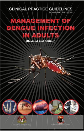
Figure 6 : CLINICAL COURSE OF DHF12
Note : Onset of defervescence usually occurs between day 3 to day 5 of illness
40
Viraemia
Course of
dengue illness FEBRILE CRITICAL RECOVERY
Shock / Bleeding Reabsorption / Fluid overloadDehydration
Days of illness
Temperature
Potential
clinical issues
Laboratory
changes
Serology
and virology
Platelet
Hematocrit
IgM/IgG
Organ Impairment
1 2 3 4 5 6 7 8 9 1 0
REVISED
JULY 2010](https://image.slidesharecdn.com/cpgdengue2010-150917092453-lva1-app6892/85/Cpg-dengue-2010-17-320.jpg)


![3 1
7.7 INTENSIVE CARE MANAGEMENT
The management of DSS in the intensive care unit (ICU) follows the general
principles of management of any critically ill patient in the ICU. However,
certain aspects which are of particular relevance to the management
of DSS are discussed here. There are several papers reviewing dengue
patients who were admitted to ICU. Several indications for ICU care were
observed as listed in the box below. 10, Level 8, 90, Level 8; 91, Level 8
Indications for referral to Intensive Care:
1. Recurrent or persistent shock
2. Requirement for respiratory support (non-invasive and invasive ventilation)
3. Significant bleeding
4. Encephalopathy or encephalitis
7.7.1 Indications for respiratory support (non-invasive and invasive ventilation)
The main objectives of respiratory support are to support pulmonary gas
exchange and to reduce the metabolic cost of breathing.
In general, respiratory support should be considered early in a patient’s
course of illness and should not be delayed until the need arises. The
decision to initiate respiratory support should be based on clinical
judgement that considers the entire clinical situation.92, Level 9
In patients with metabolic acidosis, respiratory support should be
considered despite the preservation of relatively normal arterial blood pH.
When PaCO2
is higher than expected to compensate for the acidosis, the
patient should be promptly intubated.
Formula to calculate the expected PaCO2
= 1.5 x [HCO3
-
] + 8±2 mmHg
In patients with encephalopathy and GCS of <9, intubation is often required
to protect the airway.93, Level 9 ; 94, Level 9
Indications for mechanical ventilation:
• Respiratory failure
• Severe metabolic acidosis
• Encephalopathy
7.7.2 Indications for haemodynamic support
In dengue, hypotension is usually due to plasma leakage or internal
bleeding. Fluid resuscitation is crucial and should be initiated first. However,
vasopressor (e.g. dopamine, noradrenaline) may be considered when a
mean arterial pressure is persistently <60 mmHg despite adequate fluid
resuscitation. 95, Level 9
CAUTION : While vasopressors increase the blood pressure, tissue hypoxia
may be further compromised by the vasoconstriction.](https://image.slidesharecdn.com/cpgdengue2010-150917092453-lva1-app6892/85/Cpg-dengue-2010-43-320.jpg)


![4 5
APPENDIX 1
CASE DEFINITION FOR DENGUE HAEMORRHAGIC FEVER
The following must ALL be present :
• Fever, or history of acute fever, lasting 2–7 days, occasionally biphasic.
• Haemorrhagic tendencies, evidenced by at least one of the following :
- a positive tourniquet test
- petechiae, ecchymoses or purpura
- bleeding from the mucosa, gastrointestinal tract, injection sites or other
locations
- haematemesis or melaena.
• Thrombocytopenia (100,000 cells per mm3
or less).
• Evidence of plasma leakage due to increased vascular permeability,
manifested by at least one of the following:
- a rise in the HCT equal to or greater than 20% above average for age,
sex and population;
- a drop in the HCT following volume-replacement treatment equal to
or greater than 20% or baseline;
- signs of plasma leakage such as pleural effusion, ascites and
hypoproteinaemia.
CASE DEFINITION FOR DENGUE SHOCK SYNDROME
All of the above four criteria for DHF must be present, plus evidence of
circulatory failure manifested by :
• Rapid and weak pulse, and
• Narrow pulse pressure [<20mmHg (2.7 kPa)]
OR manifested by :
• Hypotension for age, and
• Cold, clammy skin and restlessness.
Grade l : Fever accompanied by non-specific constitutional symptoms;
the only haemorrhagic manifestation is a positive tourniquet
test and / or easy bruising.
Grade ll : Spontaneous bleeding, in addition to the manifestations
of Grade l patients, usually in the form of skin or other
haemorrhages.
*Grade lll : Circulatory Failure manifested by a rapid, weak pulse and
narrowing of pulse pressure or hypotension with the presence
of cold, clammy skin and restlessness.
*Grade lV : Profound shock with undetectable blood pressure or pulse.
Note : * i. Grades III and IV are classified as Dengue Shock Syndrome
ii. The WHO classification is being reviewed and revised.](https://image.slidesharecdn.com/cpgdengue2010-150917092453-lva1-app6892/85/Cpg-dengue-2010-57-320.jpg)


![5 2
APPENDIX 7
SEARCH STRATEGY
The following free text terms or MeSH terms were used either singly or
in combination: Dengue Hemorrhagic Fever”[MeSH] OR “Dengue”[MeSH]
AND (Clinical Trial[ptyp] OR Meta-Analysis[ptyp] OR Randomized Controlled
Trial[ptyp]) AND English[lang] AND “humans”[MeSH Terms] (“Dengue
Hemorrhagic Fever/classification”[MeSH] OR “Dengue Hemorrhagic
Fever/diagnosis”[MeSH]) OR “Dengue/classification”[MeSH] OR “Dengue/
diagnosis”[MeSH]) AND English[lang] AND “humans”[MeSH Terms];
Dengue AND “viral Isolation”; Dengue AND “Laboratory test” ; Dengue
AND “Laboratory diagnosis” Dengue AND “serology diagnosis”; IgM AND
IgG; Dengue AND “molecular test”; (Dengue AND “Dengue Hemorrhagic
Fever”) AND “Fluid Management”; “Dengue Shock Syndrome “[MeSH] OR
“Dengue Hemorrhagic Fever”[MeSH]; (Dengue Hemorrhagic Fever) AND
(Fluid Management) AND Shock; dengue AND admission criteria AND
laboratory AND clinical; dengue AND admission criteria AND laboratory ;
dengue AND hospitalization; dengue AND hospitalization criteria; dengue
AND admission; Dengue AND viral load AND day; dengue & defervescence
; dengue AND viraemia AND duration; dengue AND viral load AND duration;
dengue AND “net screen”; dengue AND net AND hospital; dengue and
triage; dengue AND triage AND “emergency department”; dengue AND
ICU; Dengue AND ventilation; dengue AND “invasive procedures”; “dengue
infection in ICU”; [MESH] dengue or dengue hemorrhagic fever or dengue
shock syndrome-therapy and mortality.](https://image.slidesharecdn.com/cpgdengue2010-150917092453-lva1-app6892/85/Cpg-dengue-2010-64-320.jpg)


