Download to read offline


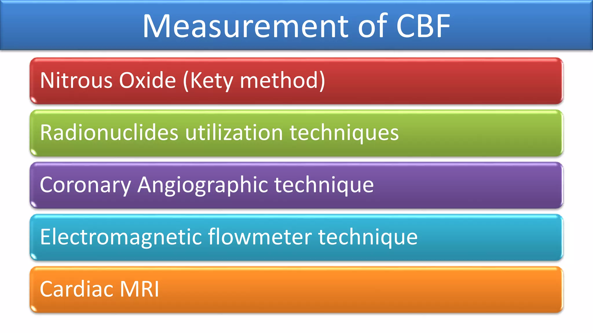


![Kety method
• Fick’s principle using 15 % N2O for 10 min
• Cerebral blood flow (CBF) =
– N2O taken by brain tissue per min
/ A-V diff of [N2O ]](https://image.slidesharecdn.com/coronarycirculation1903-200108133236/75/Coronary-circulation1903-31-2048.jpg)

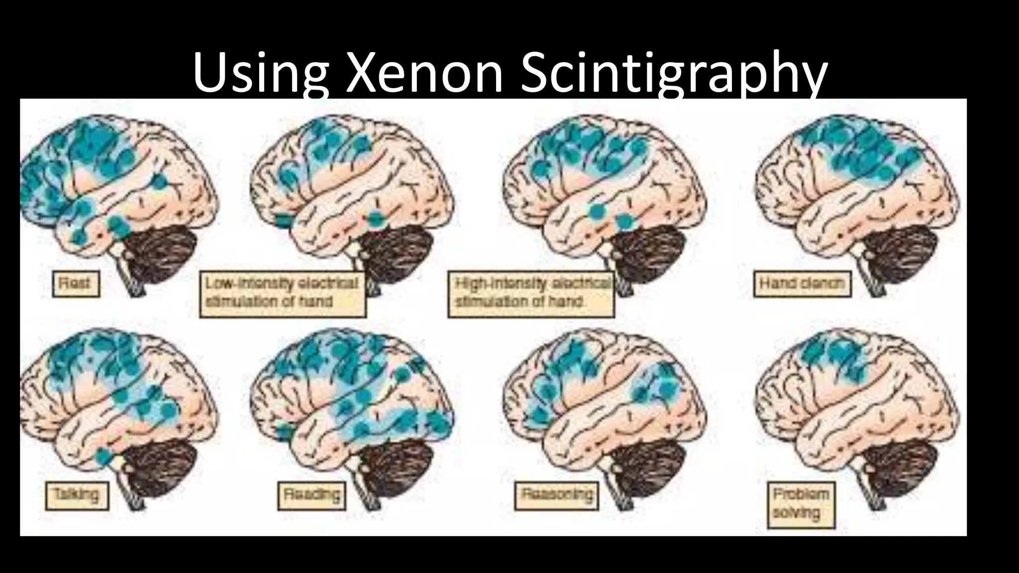

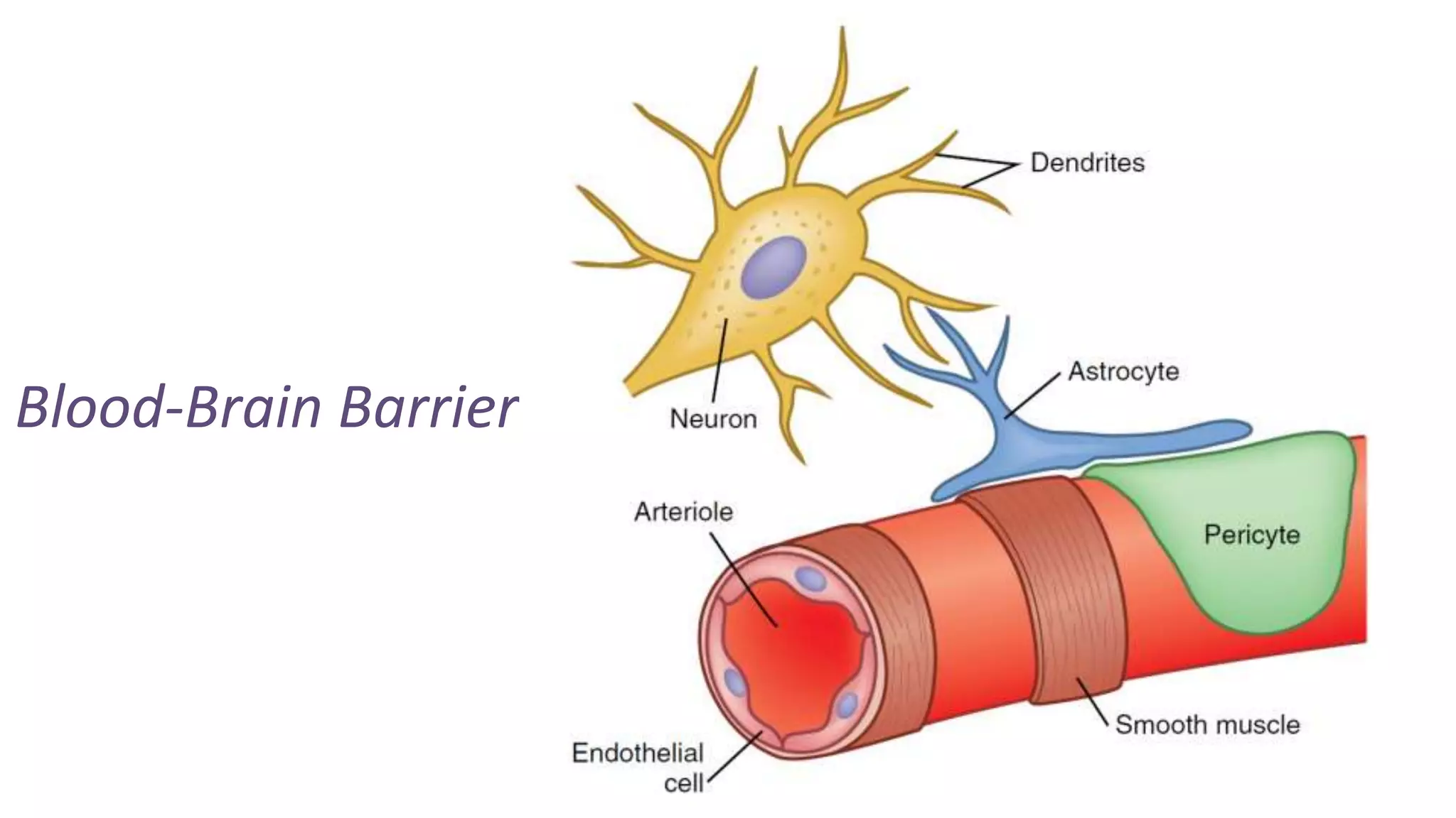
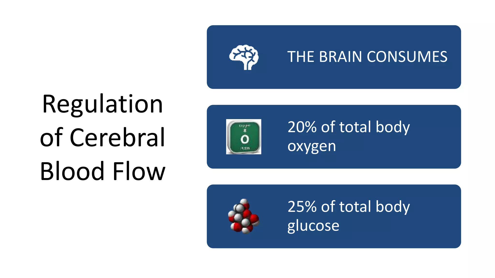

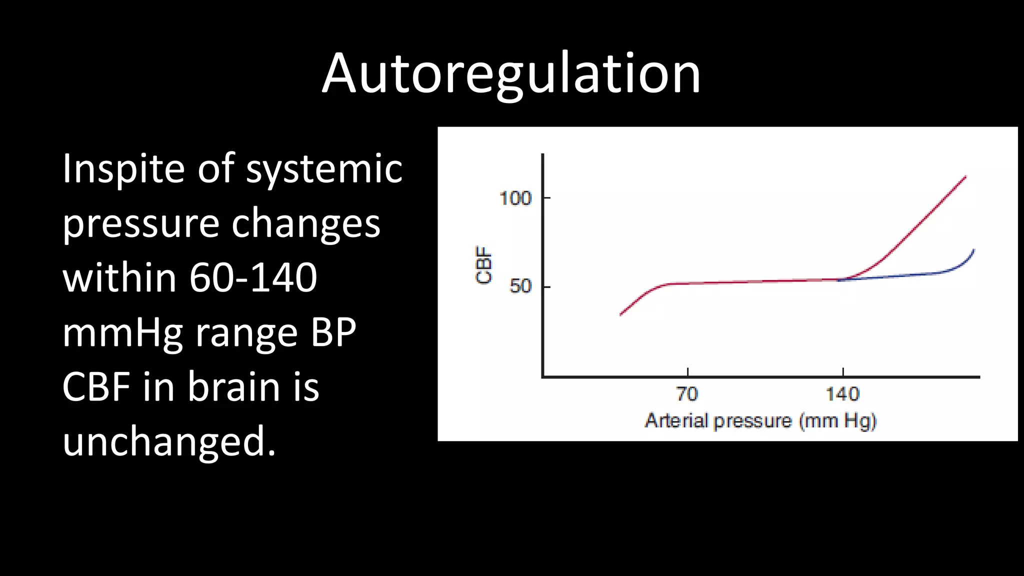


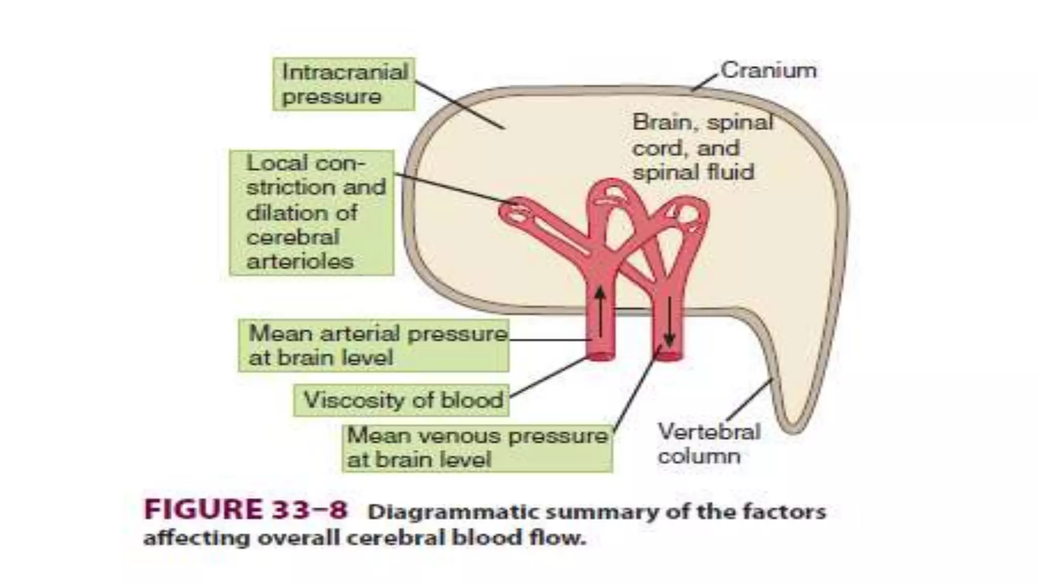

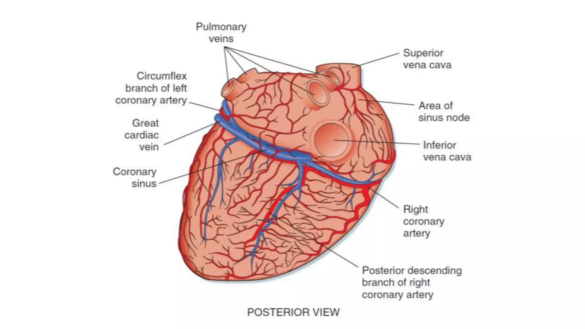
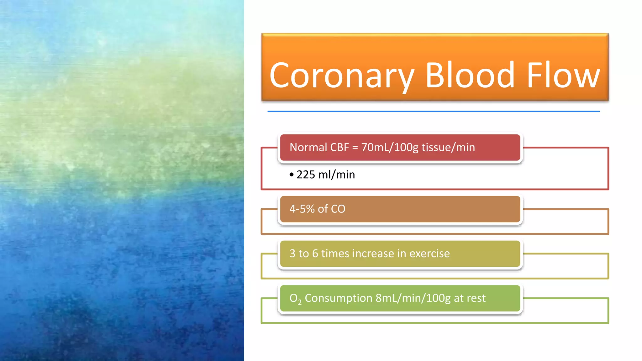
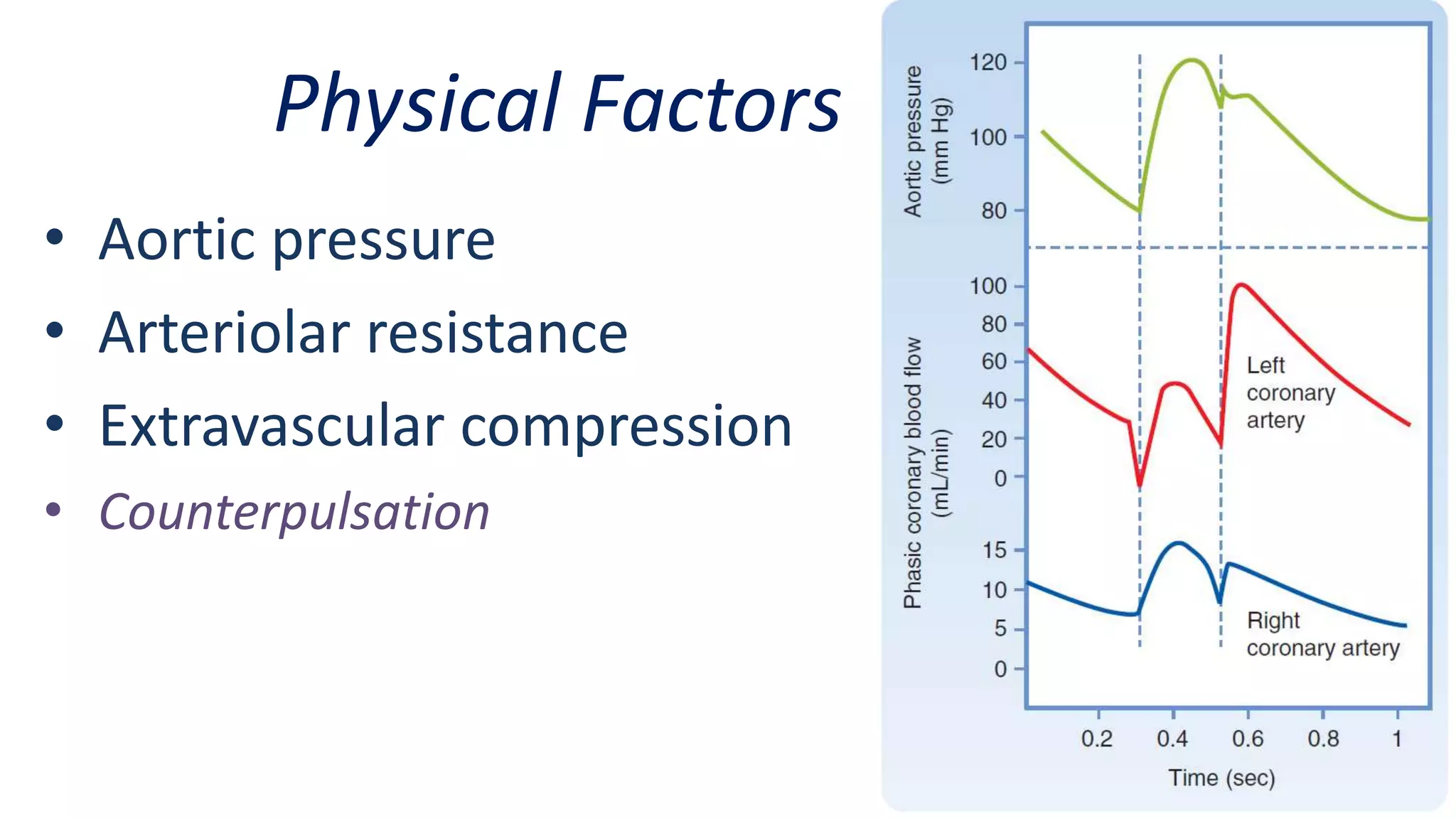
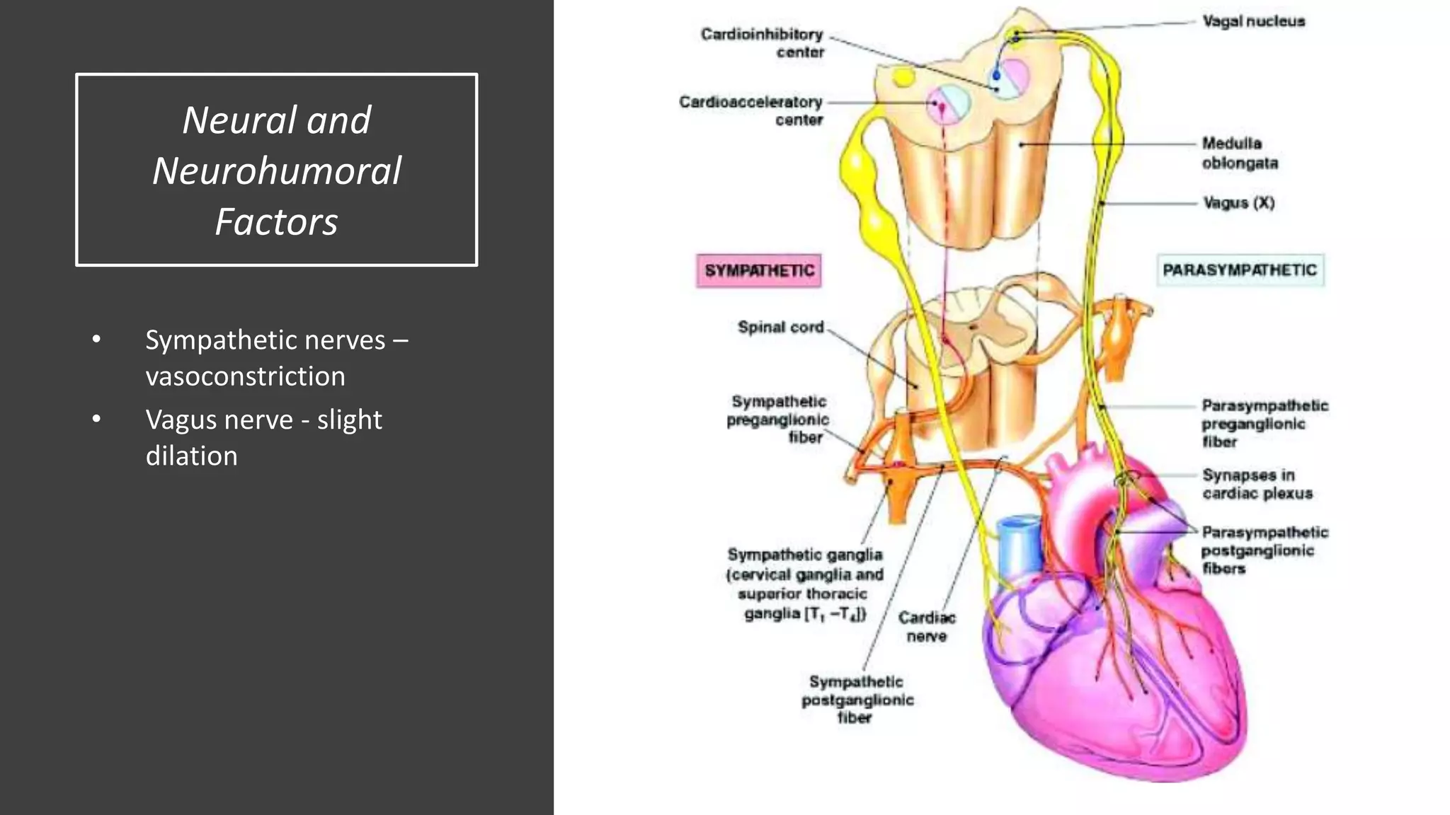
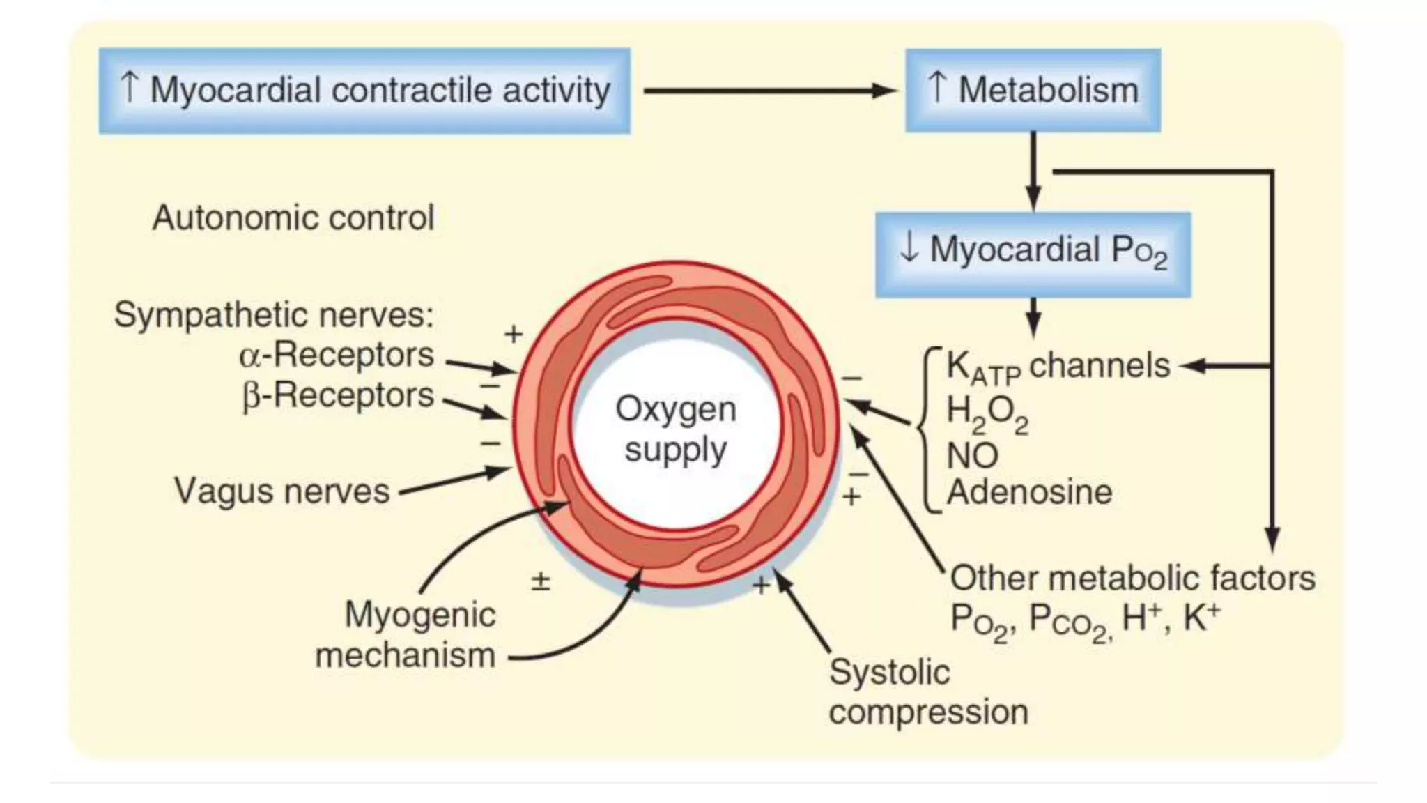
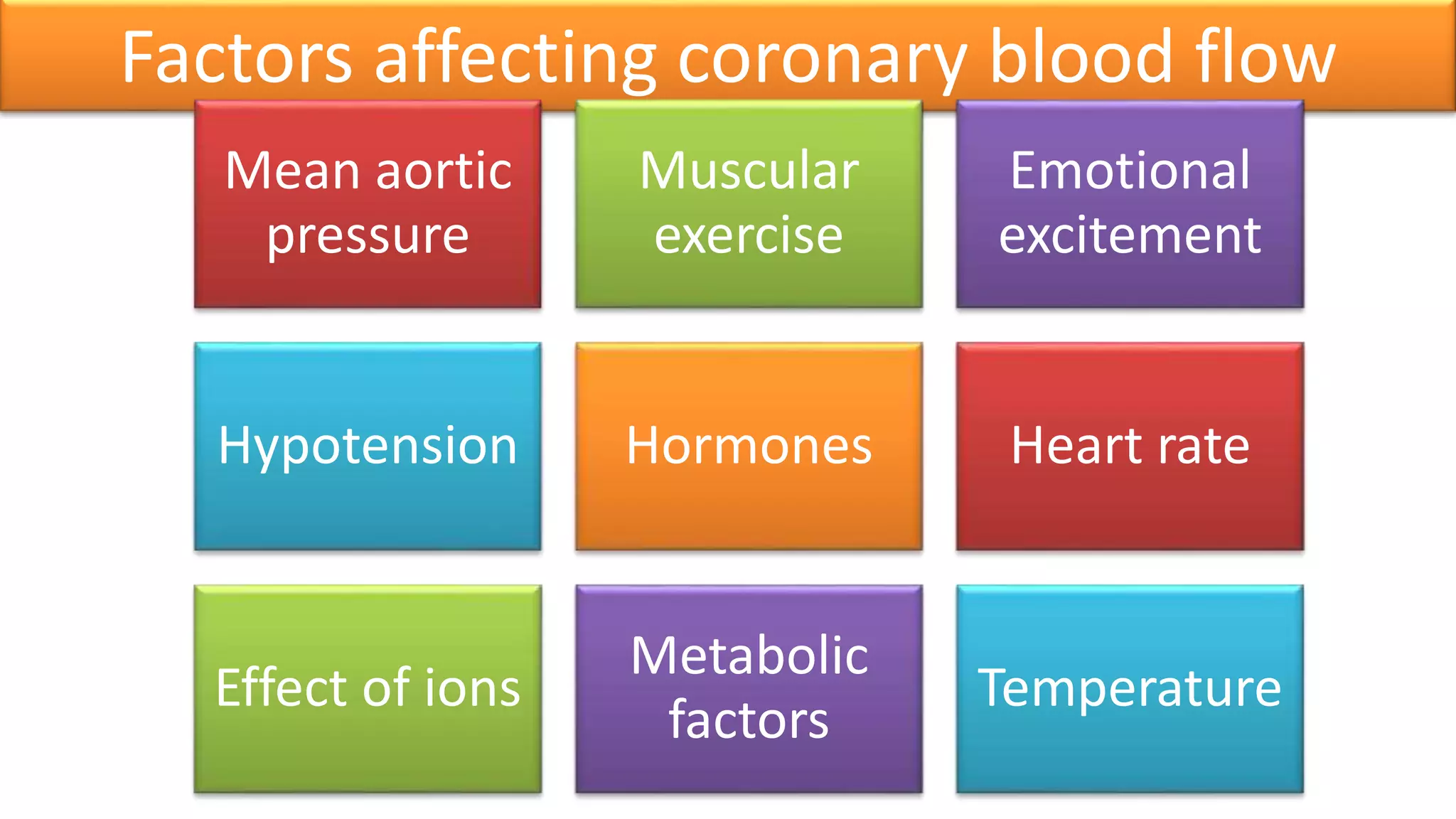
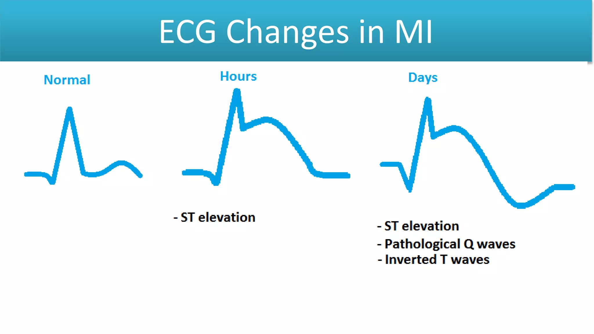
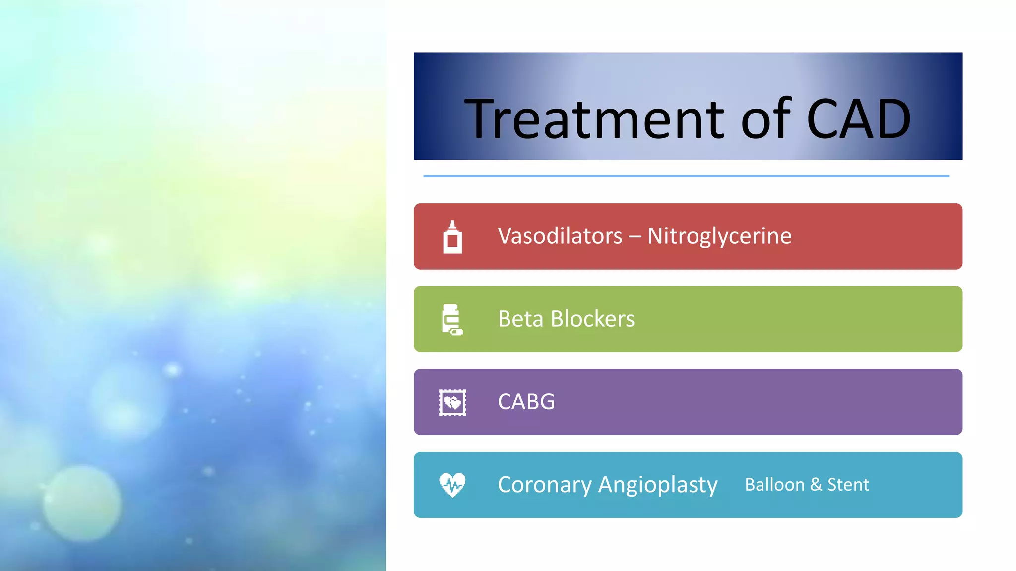
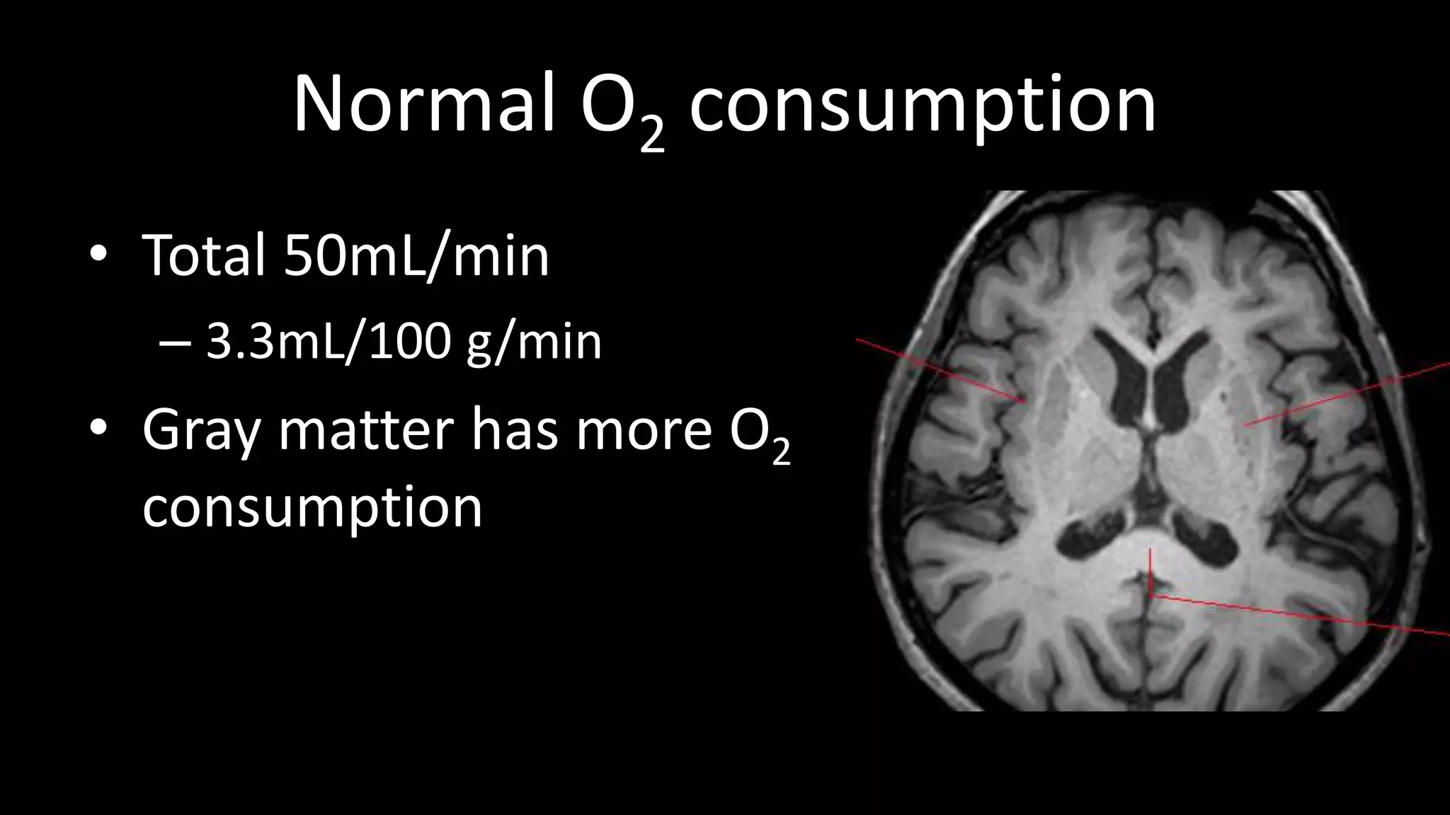
This document discusses coronary circulation and cerebral circulation. It provides details on factors that influence coronary blood flow such as physical factors like aortic pressure and metabolic factors. It also discusses measurement of coronary blood flow and conditions like angina pectoris, myocardial infarction, and their treatment. For cerebral circulation, it discusses measurement of cerebral blood flow using techniques like the Kety method and imaging modalities. It also discusses regulation of cerebral blood flow including autoregulation and the Monro-Kellie doctrine.


![Anaesthesia for intracranial aneurysm clipping[1].pptx](https://cdn.slidesharecdn.com/ss_thumbnails/anaesthesiaforintracranialaneurysmclipping1-250918124339-735bae7b-thumbnail.jpg?width=640&height=640&fit=bounds)


![Lec61[1]](https://cdn.slidesharecdn.com/ss_thumbnails/lec611-100501100520-phpapp02-thumbnail.jpg?width=640&height=640&fit=bounds)


